
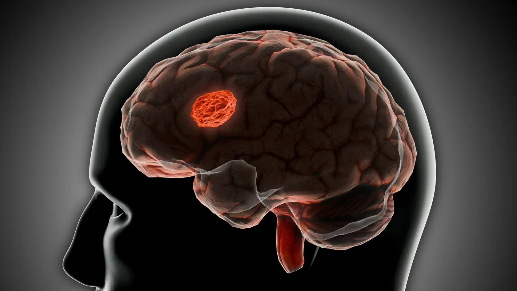
Epilepsy Surgery
Three randomized trials found that surgery stops seizures far more often than staying on medication. The average patient is still referred after about twenty years. What the evidence says, and when to ask.
About This Department
Three randomized trials have compared brain surgery against carrying on with medication for epilepsy that drugs have failed to control, and all three found the same thing by a wide margin. In the largest, 77 percent of children who had surgery were free of seizures a year later against 7 percent of those who continued on tablets. The authors of one of the adult trials also wrote that patients are typically referred for surgery after twenty years of seizures, often too late to avoid lasting disability. Those two facts sit uneasily together, and this page is about the gap between them. Nothing here argues that everyone with epilepsy should have brain surgery, since most people with epilepsy are controlled by their first or second medicine and will never need to think about any of this. The argument is narrower and it is about the minority for whom the drugs have stopped working, where the evidence says one thing and ordinary practice does another.
Free consultation
Send your records and find out whether an evaluation is worth doing
Send a list of every antiseizure medicine tried with the dose reached and why each was stopped, a description of what the seizures look like and how often they happen, every brain scan with the image files as well as the reports, any electroencephalogram reports including video monitoring, and any neuropsychology assessment. An epileptologist and a neurosurgeon review the file together and tell you whether your epilepsy meets the definition of drug resistant, whether a surgical evaluation is likely to find something treatable, and what that evaluation would involve. No fee, no obligation, and a coordinator replies in your own language, usually within the same working day.
What the trials found
Surgery is difficult to test in a randomized trial because neither the surgeon nor the patient can be blinded and because families understandably resist being allocated to a waiting list, and three teams did it anyway, and the results are among the largest treatment effects reported anywhere in neurology.
Wide table. Swipe or drag it sideways on a narrow screen, because it scrolls instead of shrinking.
| Trial and who was in it | Result | What has to be said alongside it |
|---|---|---|
| Canada, 80 adults with temporal lobe epilepsy | At one year, 58 percent of the surgical group were free of seizures that impaired awareness against 8 percent of the medical group. | One year only, single center, 80 patients. Four surgical patients had adverse effects, and the one death in the trial was in the medical group. |
| United States, 38 adults referred early | In the second year, none of the 23 on medication alone were seizure free against 11 of the 15 who had surgery. | Planned for 200 patients and stopped at 38 for slow recruitment. The quality of life endpoint failed on its main analysis. |
| India, 116 children and adolescents | At twelve months, 44 of 57 in the surgical group were seizure free, which is 77 percent, against 4 of 59 in the medical group. | Serious adverse events occurred in 33 percent and weakness of one side in 26 percent. Measured intelligence did not improve. |
| What the three share | Every one compared surgery against staying on medication, and every one favored surgery decisively. | All three were single center or nearly so, all had short primary endpoints, and none was blinded because none could be. |
| What none of them shows | None followed patients long enough to say what happens at five or ten years. That comes from cohorts instead. | And none demonstrated that surgery reduces the risk of dying, which is a claim this page does not make. |
When drugs stop working
Referral rests on a second body of evidence, which is that once two properly chosen medicines have failed, the chance a third or fourth will succeed is small. One clinic in Glasgow followed newly treated patients for thirty years and published the numbers twice, in 2000 and again in 2018, with more than a dozen new drugs licensed in between, and the second report reached the same conclusion as the first.
Another wide one. Swipe or drag it sideways on a narrow screen, because it scrolls instead of shrinking.
| Which regimen | Chance it works | Note |
|---|---|---|
| The first medicine | 906 of 1,795 patients, or 50.5 percent | Half of everyone with newly diagnosed epilepsy is controlled by the first drug they are given. |
| The second regimen | Adds 11.6 percent | Still worth trying, and the return has already fallen to less than a quarter of the first attempt. |
| The third regimen | Adds 4.4 percent | This is the point at which most guidelines say the word surgery should have been said out loud. |
| Every subsequent regimen combined | Adds 2.12 percent in total | Not zero, and not a plan. Each additional regimen carried 1.73 times the odds of not responding. |
| If the first drug failed because it did not work | 11 percent later became seizure free | From the earlier report. Failing because of side effects or an allergy is a different and much more hopeful situation. |
| What thirty years changed | Nothing, on this measure | Despite many new medicines, the authors reported that overall outcomes in newly diagnosed epilepsy have not improved. |
Who the operation is for
The definition, and the number two
An international task force settled this in 2010 and the definition has not moved since. Drug resistant epilepsy is the failure of adequate trials of two tolerated, appropriately chosen and correctly used antiseizure drug schedules, whether given singly or in combination, to achieve sustained freedom from seizures. Every word in that sentence is doing work. Adequate means the dose was pushed to the point of benefit or intolerance instead of being abandoned early. Appropriately chosen means the drug suits the seizure type, since the wrong medicine failing proves nothing. Tolerated means you were actually able to take it. If two drugs have failed under those conditions, you meet the definition, and the next step is an assessment instead of a third prescription. Meeting the definition does not mean you will be offered an operation, and it does mean the question should now be asked by somebody qualified to answer it, which in practice means a center that runs a full presurgical evaluation and sees enough cases to be good at it.
What surgery is trying to do
Most epilepsy surgery removes or disconnects the piece of brain where the seizures start. That only works if the seizures reliably start in one place, if that place can be identified, and if removing it will not cost more than the seizures do. The commonest operation removes the front part of one temporal lobe, and the commonest reason for it is scarring of the hippocampus on that side. Other operations remove a small tumor or a patch of malformed cortex, disconnect one whole hemisphere in a child whose seizures come from a hemisphere that is already badly damaged, or cut the bundle joining the two halves of the brain to stop drop attacks spreading. Those last two are not attempts at a cure but at a specific improvement, and the honest way to describe them is that they trade one problem for a smaller one. A surgeon who does not distinguish between an operation aimed at stopping seizures altogether and one aimed at stopping a particular kind of seizure has left out the most important part of the conversation.
Who it is not for
Generalized epilepsies that involve the whole brain at once are not resectable, and neither are epilepsies whose seizures arise independently from several places. Someone whose seizures start in an area responsible for language or movement may be offered a smaller operation, a device, or nothing surgical at all. And a meaningful number of people who could have surgery decide against it, which is a legitimate choice and not a failure of information. At one large German center, 44.9 percent of evaluated adults who did not have an operation had declined it themselves, and that figure rose to 53.2 percent in the last four years of the series. That is a single center and the trend may not hold elsewhere, and it is a reminder that the treatment gap is not entirely a failure of referral. Some of it is people who were told everything and decided that living with their seizures was preferable to an operation on their brain, which is a conclusion nobody else is entitled to overrule.
The twenty years
Why the wait costs you
If waiting were merely frustrating it would be a quality-of-life argument, but the stronger claim is that the wait itself lowers the chance the operation works, and three separate analyses in three different populations point the same way.
- In the 664-patient temporal lobectomy series. The risk of seizures coming back rose steadily with each band of epilepsy duration, from five to ten years, to ten to fifteen years, to more than fifteen. The authors reported hazard values without confidence intervals so the size of the effect cannot be quoted, and the direction was consistent and it was their headline finding. Their conclusion was that lost years translate into poor seizure outcome and that all drug-resistant temporal lobe epilepsy should be referred at the earliest opportunity.
- In children having surgery outside the temporal lobe. Pooling 36 studies and 1,259 children, operating at seven years of epilepsy or less instead of longer carried odds of 1.52 for seizure freedom, with a confidence interval of 1.07 to 2.14. Overall, 704 of the 1,259 children, or 56 percent, became free of seizures.
- In patients whose scan showed nothing. A meta-analysis of the hardest group of all, temporal lobe epilepsy with a normal magnetic resonance scan, found shorter epilepsy duration predicted seizure freedom with odds of 2.57 and a confidence interval of 1.21 to 5.47. Duration mattered even where there was no visible lesion to remove.
- Three populations, one direction. Adults and children, temporal and extratemporal, lesional and not. None of these is a randomized comparison of early against late surgery, so the effect could partly reflect that people with milder epilepsy get referred sooner. The consistency across such different groups is the reason the field takes it seriously.
- The one trial that tried to test it directly. The American early-surgery trial was designed precisely to answer this and enrolled 38 patients against a target of 200. It was stopped for slow recruitment, which is itself evidence about the problem, since the investigators could not find enough people who had been referred within two years of their drugs failing.
How long it lasts
Randomized trials measured one and two years. What most people actually want to know is whether the seizures stay away, and for that the best source is a London cohort of 615 adults followed annually for a median of eight years and as long as nineteen.
Apart from simple partial seizures, meaning brief auras without loss of awareness, 52 percent were free of seizures at five years with a confidence interval of 48 to 56 percent, and 47 percent at ten years with a confidence interval of 42 to 51. The authors described their own figures as providing realistic expectations, which is a polite way of noting that they sit below the short-term rates usually quoted. Resections outside the temporal lobe carried twice the hazard of recurrence compared with anterior temporal resection, with a confidence interval running from 1.1 to 3.6. Those figures are lower than the ones most people carry away from a first consultation, and they are the ones to plan around, because a realistic number that turns out to be beatable is a much better starting point than an optimistic one that does not. Half of a group of people whose seizures had defeated every drug available to them were free of seizures a decade later, and that is a remarkable result stated plainly.
Two more findings from that cohort deserve to be better known, and the first is that the pattern is not a simple decline, since relapse became less likely the longer someone stayed seizure free, and remission became less likely the longer seizures continued. The second is that late remission happened, and in 18 of 93 such cases it was associated with starting a medicine the patient had not previously tried. Surgery and drugs are not alternatives in the way patients often assume, and a good result frequently means both.
One caution about that cohort. Its 615 patients include hemispherectomies and palliative operations alongside 497 anterior temporal resections, so the 52 percent figure describes a mixed group instead of temporal lobectomy alone. Its definition of seizure freedom also allows continuing auras, which is more permissive than the strictest published standard. Comparing it directly against a series that used a stricter definition will produce a difference that is entirely an artifact of the definitions.
What the workup asks
The evaluation, step by step
When the scan looks normal
A normal scan lowers the odds without closing the door
Pooling 40 studies covering 697 patients with no visible lesion and 2,860 with one, the odds of becoming seizure free were 2.5 times higher when a lesion was present, with a confidence interval of 2.1 to 3.0. The gap was similar for temporal surgery at 2.7 and for surgery elsewhere at 2.9. Those are ratios instead of rates, and the analysis does not report what proportion of patients with a normal scan became seizure free, so no percentage for that group appears on this page. What it does establish is that a normal scan makes the operation harder and does not make it pointless. The comparison also has a subtlety worth knowing, since the lesional group in that analysis was defined by a lesion on either the scan or the tissue examined after surgery, which means some of those patients also had a normal scan beforehand and only turned out to have a lesion afterward. Your own question is narrower than the one that analysis answers.
What predicts a good result anyway
Within the group whose scan shows nothing, a meta-analysis found that an electrical recording localizing cleanly to one temporal lobe predicted seizure freedom strongly, with odds of 3.89 during seizures and 3.38 between them, and that shorter epilepsy duration predicted it too. The same analysis found the metabolic scan was not predictive in this group, with odds of 2.11 and a confidence interval from 0.95 to 4.65 that includes no effect. That is an uncomfortable finding for a test often described as the answer to a normal scan, and it is worth knowing before anyone tells you a positive metabolic scan settles the matter.
Recording from inside
Two ways to do it
When scalp recording cannot settle where seizures start, electrodes go inside the head for a week or two while you wait for seizures again, and one method opens the skull and lays sheets of electrodes on the brain surface. The other passes thin electrodes through small holes along planned trajectories, sampling deep structures that surface sheets cannot reach. Both are followed by a second operation, either to remove the tissue identified or simply to take the electrodes out.
What the international registry found
An international registry compared the two in 1,468 patients across ten centers in seven countries, matched for the characteristics that drive the choice between them. Complications, defined as infection, symptomatic bleeding or a permanent neurological deficit, occurred in 9.6 percent after surface sheets against 3.3 percent after depth electrodes. Among patients who went on to a resection, seizure freedom was 55 percent after depth-electrode evaluation against 41 percent after surface sheets, with odds of 1.66 and a confidence interval of 1.21 to 2.26. Pulling the other way, surface sheets were more likely to lead to an operation at all, with odds of 1.4. This was a registry and not a randomized trial, the two methods are chosen for different patients, and the authors called it the highest feasible level of evidence for this question, which is an honest way of saying no trial is coming. Both methods are safe in absolute terms and the difference between them is real.
The risks, in numbers
A systematic review of complications in epilepsy surgery gathered the figures that follow, and it is worth knowing how its authors defined their terms before reading them. Minor complications resolved completely within three months of surgery. Major complications persisted beyond that. Without those definitions the words mean nothing.
After resective surgery, minor medical complications occurred in 5.1 percent of patients and major ones in 1.5 percent, with leakage of spinal fluid the commonest. Major neurological complications occurred in 4.7 percent. Perioperative mortality was 0.4 percent for temporal lobe surgery and 1.2 percent for surgery elsewhere in the brain. The authors' summary was that most complications are minor or temporary, that major permanent neurological complications remain uncommon, and that death from epilepsy surgery in the modern era is rare.
Set against that, the untreated condition is not safe either, and in a cohort of people with drug-resistant epilepsy followed for 938 device-years, overall mortality ran at 6.9 deaths per thousand person-years and sudden unexpected death in epilepsy at 2.0 per thousand person-years. Nobody has shown that surgery reduces those figures, because no study has compared mortality between people who became seizure free after an operation and people who did not, so this page does not claim it. What can be said is that uncontrolled seizures carry a measurable risk of dying and that the operation carries a smaller and better-characterized one. Sudden unexpected death in epilepsy is the term for a death that follows a seizure with no other explanation found afterward, it is the commonest cause of premature death in people whose seizures are not controlled, and the risk falls as seizure control improves whatever the reason for that improvement. Nobody has demonstrated that surgery specifically produces that fall.
What can go wrong
Specific risks depend almost entirely on which part of the brain is being operated on, which is why a general consent conversation is less useful than a specific one.
Wide again. Drag the table sideways on a narrow screen, because it scrolls instead of shrinking.
| Risk | How often | Who it applies to |
|---|---|---|
| Death around the operation | 0.4 percent, or 1.2 percent | The first for temporal lobe surgery, the second for resections elsewhere in the brain. |
| A lasting neurological deficit | 4.7 percent | Anything still present beyond three months. The nature of it depends on what was removed. |
| A lasting visual field defect | 2.1 percent classed as major | Far more people have a defect detectable on formal testing. See the section on vision below. |
| Weakness of one side | 26 percent in the pediatric trial | Children having large resections and hemispheric disconnections. Anticipated and related to the region removed. |
| Complications of electrode monitoring | 7.7 percent minor, 0.6 percent major | Only those who need recording from inside the head, which is a minority of candidates. |
| Memory or word-finding difficulty | No pooled figure exists | Mainly surgery on the language-dominant temporal lobe. The individual studies are discussed below. |
The days around surgery
Memory and words
The characteristic risk of dominant-side surgery
Removing part of the temporal lobe on the side that handles language can affect verbal memory and the ability to retrieve names. In a study comparing open resection against laser ablation, 21 of 22 patients who had open surgery on the language-dominant side declined on at least one naming task afterward, and 11 of 17 who had it on the other side declined at recognizing famous faces. That study was small, was not randomized, and came from a group with an interest in the laser technique, and even allowing for all of that, 21 of 22 is a figure worth carrying into a consultation. In the American early-surgery trial, memory decline on a word-list test occurred in 4 of the 11 surgical patients assessed, which the authors reported alongside an explicit warning that their sample was too small for conclusions.
Why no single percentage appears here
No pooled estimate of verbal memory or naming decline after dominant temporal resection was available for this page, and inventing one from two small studies would be worse than saying so. What the evidence supports is a description instead of a number. Word-finding difficulty and verbal memory decline are the characteristic costs of operating on the language-dominant temporal lobe, they are common enough that a substantial proportion of patients show them on formal testing, and they are the specific reason the preoperative neuropsychology exists, which is also why a center that proposes operating on the language-dominant side without formal testing beforehand has skipped the step that would have told you what you were risking. Ask your own team for their own figures, and treat a center that cannot give you any as having answered the question.
Vision
Fibers carrying vision from the upper part of the visual field sweep forward through the temporal lobe before turning back toward the visual cortex, which puts them directly in the path of a temporal resection. The result is a defect in the upper outer quadrant of vision on the opposite side. How often that happens depends entirely on how hard you look for it.
Among 54 patients tested with formal perimetry two years after mesial temporal surgery, 49 had a visual field defect, which is 91 percent. Not one of them reported any visual change, and not one had an abnormal bedside examination. The severity of the defect was not associated with whether they were driving three years later.
Place that beside the 2.1 percent rate of major visual field defects from the complications review and the two figures look contradictory, and they are measuring different things. One counts any deviation on a sensitive machine test, the other counts deficits that persist and matter clinically. Both are true at once, and the practical summary is that almost everyone who has this operation acquires a measurable blind area that almost nobody notices. Driving regulations differ by country and are the reason formal testing is done at all, so ask specifically what your own licensing authority requires and get the perimetry result in writing before you travel home. The other point that follows from this is about consent, since a defect that ninety percent of patients acquire and almost none notice belongs in the conversation as an expected consequence and not as a rare complication, and describing it the second way makes the consent process less honest rather than more reassuring.
Questions to take with you
The first four settle whether you should be having this conversation at all. The rest settle what you are being offered.
- Have two appropriately chosen medicines been tried at adequate doses, and if so, why has surgery not been raised before now?
- How many years have passed since my seizures became resistant to drugs?
- Do my seizures appear to start in one place, and what is the evidence for that?
- Has my scan been repeated using an epilepsy protocol, or only reported from a routine scan?
- Which structures would be removed, and what does each of them do?
- Is the target on my language-dominant side, and how has that been established?
- What are this center's own figures for seizure freedom and for complications in operations like mine?
- Will electrodes need to go inside my head first, and if so, by which method and why that one?
- Does a multidisciplinary meeting review my case, and can I have its conclusion in writing?
- If I do become seizure free, when would reducing my medication be discussed?
Coming off the tablets
When resection is impossible
The devices reduce seizures and rarely stop them
If seizures start in more than one place, or in a place that cannot be removed, three implanted devices exist and all three work in the same limited sense. Stimulation of a deep thalamic nucleus was tested in 110 patients, where the blinded phase showed a median seizure reduction of 40.4 percent against 14.5 percent in the unstimulated group, and at two years 14 of the 110 had been seizure free for at least six months. A responsive device that detects and interrupts seizures at their source reduced seizures by 37.9 percent against 17.3 percent for a sham device over twelve blinded weeks in 191 adults, and over nine open-label years 47 of 256 patients, or 18.4 percent, achieved at least a year of seizure freedom. Vagus nerve stimulation, tested against low-intensity stimulation rather than against nothing, produced a 28 percent reduction against 15 percent, and across a manufacturer registry of 5,554 patients and a literature review of 2,869 more, roughly 8 percent became seizure free.
Read those numbers carefully
Almost every impressive long-term figure for these devices comes from open-label follow-up with no control group, and the blinded phases are short. An independent Norwegian trial implanted 18 patients and found no significant difference between stimulated and unstimulated groups at the end of its six-month blinded period, with its authors writing that their results were not as encouraging as those reported from many other, mainly unblinded and open studies. Several of the long-term device papers include employees of the manufacturer among their authors, which does not make them wrong and does mean the reader should know. Two of the three devices also come with a practical burden that rarely appears in the summaries, which is that they need programming visits, battery replacements over the years and a specialist willing to adjust them, and a device that nobody in your own country knows how to manage is worth considerably less than the trial figures suggest.
The comparison that matters
When a resection is possible, roughly half of well-selected patients stop having seizures altogether. When it is not, the devices commonly halve the seizures in half to three-quarters of patients and seizure freedom is uncommon, running from about 8 percent with vagus nerve stimulation to about 18 percent with the responsive device. These are not head-to-head figures and they never will be, since device patients are by definition those in whom resection was ruled out, and the definitions of seizure freedom differ between the studies. The ordering is nonetheless the right way round, and it is the reason a surgical evaluation should come before a device is discussed. A device offered without a preceding evaluation is a decision to stop looking for the operation that might have worked, and the only way to know whether that decision is right is to have done the looking first.
What to send us
Whether you are even a candidate is answerable from documents before anyone travels. This is the list that makes that possible.
- Every medicine you have tried, with the dose and the reason it stopped. This is the single most important document and it is the one almost nobody has, and whether a drug failed because it did not work, because you could not tolerate it, or because it was never pushed to an adequate dose changes everything about what happens next.
- A description of the seizures, ideally with video. What happens first, what a witness sees, how long it lasts, how often. A phone video of a typical seizure is worth more than several paragraphs of description, and families almost always have one.
- Every brain scan, as image files. Send the actual images and not only the reports. A scan reported as normal is repeated here under an epilepsy protocol, and that repeat is frequently where the answer is found.
- All electroencephalogram reports, especially any video monitoring. If you have had a monitoring admission elsewhere, the report matters and the raw recording matters more. Ask whether it can be released on a disc.
- Any neuropsychology assessment, however old. An assessment from five years ago is not out of date. It is a second point in time, and having two lets anyone see the direction of travel.
Coming to Istanbul
Epilepsy surgery is unusual among the operations described on this site because the assessment is longer than the operation and cannot be compressed. Nobody can tell you from abroad whether you are a candidate, and the monitoring admission that answers that question takes days of inpatient time. Plan around the evaluation instead of around the surgery, and expect two separate trips.
This table is wide too. Swipe it sideways on a narrow screen, because it scrolls instead of shrinking.
| Stage | Length of stay | What comes out of it |
|---|---|---|
| Review of your documents | No travel | Whether your epilepsy meets the definition of drug resistant, and whether an evaluation is worth the journey. |
| The evaluation admission | Ten to fourteen days | Scan, video monitoring until enough seizures are recorded, neuropsychology, and a multidisciplinary conclusion. |
| If electrodes are needed inside | A further two to three weeks | An implantation, a week or two of recording, and then either a resection or removal of the electrodes. |
| The operation itself | Seven to ten days | Three to five hours in theater, a night under close observation, and home within about a week. |
| Before you fly | Allow a few clear days | A wound check, the formal visual field test, and a written medication plan naming every drug and dose. |
| Follow up after you return home | Years, and measured in years | Repeat neuropsychology at six to twelve months, a named contact here, and a local neurologist who has the operative note. |
Epilepsy surgery FAQ
When should surgery be considered?
How likely is it to stop my seizures?
Is it dangerous?
Will it affect my memory?
What if my scan is normal?
Will I be able to stop my medication?
What if the seizures cannot be traced to one place?
How long would I need to be in Turkey?
References
- Wiebe S, Blume WT, Girvin JP, Eliasziw M. A randomized, controlled trial of surgery for temporal-lobe epilepsy. The New England Journal of Medicine. 2001;345(5):311-318.
- Engel J, McDermott MP, Wiebe S, Langfitt JT, Stern JM, Dewar S, Sperling MR, Gardiner I, Erba G, Fried I, Jacobs M, Vinters HV, Mintzer S, Kieburtz K. Early surgical therapy for drug-resistant temporal lobe epilepsy, a randomized trial. JAMA. 2012;307(9):922-930.
- Dwivedi R, Ramanujam B, Chandra PS, Sapra S, Gulati S, Kalaivani M, Garg A, Bal CS, Tripathi Madhavi, Dwivedi SN, Sagar R, Sarkar C, Tripathi Manjari. Surgery for drug-resistant epilepsy in children. The New England Journal of Medicine. 2017;377(17):1639-1647.
- Kwan P, Brodie MJ. Early identification of refractory epilepsy. The New England Journal of Medicine. 2000;342(5):314-319.
- Chen Z, Brodie MJ, Liew D, Kwan P. Treatment outcomes in patients with newly diagnosed epilepsy treated with established and new antiepileptic drugs, a 30-year longitudinal cohort study. JAMA Neurology. 2018;75(3):279-286.
- Kwan P, Arzimanoglou A, Berg AT, Brodie MJ, Hauser WA, Mathern G, Moshe SL, Perucca E, Wiebe S, French J. Definition of drug resistant epilepsy, consensus proposal by the ad hoc Task Force of the ILAE Commission on Therapeutic Strategies. Epilepsia. 2010;51(6):1069-1077.
- Radhakrishnan A, Menon R, Thomas SV, Abraham M, Vilanilam G, Kesavadas C, Thomas B, Cherian A, Varma RP. Time is brain, how early should surgery be done in drug-resistant temporal lobe epilepsy. Acta Neurologica Scandinavica. 2018;138(6):531-540.
- Zheng V, Gaily E, Karppinen A, Koroknay-Pal P, Lehtinen H, Metsahonkala EL. Referral patterns for pediatric resective epilepsy surgery in a publicly funded healthcare system. Journal of Neurosurgery: Pediatrics. 2025;37(1):29-41.
- de Tisi J, Bell GS, Peacock JL, McEvoy AW, Harkness WFJ, Sander JW, Duncan JS. The long-term outcome of adult epilepsy surgery, patterns of seizure remission, and relapse, a cohort study. The Lancet. 2011;378(9800):1388-1395.
- Cloppenborg T, May TW, Blumcke I, Fauser S, Grewe P, Hopf JL, Kalbhenn T, Polster T, Schulz R, Woermann FG, Bien CG. Differences in pediatric and adult epilepsy surgery, a comparison at one center from 1990 to 2014. Epilepsia. 2019;60(2):233-245.
- Willmann O, Wennberg R, May T, Woermann FG, Pohlmann-Eden B. The contribution of 18F-FDG PET in preoperative epilepsy surgery evaluation for patients with temporal lobe epilepsy, a meta-analysis. Seizure. 2007;16(6):509-520.
- Tellez-Zenteno JF, Hernandez Ronquillo L, Moien-Afshari F, Wiebe S. Surgical outcomes in lesional and non-lesional epilepsy, a systematic review and meta-analysis. Epilepsy Research. 2010;89(2-3):310-318.
- Wang X, Zhang C, Wang Y, Hu W, Shao X, Zhang JG, Zhang K. Prognostic factors for seizure outcome in patients with MRI-negative temporal lobe epilepsy, a meta-analysis and systematic review. Seizure. 2016;38:54-62.
- Englot DJ, Breshears JD, Sun PP, Chang EF, Auguste KI. Seizure outcomes after resective surgery for extra-temporal lobe epilepsy in pediatric patients. Journal of Neurosurgery: Pediatrics. 2013;12(2):126-133.
- Jehi L, Morita-Sherman M, Love TE, Bartolomei F, Bingaman W, Braun K, Busch RM, Duncan J, Hader WJ, Luan G, Rolston JD, Schuele S, Tassi L, Vadera S, Sheikh S, Najm I, Wiebe S. Comparative effectiveness of stereotactic electroencephalography versus subdural grids in epilepsy surgery. Annals of Neurology. 2021;90(6):927-939.
- Hader WJ, Tellez-Zenteno JF, Metcalfe A, Hernandez-Ronquillo L, Wiebe S, Kwon CS, Jette N. Complications of epilepsy surgery, a systematic review of focal surgical resections and invasive EEG monitoring. Epilepsia. 2013;54(5):840-847.
- Quigg M, Barbaro NM, Ward MM, Chang EF, Broshek DK, Langfitt JT, Yan G, Laxer KD, Cole AJ, Sneed PK, Hess CP, Yu W, Newman SA, Mueller S, Tripathi M, Heck CN, Miller JW, Garcia PA, McEvoy A, Fountain NB. Visual field defects after radiosurgery versus temporal lobectomy for mesial temporal lobe epilepsy, findings of the ROSE trial. Seizure. 2018;63:62-67.
- Drane DL, Loring DW, Voets NL, Price M, Ojemann JG, Willie JT, Saindane AM, Phatak V, Ivanisevic M, Millis S, Helmers SL, Miller JW, Meador KJ, Gross RE. Better object recognition and naming outcome with MRI-guided stereotactic laser amygdalohippocampotomy for temporal lobe epilepsy. Epilepsia. 2015;56(1):101-113.
- Boshuisen K, Arzimanoglou A, Cross JH, Uiterwaal CSPM, Polster T, van Nieuwenhuizen O, Braun KPJ. Timing of antiepileptic drug withdrawal and long-term seizure outcome after paediatric epilepsy surgery (TimeToStop), a retrospective observational study. The Lancet Neurology. 2012;11(9):784-791.
- Fisher R, Salanova V, Witt T, Worth R, Henry T, Gross R, Oommen K, Osorio I, Nazzaro J, Labar D, Kaplitt M, Sperling M, Sandok E, Neal J, Handforth A, Stern J. Electrical stimulation of the anterior nucleus of thalamus for treatment of refractory epilepsy. Epilepsia. 2010;51(5):899-908.
- Salanova V, Sperling MR, Gross RE, Irwin CP, Vollhaber JA, Giftakis JE, Fisher RS. The SANTE study at 10 years of follow-up, effectiveness, safety, and sudden unexpected death in epilepsy. Epilepsia. 2021;62(6):1306-1317.
- Herrman H, Egge A, Konglund AE, Ramm-Pettersen J, Dietrichs E, Tauboll E. Anterior thalamic deep brain stimulation in refractory epilepsy, a randomized, double-blinded study. Acta Neurologica Scandinavica. 2019;139(3):294-304.
- Morrell MJ. Responsive cortical stimulation for the treatment of medically intractable partial epilepsy. Neurology. 2011;77(13):1295-1304.
- Nair DR, Laxer KD, Weber PB, Murro AM, Park YD, Barkley GL, Smith BJ, Gwinn RP, Doherty MJ, Noe KH, Zimmerman RS, Bergey GK, Anderson WS, Heck C, Liu CY, Lee RW. Nine-year prospective efficacy and safety of brain-responsive neurostimulation for focal epilepsy. Neurology. 2020;95(9):e1244-e1256.
- Handforth A, DeGiorgio CM, Schachter SC, Uthman BM, Naritoku DK, Tecoma ES, Henry TR, Collins SD, Vaughn BV, Gilmartin RC, Labar DR, Morris GL, Salinsky MC, Osorio I, Ristanovic RK, Labiner DM. Vagus nerve stimulation therapy for partial-onset seizures, a randomized active-control trial. Neurology. 1998;51(1):48-55.
- Englot DJ, Rolston JD, Wright CW, Hassnain KH, Chang EF. Rates and predictors of seizure freedom with vagus nerve stimulation for intractable epilepsy. Neurosurgery. 2016;79(3):345-353.
- Chen JS, Lamoureux AA, Shlobin NA, Elkaim LM, Wang A, Ibrahim GM, Phillips HW, Weil AG, Fallah A. Comparison of hemispheric surgery techniques for pediatric drug-resistant epilepsy, an individual patient data meta-analysis. Neurology. 2023;101(4):e410-e424.
- Krucoff MO, Chan AY, Harward SC, Rahimpour S, Rolston JD, Muh C, Englot DJ. Rates and predictors of success and failure in repeat epilepsy surgery, a meta-analysis and systematic review. Epilepsia. 2017;58(12):2133-2142.
Editor's note
Written by the Biruni Hospital medical editorial team. Reviewed by Assistant Professor Fikret BAŞKAN, Neurosurgery.
Medically reviewed by
Assistant Professor Fikret BAŞKAN
Neurosurgery
Related Treatments
View All
ALIF - Anterior Lumbar Interbody Fusion Surgery
Coming in from the front restores the curve of the lower back better than any posterior route and leaves the back muscles untouched. It also carries one risk most pages will not name, and this page names it.

Brain AVM Surgery - Arteriovenous Malformation Surgery
An unruptured brain AVM bleeds at roughly 2.2 percent a year, and surgery spends its whole risk on a single morning. This page sets those two shapes of risk side by side, explains what the ARUBA trial does and does not say about your case, and shows how a Spetzler-Martin grade changes the answer.
Brain Tumor Surgery - Brain Tumor Removal
Brain surgery is one trade repeated all day, how much tumor comes out against what the brain around it still does. This page puts the published numbers on both sides of that trade.
Cervical Microdiscectomy - Minimally Invasive Neck Disc Surgery
Neck disc surgery is done from the front, through the space between your windpipe and your carotid artery, and almost everything that goes wrong that is not the disc happens in that corridor. What has been measured in it.
Chiari Malformation Surgery
A Chiari malformation is measured in millimeters and treated according to symptoms, and those two things come apart more often than patients are told. Most people with the finding never need an operation. This page sets out who does, what posterior fossa decompression removes, why surgeons still argue over opening the dura, and what the pooled studies say about both sides of that argument.