
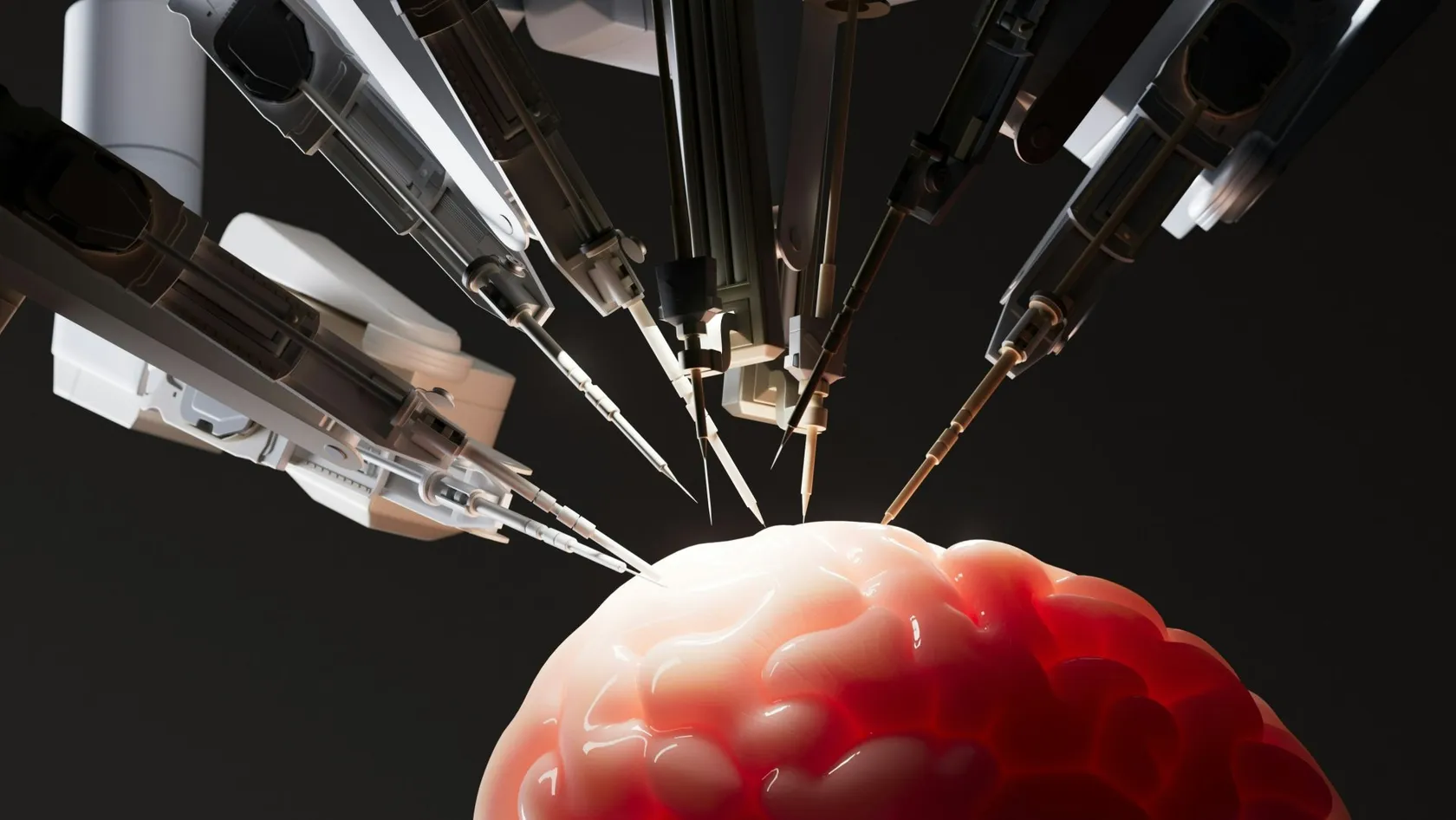
Endoscopic Brain Surgery
Three quite different operations share this name, and the one you need decides everything about your stay, your risks and the date you can fly. This page separates them and puts the published numbers on each.
About This Department
Nothing is cut on the outside of your head. The road in runs through your nose.
Two or three days on the ward, no scar anyone can see, and one complication that matters more than all the others together, which this page names in full.
Three quite different operations get filed under the same phrase, and a patient searching it ends up reading about somebody else's problem. One goes through the nostril to reach a tumor sitting under the brain. One goes through a hole the width of a pencil to work inside the fluid spaces in the middle of the brain. The third shrinks an ordinary craniotomy down, because a camera can see around corners that a microscope cannot.
They share a camera and almost nothing else.
What follows separates the three, gives the published numbers for each, and spends real space on the complication that runs through all of them, which is spinal fluid escaping from a hole that should have sealed, a risk that brings twelve weeks of restrictions with it and lands as the part patients discover late, the part a page written to sell an operation leaves out.
What the endoscope changed
A surgical microscope sits outside the patient and looks down a corridor, so everything it shows lies in a straight line from the lens, and anything tucked around a corner stays invisible. An endoscope carries its light and its camera at the tip of a rod the width of a pencil, and it goes inside the corridor. Once the lens is past the opening, the picture widens, the surgeon works close to the target instead of far from it, and an angled tip looks sideways at the parts of a tumor a straight view would have left behind.
So the opening shrinks. Everything patients like about the technique follows from that one change.
What the endoscope did not change is the operation itself. The tumor still has to come off the same vessels and nerves, judgment still decides how much can safely go, and a difficult tumor stays difficult with a better camera pointed at it. Anyone offering the technique as a reason your case has become easy is describing the equipment rather than your scan.
Through the nose, and why that road exists
The pituitary gland sits in a small bony hollow in the floor of the skull, directly behind the back wall of your nasal cavity and directly below the crossing point of the optic nerves. Reaching it from above means lifting the brain out of the way. Reaching it from the nose means passing through an air space that is already there, opening one thin wall of bone, and arriving at the tumor without touching the brain at all.
Surgeons stopped opening the skull for these tumors decades ago for exactly that reason, and an endoscope suits the route for the same one.
What actually happens in the operating room
A neurosurgeon and, in many teams, an ear nose and throat surgeon work together through both nostrils, one holding and steering the endoscope while the other uses both hands on instruments, and from there the nasal passage is widened by moving structures aside rather than cutting them away where possible, part of the septum is opened to give both nostrils access to the same corridor, and the thin bone at the back is removed. The lining of the brain comes next, and behind it sits the tumor. Fine curettes and suction take it out piece by piece, and an angled endoscope goes in at the end to look up and sideways into the corners for anything left behind. Then the hole is sealed, which is a separate operation in its own right and gets its own section below. Both nostrils serve as the only entry, and a patient looking in a mirror on day two sees nothing beyond some swelling.
That road reaches further than the pituitary. Tumors along the midline floor of the skull, including some meningiomas, craniopharyngiomas and chordomas, are approached this way in centers that do the work regularly, and the exposure required for those goes well beyond a straightforward pituitary case.
Inside the ventricles, and living without a shunt
Your brain makes spinal fluid deep inside itself, and it circulates through four connected chambers before draining away. Block one of the narrow passages between them, which a cyst or a tumor easily does, and fluid backs up under pressure. Doctors call that hydrocephalus, and it shows as headache, unsteady walking, blurred vision and, as it worsens, drowsiness.
Traditionally that is treated with a shunt, meaning a permanent tube running from the brain under the skin to the abdomen, with a valve. Shunts work. They also block, get infected, need revising, and stay in your body for the rest of your life.
What the operation involves
An endoscope offers a different answer. Through one small hole in the skull the camera is passed into the chamber above the blockage, and the surgeon makes an opening in its floor so that fluid escapes into the normal spaces around the brain and reaches its drainage route by another path. Nothing stays behind. Surgeons call it an endoscopic third ventriculostomy, it takes under an hour in straightforward cases, and when it works the patient walks out with no implant at all.
The trade, in published numbersA meta-analysis of 22 studies covering 556 adults with long standing enlarged ventricles found success rates of 81.8 percent for the endoscopic operation and 86.7 percent for a shunt, a difference that was not statistically significant. Complications told a different story, at 4.6 percent after the endoscopic route against 27.1 percent after shunting. A separate meta-analysis of five randomized trials in obstructive hydrocephalus found no difference in operative success either, with markedly lower infection and blockage rates in the endoscopic group. Read together, the endoscope does not work better. It fails in a similar share of patients and causes far less trouble in the ones where it works.
Success depends heavily on why the fluid is blocked, on age, and on whether there has been meningitis or bleeding in the past, so the honest conversation before this operation is about your own probability rather than the pooled one. Where it fails, a shunt is still available and nothing has been lost except time.
Surgeons use the same access to take out colloid cysts and small tumors sitting inside the chambers, and to open a window between the two sides when only one is obstructed.
Keyhole openings in the skull
Version three shrinks an ordinary craniotomy. An incision hidden in an eyebrow or behind an ear, an opening in the bone a few centimeters across, and an endoscope taken in alongside the microscope so the surgeon can look around the corners the small window would otherwise hide. Surgeons call it endoscope assisted surgery, and the endoscope is a second pair of eyes rather than the main instrument.
Smaller openings mean less muscle divided and less bone removed, which shows up as less pain and a shorter stay. The limits come from geometry. A small window gives few angles, so a tumor that turns out bigger or more vascular than the scan suggested can force the surgeon to extend the opening during the operation, and a patient consenting to a keyhole approach should be consenting to that possibility in the same conversation.
Which route suits which problem
Position decides almost everything. A tumor in the midline floor of the skull is reachable from below, a blockage in the fluid pathways is reachable from a small hole above, and a tumor out on the surface of the brain is neither.
This table scrolls sideways on a narrow screen. Swipe or drag to see every column.
| Problem | Usual endoscopic route | What decides against it |
|---|---|---|
| Pituitary tumor pressing on the optic nerves | Through both nostrils | Growth spreading widely sideways, or a very firm tumor wrapped around vessels |
| Blocked fluid pathways causing pressure | One small hole, opening the floor of the third chamber | Previous meningitis or bleeding, very young age, unusual anatomy on the scan |
| Colloid cyst in the middle of the brain | Endoscope through the chamber | Small ventricles leaving no working space, or a cyst too solid to aspirate |
| Midline skull base meningioma or craniopharyngioma | Extended approach through the nose | Tumor extending out to the sides, past the carotid arteries |
| Small tumor at the front of the skull base | Keyhole opening through an eyebrow incision | Size or blood supply demanding more angles than a small window allows |
| Glioma in the substance of the brain | None of them | This needs an open craniotomy with mapping, and no camera changes that |
Read the final row twice, because searches for endoscopic brain surgery are full of people whose tumor cannot be treated this way at all.
The spinal fluid leak
Everything above depends on making a hole in the floor of the skull and then closing it well enough that spinal fluid stays inside. Fail, and fluid runs down into the nose. That sounds minor. It is anything but, because the same open channel lets bacteria travel the other way, and meningitis after skull base surgery is a serious illness. Sealing that hole is a genuine reconstructive operation performed at the end of the tumor removal. Fat or synthetic material fills the cavity, a graft goes underneath the bone edge, and in most cases a flap of the lining of the nasal septum, with its own blood supply left attached, is rotated over the whole repair. That flap changed the field. Before it came into use, leak rates after extended approaches through the nose were high enough to limit how far surgeons were willing to go, and a living flap that heals rather than a patch that merely covers is the reason those approaches are now routine.
Research continues on what goes underneath it, including injectable materials that set hard against the bone, and a 2024 review of the published comparisons found lower leak rates when such a material was combined with the flap rather than using the flap alone, on a small number of studies.
What you do for twelve weeks
Your part in this is mechanical and it lasts three months.
Anything that raises pressure inside your head pushes against the repair while it is healing, which is why the instruction sheet forbids blowing your nose, straining, heavy lifting and bending forward for twelve weeks, and patients who treat those rules as advice rather than as part of the operation are the ones who come back with a leak, which usually means going back to the operating room to redo the repair.
The weeks after nasal surgery
Admission for a straightforward pituitary case runs two to three days at large centers, with one night in intensive care and one or two on the ward afterward. No operation described anywhere on this site has a shorter admission, and that gap makes the single biggest practical difference between this route and opening the skull.
This table scrolls sideways on a narrow screen. Swipe or drag to see every column.
| When | What happens |
|---|---|
| Day 1 | Awake and eating, blocked nose, hormone and fluid balance watched closely, out of intensive care |
| Days 2 to 3 | Home or hotel, breathing through the mouth, sleeping propped up, mild headache |
| Week 2 | Nasal review and any stitches out at ten to fourteen days, hormone bloods repeated |
| Weeks 3 to 4 | Bloody discharge settling, saline rinses started once the team says so, desk work possible |
| Weeks 4 to 6 | Back to a sedentary job by four weeks and to an active one by six, smell still dulled |
| Week 12 | The repair is mature, and nose blowing, lifting and bending come back |
The six rules that protect the repair
Six rules govern the first twelve weeks, and every one of them protects the same repair.
- No nose blowing at all. Sneeze with your mouth wide open.
- Nothing heavier than about five kilos, and no bending forward past your waist to pick something up.
- No straining on the toilet, which means taking the laxative you are sent home with.
- No firm pressure on the face for the first month, which includes glasses frames that press hard on the bridge of your nose.
- Saline rinses only when your team tells you to start, since starting too early disturbs the repair.
- Clear fluid dripping from one nostril, especially leaning forward, means calling that day.
Hormones and vision, the two things checked
Surgery on the pituitary is surgery on the gland that instructs most of the others, so the follow up is chemical rather than surgical and it belongs to an endocrinologist as much as to the surgeon.
Water balance, in the first week
The back of the pituitary makes the hormone that tells your kidneys to hold on to water. Handle that area and it can stop working temporarily, occasionally permanently, and the result is enormous volumes of very pale urine and a thirst that cannot be satisfied, a condition doctors call diabetes insipidus, which has nothing to do with diabetes as most people know it and which a nasal spray or tablet replacing the missing hormone treats. Most cases in the first week settle within days.
The one thing to watch on the flight homeUrine that is almost colorless, in large volumes, with constant thirst, can appear a week or more after the operation, sometimes after a period of everything seeming normal. Untreated it dehydrates a person quickly, and a long flight with limited access to water makes that worse. Learn the signs before you leave the ward, carry the medication if your team has prescribed it, and drink to thirst rather than to a schedule.
The other hormones, and your eyes
Cortisol matters most, because a body that cannot make it copes badly with illness or stress. Blood tests around the operation and again at follow up show which hormones the gland is still producing, replacement is started where it is needed, and some patients need a small dose for life while others recover completely. Thyroid and sex hormones are checked on the same schedule, and a functioning tumor that was producing too much of something is judged by whether those levels fall into the normal range afterward. Vision gets measured separately. Pressure upward on the crossing of the optic nerves takes away the outer edges of both visual fields, often so gradually that the patient never noticed, and taking that pressure off frequently restores them, with recovery running quickest in the first weeks and continuing for months, going better the shorter the time the nerves spent compressed, which is one of the few genuinely time sensitive reasons to get on with the operation.
When the open operation is the better one
Plenty of patients arrive having decided on an endoscopic operation because it sounds gentler, and part of a useful consultation is being told when it is the wrong choice.
A tumor that has grown out sideways beyond the carotid arteries cannot be reached safely from the midline, and a very firm or heavily calcified tumor resists the fine instruments a narrow corridor allows. Tumors wrapped around major vessels need the room and the angles that only an open approach provides. Active sinus infection has to be treated before anyone opens a channel between the nose and the brain. And a glioma in the substance of the brain has never been an endoscopic problem at all, whatever a search result implies.
Sometimes both are needed, in stages, and hearing that plan is a good sign.
What is honestly worse this way
Any page describing only the advantages of a technique is a brochure. Here come the entries on the other side of the ledger.
Set against those, the gains are real and measurable. The same pooled analysis of repeat pituitary operations found vision improved in 73.2 percent of the endoscopic group against 49.6 percent of the microscopic group, with modestly higher rates of complete removal and hormonal cure, in tumors that were on average harder.
Flying home, and the six week number
Short ward stay, long wait before the flight. That combination catches out almost every international patient who books this operation, so plan around it from the beginning.
Cabin pressure at cruising altitude is lower than at ground level, so gas trapped in the body expands and the pressure difference across the fresh repair in your skull base rises exactly when you want it left alone. A 2026 survey of attending neurosurgeons, using case based scenarios, produced a mean recommended wait of 4.4 weeks after a straightforward operation through the nose and 6.0 weeks where a spinal fluid leak had been repaired during the surgery. The same survey found wide disagreement between individual surgeons, which is an argument for a written opinion about your own operation rather than a rule of thumb from a forum. So the plan looks like this. Two or three days before surgery for tests, imaging and the endocrine review, two or three nights in hospital, then a nasal and hormone check at around ten to fourteen days, and a return flight booked from roughly the fifth week onward for a routine case and later where the floor of the skull was repaired. Some patients go home earlier with their surgeon's written clearance and complete the follow up locally, and that is a decision to take with the team rather than with a booking website.
On the aircraft itself, chew or swallow during descent to help your ears, keep drinking water, and keep the hormone medication in your hand luggage, and ask before you fly whether you should carry a short course of the water tablet or spray in case the thirst starts on the way home.
What moves the cost
No price appears on this page, and with three different operations sharing one name a single figure would be meaningless. An hour long ventriculostomy and a four hour extended approach through the nose for a skull base meningioma are not comparable pieces of work.
Six things carry the difference. Which of the three operations you are actually having, and how long it takes. Whether an ear nose and throat surgeon operates alongside the neurosurgeon, which many teams treat as standard for the nasal route. How the skull base is reconstructed, since a septal flap plus graft material costs more than a simple closure and prevents more leaks. Intensive care nights and ward nights. The endocrine work, meaning hormone panels before and after surgery and the specialist reviewing them. And the pathology, which for these tumors increasingly includes molecular tests.
Five questions that make quotes comparable
Which of the three operations does this figure assume, and for how long in the operating room? Is a second surgeon for the nasal access included in the price? Which reconstruction is planned, and does the quoted figure include the graft material and the septal flap work? Are the hormone tests and the endocrinologist reviews included, before and after? If the approach has to be converted to an open operation during surgery, what happens to the quote?
Published packages in this market normally cover transfers, pre-operative tests, surgeon and anesthesia fees, a stated number of nights, the post-operative scan, an interpreter and a set number of hotel nights, while leaving out flights, insurance, nights beyond the plan, and the treatment of a complication, so for this operation check the endocrine follow up in particular, because it is the item assumed most and written down least.
Send the MRI, the hormone results and any visual field test you already have. A surgeon reading them will tell you which route your tumor allows, and that opinion costs nothing and obliges you to nothing.
Planning the trip
Short admissions tempt people into traveling alone. Do not.
For the first two weeks you are breathing through your mouth, sleeping badly, sometimes on new hormone replacement, and under instructions not to lift or bend, which rules out a suitcase and most of what a hotel stay demands. Patient rooms carry a second bed so one person stays with you through the admission, and accommodation for you both on the nights either side, the airport transfers and every trip between hotel and hospital are arranged by the international patients office. On a pituitary case that transport matters for a reason particular to this operation, since the hormone and fluid checks bring you back to the hospital more often than a tumor patient would expect. The international patients team works in English, Arabic, French, Russian, Serbian, Romanian and Spanish, other languages are interpreted on request, and one coordinator holds your case from your first message until discharge. Ask for a woman surgeon or physician at the start and the request goes to the department, which meets it wherever the rota allows. An appointment confirmation and an invitation letter naming the hospital and your doctor, the paperwork most consulates ask for with a medical visa, are issued about ten days before you travel. Meals from the hospital kitchen cover halal, vegetarian and diabetic requirements, and there is a prayer room on site.
Send your scans as original files on a disc or a drive, and bring the hormone results and any visual field test with the dates on them, because comparing before and after is the whole point of both.
Follow up once you are home
Leave with a file that any endocrinologist can act on. The operation note naming the approach and the reconstruction, the discharge summary, the imaging, the full pathology, every hormone result with its date, the replacement doses with review dates, and the schedule for the next scan and the next blood tests, and arrange the local endocrinologist before you travel, since the first review falls due within weeks.
Your coordinator stays reachable on the same WhatsApp number once you are home, so a photograph, a laboratory report or a question about a dose gets an answer quickly, and local scans can be sent back for review by the team that operated.
Some things need attention the same day, wherever you are.
Clear watery fluid dripping from one nostril, particularly when you lean forward or strain. Fever with a headache and a stiff neck, which after this operation is treated as meningitis until proven otherwise. Very large volumes of pale urine with unquenchable thirst, or the opposite picture of confusion and drowsiness with no thirst at all. Sudden worsening of vision. Heavy bleeding from the nose. Severe headache different from the one you were sent home with. For any of these the nearest emergency department comes first, with your operation note in your hand, and a message to your coordinator afterward.
Endoscopic brain surgery FAQ
How long do I have to stay before flying home?
Will I have a scar on my face or head?
Why can I not blow my nose for three months?
Is the endoscopic operation better than the open one?
Will I need hormone tablets for life?
Can someone stay with me in the room?
Will my sense of smell come back?
Written by the Biruni Hospital medical editorial team. Reviewed by Dr Yunus Emre Yavuz, Neurosurgery.
References
- Esquenazi Y, Essayed WI, Singh H, Mauer E, Ahmed M, Christos PJ, Schwartz TH. Endoscopic endonasal versus microscopic transsphenoidal surgery for recurrent and residual pituitary adenomas. World Neurosurgery. 2017;101:186-195.
- Gillespie CS, Fang WYS, Lee KS, Clynch AL, Alam AM, McMahon CJ. Long-standing overt ventriculomegaly in adults. A systematic review and meta-analysis of endoscopic third ventriculostomy versus ventriculoperitoneal shunt as first-line treatment. World Neurosurgery. 2023;174:213-220.
- Pasqualotto E, Schmidt PHS, Ferreira ROM, Chavez MP, da Silva FFS. Endoscopic third ventriculostomy versus ventriculoperitoneal shunt in patients with obstructive hydrocephalus. An updated systematic review and meta-analysis. Asian Journal of Neurosurgery. 2023;18(3):468-475.
- Shin DW, Yee GT. Skull base reconstruction using hydroxyapatite and nasoseptal flap versus nasoseptal flap alone. Meta-analysis and systematic review. Scientific Reports. 2024;14(1):28433.
- Mampre D, Rupp C, Wilson J, Kelbert J, Riha G, Richardson M, et al. Postoperative recommendations for commercial air travel after neurosurgical intervention. Aerospace Medicine and Human Performance. 2026;97(6):411-418.
Editor's note
Written by the Biruni Hospital medical editorial team. Reviewed by Assoc. Prof. Dr. Melih ÜÇER, Neurosurgery.
Medically reviewed by
Assoc. Prof. Dr. Melih ÜÇER
Neurosurgery
Related Treatments
View All
ALIF - Anterior Lumbar Interbody Fusion Surgery
Coming in from the front restores the curve of the lower back better than any posterior route and leaves the back muscles untouched. It also carries one risk most pages will not name, and this page names it.

Brain AVM Surgery - Arteriovenous Malformation Surgery
An unruptured brain AVM bleeds at roughly 2.2 percent a year, and surgery spends its whole risk on a single morning. This page sets those two shapes of risk side by side, explains what the ARUBA trial does and does not say about your case, and shows how a Spetzler-Martin grade changes the answer.
Brain Tumor Surgery - Brain Tumor Removal
Brain surgery is one trade repeated all day, how much tumor comes out against what the brain around it still does. This page puts the published numbers on both sides of that trade.
Cervical Microdiscectomy - Minimally Invasive Neck Disc Surgery
Neck disc surgery is done from the front, through the space between your windpipe and your carotid artery, and almost everything that goes wrong that is not the disc happens in that corridor. What has been measured in it.
Chiari Malformation Surgery
A Chiari malformation is measured in millimeters and treated according to symptoms, and those two things come apart more often than patients are told. Most people with the finding never need an operation. This page sets out who does, what posterior fossa decompression removes, why surgeons still argue over opening the dura, and what the pooled studies say about both sides of that argument.