Ten times the local recurrence without radiotherapy. The same survival at ten years. Both figures come from one trial, and together they say what this treatment is actually for.
About This Department
RADIATION ONCOLOGY
Leaving radiotherapy out raised local recurrence tenfold. Ten year survival came out identical.
PRIME II randomized 1,326 women aged 65 and over, all with small hormone-receptor-positive node-negative tumors and all taking endocrine therapy, between whole breast radiotherapy and none at all. Local recurrence inside ten years was 9.5 percent without it and 0.9 percent with it. Overall survival at ten years was 80.8 percent and 80.7 percent. Both numbers are true and they mean different things.
9.5% vs 0.9%
Ten year local recurrence without radiotherapy against with it, in low-risk women over 65
26 Gy in 5
One week of treatment, matching three weeks across 4,096 randomized patients
7.4% per gray
Rise in major coronary events for each gray of mean heart dose, with no threshold found
Free
Written opinion on your scans and reports before you travel
Thirty years of this field has moved in one direction
Almost every change to breast radiotherapy since the 1990s has been a reduction. Fewer sessions. Smaller volumes of tissue treated. Lower total doses. In a defined group of patients, no radiotherapy at all.
Each of those reductions was tested against the longer or larger version in a randomized trial, and in each case the shorter or smaller option held its ground. That is unusual. Most of medicine moves by adding things.
What got shorter
Six weeks became three weeks, and three weeks became one. A 2020 trial of 4,096 patients found 26 grays in five sessions over one week not inferior to 40 grays in fifteen over three.
What got smaller
Whole breast treatment became partial breast treatment for lower-risk tumors, with local relapse at five years of 0.5 percent against 1.1 percent in the whole breast group.
That history matters when you are handed a plan, because a six-week course offered in 2026 for an ordinary early breast cancer is a course that was superseded twice. Ask why, and accept a specific answer about your own case. Real reasons exist. So does inertia.
What the treatment is buying, stated precisely
Radiotherapy after breast-conserving surgery reduces the chance the cancer comes back in the same breast. That is its first and most reliable effect. Whether it also extends life depends entirely on who is being treated, and conflating the two is how patients end up either frightened or falsely reassured.
Local control and survival are separate questions
PRIME II answers this as cleanly as any trial in oncology. Among 1,326 women aged 65 and over with hormone-receptor-positive, node-negative tumors of three centimeters or less, excised with clear margins and treated with endocrine therapy, omitting radiotherapy raised local recurrence within ten years from 0.9 percent to 9.5 percent, a hazard ratio of 10.4. Distant recurrence as a first event was no higher without radiotherapy, at 1.6 percent against 3.0 percent. Overall survival at ten years was 80.8 percent without and 80.7 percent with. Breast cancer specific survival did not differ either.
Where your own case sits on that spectrum
So in that population the treatment prevented recurrences in the breast and did not change how long women lived. Younger patients, larger or higher-grade tumors, involved nodes and hormone-receptor-negative disease are a different situation, where local control and survival are more tightly linked and where the case for treating is much stronger. Where you sit on that spectrum is the first thing to establish, and it is decided by your pathology report rather than by a general policy.
When leaving it out is a genuine option
Very few patients are told this is on the table. A narrow group of them should be.
Drag the table sideways on a narrow screen.
The conditions that were met by the women in whom omission proved reasonable
Condition
What the trial required
Age
Sixty-five years or older
Tumor size
Three centimeters or less in the largest dimension
Nodes
No involved lymph nodes
Receptors
Hormone-receptor-positive disease
Surgery
Breast-conserving surgery with clear excision margins
Endocrine therapy
Taken as adjuvant treatment, which is doing part of the work
What you accept
Roughly one chance in ten of a recurrence in that breast over ten years, against one in a hundred with radiotherapy
Why the endocrine therapy clause is not a detail
The women in PRIME II who avoided radiotherapy were all taking endocrine therapy, and that treatment is part of the reason their recurrence rate was as low as it was. Omitting radiotherapy and then stopping the tablets because of side effects is not the arrangement that was tested, and it is a common sequence in practice. Anyone considering this route should treat the endocrine therapy as the thing they have committed to, since a recurrence in the breast means a second operation and usually radiotherapy afterwards anyway, which is a worse year than the one the omission was meant to avoid.
The trade belongs to you and the decision does too. No department should make it quietly on your behalf in either direction.
One week instead of three, and which schedule to ask about
FAST-Forward recruited 4,096 patients across 97 UK centers after breast-conserving surgery or mastectomy, and randomized them three ways.
Forty grays in fifteen sessions over three weeks, which was the international standard at the time and had itself replaced a fifty-gray, twenty-five-session, five-week course.
Twenty-seven grays in five sessions over one week.
Twenty-six grays in five sessions over one week.
What separated the two five-session arms
At a median follow-up of just under six years, the primary endpoint of relapse in the treated breast had occurred in 31, 27 and 21 patients across the three groups, and both one-week schedules met the non-inferiority criterion against three weeks. Normal tissue effects separated them. Moderate or marked clinician-assessed effects in the breast or chest wall at five years were reported in 9.9 percent of the three-week group, 15.4 percent of the 27-gray group and 11.9 percent of the 26-gray group. Across all assessments from one to five years the odds ratio against three weeks was 1.55 for 27 grays and 1.12 for 26 grays, and patient and photographic assessments told the same story. The higher of the two one-week doses cost cosmetic outcome. The lower one did not. That distinction has practical consequences, because both five-fraction doses will appear in published protocols and in quotations, and a department quoting 27 grays is quoting the arm of the trial that came out worse on the outcome patients notice most.
So the answer that came out of the trial was specific. Twenty-six grays in five fractions rather than twenty-seven, and that one-gray difference is the whole point of running the trial with three arms.
Travel changes what this evidence is worth. It turns a three-week stay into a one-week stay for a large share of early breast cancer, so the five-fraction dose being proposed is the first thing to establish before anything else is booked.
Treating part of the breast, and whether you need a boost
The other reduction was in volume rather than time.
IMPORT LOW randomized 2,018 women aged 50 and over, with unifocal invasive ductal cancer of three centimeters or less, none to three positive nodes and margins of at least two millimeters, between whole breast treatment, a reduced dose to the whole breast with full dose to the tumor area, and partial breast treatment alone. All three used fifteen fractions. Five-year local relapse was 1.1 percent in the whole breast group, 0.2 percent in the reduced-dose group and 0.5 percent in the partial breast group, and both experimental approaches were non-inferior. Patients in the partial breast group reported significantly less change in breast appearance and significantly less hardening or firmness of the breast. What made that trial unusually easy to copy is that the partial breast technique used nothing exotic. It used the standard tangential beams shortened in length, which is why the authors could say the approach was implementable in radiotherapy centers worldwide, and why a department declining to offer it cannot blame its equipment.
The tumor bed boost, which is a separate question
Boost means extra dose to the area the tumor came from, added after the main course. It reduces local recurrence and it costs cosmetic outcome, so it belongs to patients whose recurrence risk is high enough to be worth that, which generally means younger age, higher grade, close margins or extensive in-situ disease alongside the invasive tumor. An older patient with a small low-grade tumor and wide margins is being offered a boost for no clear reason, and a boost added by default across a clinic is a habit rather than a decision. It also lengthens the course, which matters if you have traveled.
Whether the lymph node areas get treated
Treating the breast is one decision. Treating the nodal areas above and behind it is another, with its own trial and its own trade-off.
MA.20 randomized 1,832 women with node-positive or high-risk node-negative disease, all having had breast-conserving surgery and systemic therapy, between whole breast radiotherapy alone and whole breast radiotherapy plus regional nodal irradiation covering the internal mammary, supraclavicular and axillary nodes. At a median follow-up of 9.5 years, ten-year overall survival was 82.8 percent with nodal treatment and 81.8 percent without, which is no difference. Ten-year disease-free survival was 82.0 percent against 77.0 percent, a hazard ratio of 0.76 that was statistically significant. The added treatment also added harm. Grade 2 or worse acute pneumonitis rose from 0.2 percent to 1.2 percent, and lymphedema from 4.5 percent to 8.4 percent.
What nodal treatment buys
Fewer recurrences. Five fewer disease-free survival events per hundred women at ten years, which is a real and meaningful reduction.
What it costs
Roughly four extra cases of lymphedema per hundred, one extra case of pneumonitis per hundred, more dose to lung and heart, and a permanent risk to the arm.
Lymphedema, which deserves naming rather than listing
Lymphedema deserves more than a place on a list. Swelling of the arm after nodal treatment is often permanent, it is managed rather than cured, and for someone whose work or life depends on that arm it is a serious cost. That does not make nodal treatment wrong. It makes it a decision that should be discussed with the arm in the room.
Radiotherapy after mastectomy
Removing the whole breast does not always remove the need for radiation.
Chest wall radiotherapy gets recommended when the risk of recurrence there or in the nodes stays high after surgery, which in practice means involved lymph nodes, a large tumor, or cancer reaching the surgical margin. The target becomes the chest wall and usually the nodal areas too, so the planning problem here resembles the nodal decision above far more closely than it resembles whole breast treatment.
Where reconstruction complicates things
Radiation and reconstruction interact, and the sequence matters. An implant placed before radiotherapy faces a higher rate of capsular contracture, infection and reconstruction failure, and the cosmetic result is less predictable than in an unirradiated reconstruction. Tissue from your own body tolerates radiation better than an implant does, and is still usually placed afterwards. None of that makes reconstruction impossible. The practical consequence is that the plastic surgeon and the radiation oncologist have to agree the order before the first operation, which requires somebody to have asked whether radiotherapy is likely while the surgical plan was still being drawn. A patient who has an implant placed and is then told radiotherapy is needed has been let down by a planning failure and not by the treatment itself, and that sequence is common enough to be worth guarding against personally.
Raise reconstruction at the first consultation, even if it feels premature.
The heart dose, and what lowers it
A population-based study of 2,168 women treated in Sweden and Denmark between 1958 and 2001 estimated each patient mean heart dose from her own radiotherapy chart and compared those who had major coronary events with those who did not. The rate of major coronary events rose by 7.4 percent for every gray of mean heart dose, with no apparent threshold. The increase began within five years and continued into the third decade. The average mean heart dose across that cohort was 4.9 grays, and the proportional increase per gray was the same in women with and without cardiac risk factors, which means women who already had heart disease carried the larger absolute risk. The study covered 963 women who had a major coronary event and 1,205 who did not, and the dose for each of them was reconstructed from her own treatment chart rather than estimated from an average, which is the reason the dose-response curve it produced has held up as well as it has.
Those women were treated with older techniques. Modern planning achieves far lower heart doses, and the finding is what drove that change.
Scroll the table sideways if your screen is narrow.
The techniques that reduce heart dose, and what each one asks of the patient
Technique
How it works
What it requires of you
Deep inspiration breath hold
You hold a deep breath during delivery, which moves the chest wall away from the heart
Holding still for roughly twenty seconds at a time, practiced beforehand
Prone positioning
Treatment lying face down, so the breast falls away from the chest wall
Lying prone comfortably on a dedicated board
Partial breast treatment
A smaller volume means less incidental dose to everything nearby
Eligibility on tumor size, grade, margins and nodes
Field-in-field and intensity modulation
Shaping the dose so less of it reaches the heart and lung for the same breast coverage
Nothing extra, beyond a department that plans this way as standard
What to ask for if your cancer is on the left
Left-sided cancers carry the most heart dose, so anyone with a left-sided tumor should ask directly what their mean heart dose in grays comes to and which of these techniques the department uses. A department that treats left breasts the same way it treats right ones has not engaged with the last decade of its own specialty.
What the sessions and the skin actually involve
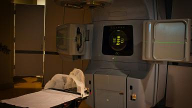
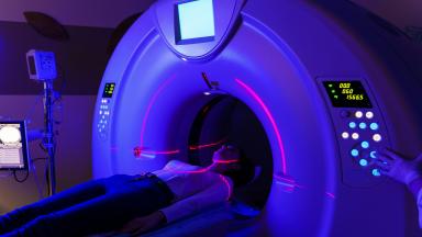
Most of an appointment is spent getting you into exactly the position the plan was built around. The beam itself runs for a minute or two, and you feel nothing while it does. Fifteen minutes in the room is typical, and breath-hold technique adds a little to that.
During a one-week course
Little happens while you are still attending. Skin changes and tiredness arrive after the last session rather than during, which surprises patients who expected to feel it sooner.
During a three-week course
Skin redness usually starts in the second or third week, and tiredness builds gradually. Both peak around a week to ten days after the final session.
The skin
Redness, warmth, itching and sometimes peeling in the treated area, worst in the fold under the breast and in the armpit. It settles over two to four weeks. Simple unperfumed moisturizer, loose cotton and keeping the area dry cover most of it.
Later changes
Some firmness of the breast tissue, occasional aching, sometimes a small permanent change in size or shape. Shoulder stiffness is worth watching for and responds to early movement.
Two things get missed. Radiation-induced fatigue behaves unlike ordinary tiredness and does not resolve with a good night of sleep, so light regular activity beats rest, and it fades over weeks to a couple of months. And breast tenderness or a firm area appearing months later needs examining rather than assuming, because distinguishing treatment change from recurrence is exactly what follow-up exists for.
The four questions that set your plan
Everything above reduces to four decisions. Each one has a trial behind it and an answer that depends on your pathology.
Four decisions, in the order they get made
Radiotherapy at all, or endocrine therapy alone. Relevant if you are over 65 with a small hormone-receptor-positive node-negative tumor, clear margins and a willingness to keep taking the tablets.
How long. One week at 26 grays in five fractions for most early disease after breast-conserving surgery, three weeks at 40 grays in fifteen where that suits the case better.
How much of the breast. Whole breast, or partial breast for lower-risk tumors in women over 50 who meet the size, grade, margin and node criteria.
Whether the nodal areas are included, which buys fewer recurrences and costs lymphedema and lung dose, and which applies to node-positive and high-risk node-negative disease.
Answering all four with reference to your own report makes it a plan. Answering all four the same way for every patient in the clinic makes it a protocol dressed up as one.
Four things on a plan that deserve a harder question
Each of these is common and none of them is automatic
A six-week course for ordinary early disease
Superseded twice by randomized evidence. There are cases where a longer course is right, and each of them has a reason that can be stated in a sentence.
A boost added without discussion
It reduces recurrence and costs cosmetic outcome, so it belongs to patients whose risk earns it. Applied to everyone, it is a default and not a judgment.
Nodal treatment offered as routine
It carries a real risk to the arm. The indication should be named, and the lymphedema figures should be part of the conversation.
No heart dose figure for a left-sided cancer
The dose-response has no threshold and begins within five years. A department that cannot tell you the number is not managing it.
One more, which cuts the other way. Be equally skeptical of anyone offering to omit radiotherapy in a patient who does not match the trial population, because the omission evidence is narrow and specific and does not stretch to cover younger women, larger tumors or involved nodes.
Having the course somewhere other than home
Of all the radiotherapy courses, breast treatment has become the one where traveling for it makes practical sense, and the five-fraction schedule did that.
How long you actually need to be here
Five-fraction treatment needs simulation, a planning gap of a few days, and then five consecutive weekday sessions, so around two weeks covers it comfortably. A three-week course needs closer to four weeks. Adding a tumor bed boost extends either one. Nodal treatment leaves the length alone and changes the planning, while post-mastectomy treatment follows the longer schedules far more than the shorter one. Get the intended total in writing before you book anything, because the difference between a two-week stay and a five-week stay changes the accommodation, the visa duration, the time you need away from work and whether somebody has to come with you, and none of those are things you want to rearrange from a hotel room in week two.
What to send first
The pathology report including receptor status and margins, the operation note, imaging, and the systemic therapy plan. The four decisions above cannot be discussed without those.
Fitness to fly
Flying after breast surgery or radiotherapy rarely causes trouble. Recent surgery, a seroma being drained, or arm swelling deserve a check before booking, and anyone with lymphedema should ask about compression for the flight.
Accommodation
Proximity matters even for five sessions, since the appointments are daily and consecutive.
Language and the paperwork
Seven languages are covered directly, namely English, Arabic, French, Russian, Serbian, Romanian and Spanish. Anything else is interpreted, arranged ahead of the first appointment. The same coordinator handles your dates, and a letter supporting a visa application follows once the schedule is set.
Follow-up once you are back home
Surveillance after breast radiotherapy runs for years, so the handover matters more here than the length of the stay does. Leave with the dose report including the mean heart dose, the treated volumes, the boost details if one was given, and a written schedule for mammography and clinical review, all in a form the person who follows you where you live can read without telephoning anyone. The plan should also name the new symptoms that warrant contact before the next appointment, which for this treatment means a new lump, skin change or nipple change in the treated breast, swelling or heaviness in the arm, and chest pain or breathlessness on exertion given what the dose-response above says about the heart. Write those down before you fly rather than trying to remember them, and ask for a copy to be sent to whoever will be seeing you at home, since a report that exists only in your own luggage tends to go missing in the year it is needed.
The six things that move the figure on a quotation
No prices appear on this page. A figure quoted before the pathology has been read amounts to a guess, and the variables behind the real total are straightforward enough to set out here, so that a quotation makes sense when one arrives.
Reading a quotation properly
Where the money actually goes
This table slides left and right on a small screen.
Six cost drivers in breast radiotherapy, and what each one is paying for
Cost driver
What it pays for
Number of fractions
Five sessions and fifteen sessions are different products, and the accommodation cost follows the same multiplier.
Treated volume
Whole breast, partial breast, chest wall and nodal areas differ in planning work and in delivery time.
Tumor bed boost
Extra sessions and extra planning, priced separately in most quotations.
Motion management
Deep inspiration breath hold or prone positioning needs equipment, coaching and longer sessions, and for a left-sided cancer it earns its cost.
Planning technique
Field-in-field, intensity modulation and daily image guidance differ in physics time and machine time.
Length of stay
Two weeks against five weeks of accommodation and living costs, which is frequently the larger part of a travel budget.
Request a quotation that separates planning, delivery, the boost if any, motion management and living costs. A single combined figure hides the one thing worth comparing, which is how many sessions you are actually paying for.
Breast cancer radiation therapy FAQ
Can I really finish in one week?
Yes, for a large share of early breast cancer treated with breast-conserving surgery. Twenty-six grays in five fractions over one week was not inferior to forty grays in fifteen over three weeks across 4,096 randomized patients, with the same cosmetic outcome at five years. Post-mastectomy treatment and cases needing nodal coverage or a boost go over three weeks or longer more of the time.
Will radiotherapy change how my breast looks?
Usually mildly. Some firmness, sometimes a small change in size or shape, occasionally a lasting difference in color. The trials measured this carefully, and moderate or marked change at five years affected around one woman in ten on the standard three-week schedule, with the 26-gray one-week schedule coming out no worse than that and the 27-gray version coming out worse. A boost increases it. Partial breast treatment reduced it.
Am I radioactive, and can I be around my grandchildren?
No and yes. External beam radiotherapy leaves nothing in your body, so there is no restriction on contact with anyone at any point during or after the course.
Can I skip radiotherapy if I am taking hormone tablets?
For some women over 65 this is a real option, and it has been tested properly. It requires a small hormone-receptor-positive node-negative tumor, clear margins, and continuing endocrine therapy. What you accept is roughly a one-in-ten chance of the cancer returning in that breast over ten years against one-in-a-hundred with treatment, and the trial found survival at ten years the same either way. Outside that group it is not an option at all.
What about my heart?
Major coronary events rise by about 7.4 percent for every gray of mean heart dose, with no threshold, and that finding is why modern planning works hard to keep the dose low. Breath-hold technique, prone positioning and smaller treated volumes all reduce it. Your mean heart dose in grays is a number the department holds, and for a left-sided cancer it is the number to ask for.
Will I get arm swelling?
Only if the lymph node areas are treated, and then in roughly eight women per hundred against four and a half per hundred without nodal treatment. Swelling gets managed and not cured, which puts it in the conversation before the plan is agreed and not afterwards.
How soon after surgery does it start?
Three to eight weeks, once the wound has healed, and after chemotherapy where chemotherapy is part of the plan. Chase a long unexplained delay. The interval does affect outcome, while a few weeks either side of the usual window is no crisis.
References
Murray Brunt A, Haviland JS, Wheatley DA, Sydenham MA, Alhasso A, Bloomfield DJ, et al. Hypofractionated breast radiotherapy for 1 week versus 3 weeks (FAST-Forward). 5-year efficacy and late normal tissue effects results from a multicentre, non-inferiority, randomised, phase 3 trial. Lancet. 2020;395(10237):1613-1626.
Kunkler IH, Williams LJ, Jack WJL, Cameron DA, Dixon JM. Breast-conserving surgery with or without irradiation in early breast cancer. New England Journal of Medicine. 2023;388(7):585-594.
Coles CE, Griffin CL, Kirby AM, Titley J, Agrawal RK, Alhasso A, et al. Partial-breast radiotherapy after breast conservation surgery for patients with early breast cancer (UK IMPORT LOW trial). 5-year results from a multicentre, randomised, controlled, phase 3, non-inferiority trial. Lancet. 2017;390(10099):1048-1060.
Whelan TJ, Olivotto IA, Parulekar WR, Ackerman I, Chua BH, Nabid A, et al. Regional nodal irradiation in early-stage breast cancer. New England Journal of Medicine. 2015;373(4):307-316.
Darby SC, Ewertz M, McGale P, Bennet AM, Blom-Goldman U, Brønnum D, et al. Risk of ischemic heart disease in women after radiotherapy for breast cancer. New England Journal of Medicine. 2013;368(11):987-998.
Editor's note
Written by the Biruni Hospital medical editorial team. Reviewed by Prof. Dr. Metin GÜDEN, Radiation Oncology.