
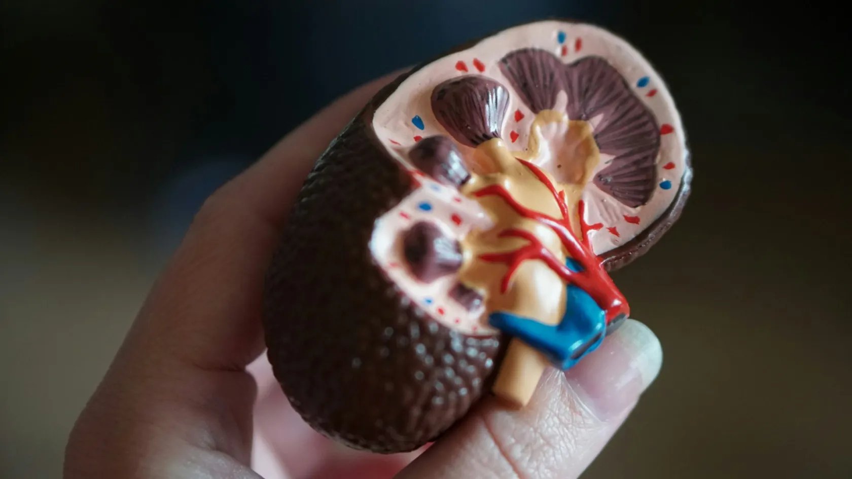
TURBT - Transurethral Resection Of Bladder Tumor
Almost every cancer operation follows a diagnosis. This one produces it, which makes the tissue that reaches the laboratory more important than anything you will feel afterwards.
About This Department
Almost every cancer operation follows a diagnosis. This one produces it. The tissue the surgeon shaves out of your bladder is not only the treatment, it is the specimen the pathologist reads to establish what you have and how deep it goes, which means the operation is graded on what reaches the laboratory, not on how it felt to you afterwards. Nobody tells patients this. A large European audit shows why they probably should be.
Free consultation
Send the cystoscopy findings and any previous resection report
Where a tumour sits inside the bladder changes the operation considerably, so a description of its position, size and number matters as much as the fact that it is there. Include the cystoscopy report, any imaging of the urinary tract, the histology from any previous resection, a list of blood thinners and a note of previous pelvic radiotherapy or bladder surgery. Say whether you have had a transurethral operation before, because scarring in the urethra changes what instrument fits. What comes back is an opinion on what the resection would involve, whether enhanced imaging would add anything and whether a second look is likely to be needed. The review costs nothing and carries no obligation. Include the previous histology in particular, because the single most useful thing anybody can tell you before a repeat resection is whether the first one answered the depth question or left it open, and that is visible only in the report itself.
An operation that is also a test
Everything that follows a bladder tumour diagnosis depends on two facts, and both come from this operation, not from any scan. How abnormal the cells look, which is grade, and how deep the tumour goes, which is stage. A CT scan can show a mass and cannot answer either question, and no blood test exists that will. Which runs contrary to how cancer usually works. In most cancers the diagnosis arrives first and the operation follows it, whereas here the operation is what produces the diagnosis in the first place.
How depth gets answered
Answering depth means including the layer beneath the tumour in what gets removed, so that the pathologist can see whether cancer has reached it. Take only the visible growth and the report will say the depth cannot be assessed, which is not a diagnosis at all but a description of an incomplete specimen, and it leaves everybody involved guessing at the one fact that determines what happens next.
Three ways the specimen fails
No muscle included, so depth cannot be established. The tissue burned instead of cut, so the architecture the pathologist needs to read has been destroyed by heat. Or the tumour removed in so many fragments that nobody can tell which piece came from the base and which from the surface.
All three are matters of technique, and none of them hurts more, takes longer or feels any different to the patient lying on the table.
Hence this page taking the shape it does. Ask for the report. Ask what it says about muscle. Every other article on this site can reasonably describe what to expect. This one has to describe what to ask for, because the difference between a thorough resection and a hurried one is invisible from the recovery bed and is not always stated plainly in the report that follows. Every other article on this site can reasonably tell you what to expect, whereas this one has to tell you what to ask for, because you cannot feel the difference between a thorough resection and a hurried one and the report that follows will not always say so plainly.
What actually happens
Twenty to sixty minutes under general or spinal anaesthetic, with nothing cut on the outside of your body at any point.
Position and examination
Legs supported in stirrups, and an examination of the abdomen and pelvis performed under anaesthetic, which is far more informative than the same examination on somebody awake and tense. A large tumour that can be felt as a mass at this point tells the surgeon something before the telescope goes anywhere.
Looking at everything
The telescope goes in and the entire lining is inspected in a systematic order before anything is removed, because these tumours are frequently multiple and the one that brought you here may not be the only one. The surgeon draws a map of what sits where, and that map is what every future inspection gets compared against, and a resection performed without one leaves the next surgeon guessing at whether a lesion is new or was simply never recorded. Skipping this step is how second tumours are discovered a year later.
The resection
A wire loop carrying an electrical current shaves the tumour away, and the pieces are washed out at the end. The surgeon works from the surface downwards and then takes the base separately, labelling it, so that the pathologist knows exactly which fragment came from the deepest point.
Stopping the bleeding and closing up
Bleeding points are sealed, the bladder is emptied and refilled to check nothing is leaking, and a catheter goes in. Irrigation fluid may run through it for some hours to keep clots from forming, so some patients wake up attached to two bags rather than one.
Where the difficulty actually sits
All of it finishes quickly and the recovery stays genuinely easy, because everything difficult about this operation happens either inside those forty minutes or in the pathology laboratory a week later.
The leg that kicks
One nerve controlling the muscles that pull your thigh inwards runs immediately outside the side wall of the bladder. Electrical current applied to that wall can stimulate the nerve directly, and the leg jerks violently at the exact moment the surgeon is holding a live wire loop against tissue a few millimetres thick. It sounds absurd. That accounts for more holes in the bladder than anything else.
What it costs beyond the perforation
Jerking also causes incomplete resection, because a surgeon who has just felt the patient move will not press as deep on the next pass, and that hesitation is precisely the moment at which the muscle sample fails to get taken. Tumours on the side walls are therefore the ones where technique matters most, and there are three ways of handling them.
Eighty patients, side wall tumours, randomised
Patients with tumours on the lateral wall were randomly assigned to conventional or bipolar resection, all of them receiving a nerve block alongside spinal anaesthesia. A jerk occurred in 2 of the 40 conventional cases, which is 5 percent, and in none of the bipolar cases, a difference that did not reach statistical significance in a trial this size. No bladder was perforated in either group. Complete resection was achieved in every patient, and muscle was sampled in 67.5 percent against 72.5 percent, again without significant difference. The authors conclude that either technique is safe when combined with a nerve block, while noting more minor bleeding after conventional resection. Eighty patients makes a small trial, and the sensible reading is that the nerve block did the heavy lifting and not that one current beat the other. Bigger trials have looked at this question and reached broadly similar conclusions, which is that both approaches are acceptable in trained hands and the surrounding measures matter more than the equipment.
Notice the intervention that actually mattered in that trial, which is that every patient received a nerve block and no bladder was perforated in either arm. The block does the work, and the choice of current refines it secondarily.
If a hole does occur
Most perforations are small, sit below the lining of the abdominal cavity and heal on their own with nothing more than a catheter left in place for a week. The rare serious version opens into the abdominal cavity itself and needs an operation to repair. Either way the single instillation described further down is cancelled, because putting a chemotherapy drug through a hole is exactly as bad as it sounds.
Current, fluid and the old danger
Two kinds of electrical current are used for this operation and the difference is more interesting than it sounds, because it determines what fluid can be used to fill the bladder.
This table scrolls sideways on a narrow screen. Swipe or drag to see every column.
| Aspect | What it means for you |
|---|---|
| The older arrangement | Current travels through the body to a pad on the thigh. It cannot work in salt water, so the bladder is filled with a sugar or alcohol solution instead, and absorbing enough of that during a long resection dilutes the blood and drops the sodium. |
| What that caused | Confusion, nausea, visual disturbance and in severe cases far worse, arriving during or shortly after the operation. This is the syndrome older urologists watched for constantly and the reason resection times were kept short. |
| The newer arrangement | The circuit completes between two points at the tip of the instrument, so no current passes through you and ordinary saline can be used. Absorbing saline does not disturb blood chemistry in the same way, which removes that complication from the picture. |
| The side benefit | Running cooler means less heat damage to the tissue, which matters because the pathologist has to read architecture that heat destroys. A specimen cooked at the edges answers fewer questions than one cut cleanly. |
Find out which is being used and what the bladder gets filled with. A fair question, and one that takes ten seconds to answer.
What white light misses
Some bladder cancers do not form a visible growth. They spread as a flat velvety patch across the lining that looks very like ordinary inflammation under a standard light, and they are among the more aggressive forms this disease takes. A resection that removes every lump and leaves those patches behind has missed the important part.
The two ways of seeing more
Two technologies address this. One puts a substance into the bladder beforehand that tumour cells absorb and that then glows pink under blue light, while the other manipulates the colour of the ordinary light so that blood vessels stand out sharply and tumours, being full of new vessels, become obvious.
Seventeen trials, compared against ordinary light
Researchers screened 1,087 studies and included 17 prospective and randomised trials comparing fluorescence imaging and narrow band imaging against standard white light cystoscopy, analysing results lesion by lesion rather than patient by patient. Resection performed with either technology produced lower recurrence rates and greater diagnostic sensitivity than white light alone. Narrow band imaging showed superior sensitivity and specificity against white light and the better overall diagnostic performance of the two. The authors describe the findings as consistent with emerging guidelines and argue for integrating these technologies into standard care. Analysing lesion by lesion rather than patient by patient is what gives the comparison its precision, since one patient can carry several tumours and only some of them show up under ordinary light. Counting patients would have obscured that entirely, and it is the kind of methodological choice that separates a useful comparison from a decorative one.
Why it is not offered everywhere
Neither has become universal, because both cost money and one of them requires putting a drug into the bladder an hour beforehand. Both deserve a question where the suspicion is a flat tumour, where cells have been found in the urine without an obvious growth, or where disease has come back repeatedly.
Taking it in one piece
Shaving a tumour away in fragments has been the standard technique since the operation was invented, and it produces exactly the specimen problem described at the top of this page, which is that nobody can afterwards tell which piece came from the base. A newer approach cuts around the tumour and lifts the whole thing out in a single piece with its base attached.
Thirty-one trials comparing the two
A meta-analysis of 31 trials compared removing the tumour whole against the conventional piecemeal technique. The whole-specimen group had significantly higher identification of muscle in the specimen and significantly less residual tumour left behind. Recurrence was lower at three months and at twenty-four months, and recurrence at the same site was lower. Complications were fewer, hospital stay was shorter and the catheter came out sooner. Operating time showed no significant difference between the two approaches at all, which is the finding that removes the usual objection, since surgeons adopting a slower technique is the argument that normally stops a change like this. Surgeons adopt slower operations reluctantly and reasonably, since theatre time is finite and every extra minute displaces somebody else on the list, so a technique that improves the specimen without costing time removes the main practical objection to it.
Better specimen, less tumour left, fewer complications, same operating time. That combination occurs rarely enough to state plainly.
Where it does not apply
Limits exist and they are real. Large tumours cannot pass out through the instrument in one piece without being divided, which defeats the entire purpose, so the technique suits growths up to around three centimetres and no more. The technique also demands practice, and not every unit performs it. If your tumour is small, ask whether it is available. If not, that counts as a reasonable answer rather than a red flag.
The dose in the recovery room
Cancer cells floating loose in the bladder after a resection can settle on the raw surface and grow there, which is one of the reasons these tumours recur as persistently as they do. Running a dose of chemotherapy into the bladder through the catheter within a few hours kills those floating cells before they take hold. A single dose rather than a course, and the evidence behind it is more precise than most people repeating the recommendation realise.
Who it helps, and who it does not
Individual data were pooled from 11 randomised trials covering 2,278 patients, half receiving a single instillation after resection and half receiving resection alone. The instillation reduced the risk of recurrence by 35 percent and cut the five year recurrence rate from 58.8 percent to 44.8 percent. It produced no benefit in patients who had been recurring more than once a year, or whose recurrence risk score was high. It did not delay progression or death from bladder cancer. In the high-risk group where it did not work, overall deaths were slightly higher in those who received it, at a hazard ratio of 1.26. That last finding is the reason this is a targeted treatment and not a universal one, and it is the part left out most consistently when the recommendation gets repeated in guidelines, in leaflets and in consultations, where the instruction to give a single instillation arrives stripped of the conditions attached to it.
Who should not receive it
So not everybody should have this treatment, and the researchers say so explicitly. It belongs to patients at low and intermediate risk of recurrence, and giving it to somebody who is already recurring several times a year achieves nothing at all while exposing them to a drug for no return. The dose is also withheld where the bladder has been perforated or where the resection was extensive, since absorption through a damaged wall is a genuine hazard rather than a theoretical one.
Why the timing is specific
Timing matters. The benefit falls away the longer the delay, so it belongs in the recovery room and not on the ward the following morning. Six hours is the usual outer limit quoted. Beyond that the cells have had time to settle and the opportunity has largely gone.
What happens in practice
Everything above describes how this operation should be done. A Belgian hospital network spent seven years measuring how often it actually was, which is a rarer and more useful thing to publish.
4,744 resections, seven hospitals, measured against guidelines
Seven Flemish hospitals built a prospective quality registry, and analysed 4,744 resections performed on 2,237 patients between 2013 and 2020, deliberately auditing adherence to European guidelines. Bladder perforation occurred in 7.5 percent and bleeding in 2.4 percent. The single instillation of chemotherapy was given to 56.9 percent of the 1,533 patients for whom it was indicated, at a median of 4.7 hours after the operation. A repeat resection was scheduled according to guidelines in 46 percent of cases. Among high-risk patients, 60.7 percent received adequate induction immunotherapy and 39.4 percent received adequate maintenance. Median time from diagnosis to resection was 19 days. All of that was measured deliberately, published openly, and used to benchmark seven hospitals against each other, which takes considerably more nerve than publishing a series of consecutive successes from one enthusiastic surgeon.
How to read those numbers
Read those figures as what they are. Here was a network that cared enough about quality to build a registry and publish its own numbers, which places it well above average rather than below it. And even in that network, four in ten eligible patients did not receive the instillation, while six in ten high-risk patients did not complete their maintenance treatment. The gap between what guidelines recommend and what happens on a Tuesday afternoon is not a failure of any individual hospital. That gap describes the ordinary condition of medicine, and knowing it exists is what allows a patient to ask a specific question rather than assume the box has been ticked. Ask three things after your resection. Was muscle present in the specimen. Did I receive the immediate instillation, and if not why not. Is a repeat resection indicated in my case.
The first two weeks
Physically this ranks among the easier operations described anywhere on this site, and the discomfort that does occur is concentrated in a few predictable places.
- The catheter usually comes out the morning after, sometimes the same evening for a small resection, and passing urine afterwards stings for the first few times.
- Blood appears in the urine on and off for around two weeks, and it can look alarming without meaning anything, because a small amount of blood colours a lot of water.
- Around days seven to fourteen there is frequently a burst of fresh bleeding as the scab inside the bladder separates, which is expected and settles with rest and extra fluid.
- Frequency and urgency are common for a week or two and improve steadily.
- Avoid heavy lifting, straining, hot baths and alcohol for two weeks, since all of them promote bleeding from a raw surface.
When bleeding is not routine
Urine the colour of dark red wine, clots the size of a fingernail, or being unable to pass urine at all are the three signs that need attention the same day rather than at the next appointment. Clot retention, meaning a bladder blocked by clot, is the commonest reason somebody returns to hospital after this operation, and it is fixed quickly by washing the bladder out. Uncomfortable but not dangerous, and far easier to deal with in the first week where somebody knows your case than at home three thousand kilometres away. Drink considerably more than usual for the first fortnight. Dilute urine clots less readily and it is the single most useful thing you can do for yourself in the fortnight after this operation, and it costs nothing beyond the inconvenience of getting up more often at night.
Why you come back
Two different return visits get confused with each other constantly, and they mean opposite things. One of them is a planned second resection a few weeks later, done because the pathology from the first showed features that make residual disease likely. Nothing has gone wrong when this is arranged. A deliberate second look at a site where roughly half of patients in this category still have cancer, and it exists precisely because one operation is frequently not enough to be sure. Hearing that a second resection is planned is disconcerting. Hearing it explained as routine rather than remedial makes the difference between anxiety and understanding, and you should expect it from whoever explains the plan to you.
The one that never ends
Surveillance is the second, and it continues indefinitely. A telescope inspection every three months at first, stretching to six months and then annually if nothing recurs, and it does not stop after five years the way follow-up for most cancers does. This disease keeps its habit of coming back for decades. Many find the surveillance harder than the operation itself. Say that out loud at the beginning rather than discovering it at the fourth appointment.
How long you stay
Seven to ten days, with the pathology rather than the recovery setting that figure. The operation itself would let most people fly within three or four days, and waiting for the report means you can be told what you have and what happens next by the people who removed it.
What the assessment covers
Assessment takes one to two days. Urine culture, because operating through infected urine is asking for trouble, kidney function, imaging of the upper urinary tract if it has not already been done, and a plan for any blood thinners you take. Hospital stay runs one night in most cases and two where the resection was extensive or bleeding was troublesome. Then five to eight days nearby, covering catheter removal if it stayed in, the fortnight bleeding risk beginning, and the pathology discussion that determines whether you need a repeat resection, bladder instillations or nothing further at all. That conversation justifies the extra days. Flying home before it happens means receiving the most important news of the episode by email.
When you are cleared to fly
Around seven days, once the urine has cleared substantially and passing it has become comfortable again. Flying earlier remains possible and carries the specific inconvenience that the scab separation bleed can arrive at thirty thousand feet, which is not dangerous and is deeply unpleasant without a bathroom queue to spare. Where a repeat resection is indicated it happens two to six weeks later, so decide before leaving whether you will return for it or have it done at home.
What drives the cost
Among cancer operations this one is inexpensive, which changes what the questions should be about. Six things move the figure.
- Enhanced imaging, since a fluorescence agent instilled beforehand carries a drug cost that ordinary light does not.
- Which resection technique, as taking a tumour out whole uses different instruments from conventional shaving.
- The size and number of tumours, which changes theatre time and how long the catheter stays.
- Whether an obturator nerve block and the associated anaesthetic input are needed.
- The pathology, which for a resection specimen involves multiple labelled fragments rather than one block.
- The single instillation, and the drug itself.
The part that is not a one-off
Single resections cost little and this disease does not, because what follows is years of telescope inspections and, for many people, courses of bladder instillations. A repeat resection within six weeks is common enough to be worth planning for from the outset. Anybody comparing quotations for this operation alone is comparing the smallest item in a much longer account. Packages here ordinarily cover the transfers, the assessment, the operating fees, the stated hospital night, an interpreter, accommodation and the appointments before you fly. Outside them sit the flights, insurance, extra nights, any repeat resection, subsequent instillation courses and long-term surveillance, all of which happen at home and all of which belong in the arithmetic before anybody compares one figure against another.
Five questions to ask before you accept a figure
Is enhanced imaging included or quoted separately. Is the single instillation inside the figure. Is the accommodation across the week after discharge covered. What a repeat resection would cost if the pathology calls for one. And what happens financially if bleeding requires readmission, given that clot retention is the commonest reason people come back. Nothing here means anything until a urologist has seen where the tumour sits. That review costs nothing.
Once you are home
Your surveillance schedule is the whole of the follow-up and it has to be arranged with somebody at home before you leave, because a telescope inspection cannot be done remotely and no scan replaces it.
What to take with you
Four documents should travel with you, in English, because the person doing that first inspection was not in the operating theatre.
- The operative note, describing where each tumour was, how many there were, what was resected and whether the bladder was perforated.
- The pathology report in full, stating grade, stage and explicitly whether detrusor muscle was present.
- Confirmation of whether the immediate instillation was given, and if it was withheld, the reason.
- Your risk category and the surveillance schedule built from it, with intervals written as dates rather than as guidance.
Get in touch here for heavy bleeding or clots, inability to pass urine, fever, or burning that is worsening rather than settling after the first week. Send a photograph of the urine against a white background if you are unsure, because the difference between pink and dark red decides the answer and neither word means the same thing to any two people describing it over a message. Smoking causes most bladder cancer, and continuing raises the chance of recurrence in a bladder that is already prone to it. Nobody wants to hear that at this point, and it remains the intervention with more evidence behind it than any drug named on this page. Stopping takes real effort. Stopping after a cancer diagnosis is, for a great many people, the only moment at which it becomes possible.
Frequently asked questions about TURBT
Why does it matter whether muscle was in the specimen?
What is the leg jerk I have read about?
Should the tumour be taken out in one piece?
Should I have the chemotherapy dose straight after the operation?
How much bleeding afterwards is normal?
How long do I need to stay, and when can I fly?
References
- Wang CW, Lee PJ, Wu CW, Ho CH. Comparison of pathological outcome and recurrence rate between en bloc transurethral resection of bladder tumor and conventional transurethral resection, a meta-analysis. Cancers. 2023;15(7):2055.
- Russo GI, Sholklapper TN, Cocci A, et al. Performance of narrow band imaging and photodynamic diagnosis fluorescence imaging compared to white light cystoscopy in detecting non-muscle invasive bladder cancer, a systematic review and lesion-level diagnostic meta-analysis. Cancers. 2021;13(17):4378.
- Bolat D, Yarimoglu S, Aydin ME. Monopolar versus bipolar transurethral resection of lateral wall-located bladder cancer under obturator nerve block, a single center prospective randomized study. International Brazilian Journal of Urology. 2021;47(3):584-593.
- Sylvester RJ, Oosterlinck W, Holmang S, et al. Systematic review and individual patient data meta-analysis of randomized trials comparing a single immediate instillation of chemotherapy after transurethral resection with transurethral resection alone. European Urology. 2016;69(2):231-244.
- Akand M, Veys R, Ost D, et al. Cohort profile, VZNKUL-NMIBC quality indicators program, a Flemish prospective cohort to evaluate the quality indicators in the treatment of non-muscle-invasive bladder cancer. Cancers. 2024;16(21):3653.
Editor's note
Written by the Biruni Hospital medical editorial team. Reviewed by Prof. Dr. Metin GÜDEN, Radiation Oncology.
Medically reviewed by

Prof. Dr. Metin GÜDEN
Radiation Oncology
Related Treatments
View All-
Bilateral Mastectomy
Whether you carry a BRCA mutation, have cancer in one breast or in both, bilateral mastectomy removes both breasts in one operation, and how much it helps depends on which of those three paths brought you to it.
-
Breast Conserving - Lumpectomy Surgery
Keeping the breast has never been the less thorough choice, and the largest recent studies now point the other way. This page explains who can have breast conserving surgery, what the margin report decides, and how long a woman traveling for it needs to stay.
-
Cytoreductive - Tumor Reduction Surgery
Patients cleared of every visible deposit lived 30 months in one gastric series. Those left with anything at all lived 7.3 months, however little remained.
-
Debulking - Ovarian Cancer Tumor Reduction
Two women with the same diagnosis on the same morning can get operations that share almost nothing. What decides it is who measured the disease first.
-
Debulking Surgery
Surgeons removed a median of 57 lymph nodes from women already cleared of visible disease. It bought no extra months, and tripled deaths within 60 days.