
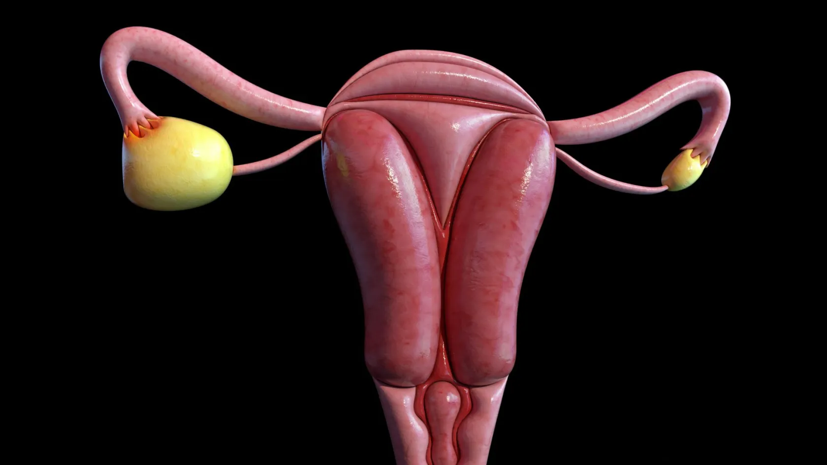
Debulking - Ovarian Cancer Tumor Reduction
Two women with the same diagnosis on the same morning can get operations that share almost nothing. What decides it is who measured the disease first.
About This Department
Debulking is not one operation with a fixed shape. It is a list of separate procedures assembled on the day, and two women with the same diagnosis on the same morning can end up with lists that share almost nothing. What decides which list you get is how carefully the disease was measured before the incision and how far into the abdomen the team is equipped to go.
Neither of those things is visible on a brochure. Both are askable.
Free consultation
Send the images themselves, and the radiology report second
Judging how extensive this operation would be means looking at the scan itself, so send the study on a disc or by file transfer. The places that decide it are the undersurface of both diaphragms, the small bowel mesentery and its root, the area around the stomach and spleen, the surface and hilum of the liver, and whether the peritoneum looks studded or smooth. Add the CA125 with its date, the albumin and the blood counts, and any operation note from a previous abdominal procedure of any kind, including a caesarean or an appendicectomy, since adhesions change what is achievable. Tell us whether fluid is collecting and how fast, how much weight you have lost and over what period, and how far you can walk without stopping. That last question is not small talk, since fitness for a long operation is part of the decision.
A word that has outlived its meaning
Debulking entered the language when the ambition really was to reduce bulk, on the theory that a smaller volume of remaining tumour would respond better to the chemotherapy that followed. Nobody aims at reduction any more. The aim is nothing visible left, and the older target of leaving deposits under a centimetre has been retired, a shift covered in more detail on our page about ovarian cancer surgery generally. What this page is about is the part that follows from that change, which is the operation itself. Once the target becomes zero, the operation stops being a standard procedure with an agreed extent and becomes a variable list assembled around wherever the disease has settled. One woman needs the ovaries, uterus and omentum and nothing else. Another needs those plus the peritoneum peeled off the pelvic sidewall, a segment of colon, the spleen and a patch of diaphragm. The second operation takes twice as long, carries several times the risk, and is the only one of the two that reaches the target in that patient. Calling both of them debulking hides more than it explains. One word, two operations.
Measuring the disease before you cut
Surgical teams do not decide this by eye.
Two formal measurements exist and both are worth knowing by name, because a unit that uses them is telling you something about how it works and a unit that has never mentioned them is telling you something too. Ask by name.
This table scrolls sideways on a narrow screen. Swipe or drag to see every column.
| Measurement | What it counts, and what it is used to decide |
|---|---|
| Peritoneal cancer index | The abdomen is divided into thirteen regions and each is scored from zero to three by the size of the largest deposit in it, giving a total out of thirty-nine. It quantifies how much disease there is and where, and it predicts both how radical the operation will need to be and how likely complete clearance is. |
| Laparoscopic predictive index | Six features are checked through a keyhole camera before committing to a large incision, covering the omentum, the liver surface, the lesser omentum and stomach and spleen, the parietal peritoneum, the diaphragms and the bowel. Developed in 2006 and updated in 2015, it has reported accuracy between 77 and 100 percent for predicting whether complete clearance is achievable. |
| Completeness of cytoreduction score | Recorded at the end rather than the beginning, this states what was left. Zero means nothing visible. It is the outcome the other two are trying to predict, and it belongs explicitly in your operation note as a number rather than as an adjective. |
| Surgical complexity score | A tally of how many and how demanding the procedures performed were, which is used to compare operations honestly between units. High complexity is where complete clearance in advanced disease usually lives, and it independently predicts serious postoperative complications. |
A prospective study of 354 women with stage IIIC or IV disease undergoing primary surgery shows how tightly these link together.
Splenectomy was performed in 206 of them, which is 58 percent, and spleen metastases were confirmed in 170 of those, meaning 82.5 percent of the spleens taken did contain disease. A high peritoneal cancer index independently predicted the need for splenectomy, with an adjusted odds ratio of 1.27, and a cut-off of 16 identified it with an area under the curve of 0.884. Splenectomy, the peritoneal cancer index and the surgical complexity score were all independent predictors of high-grade complications afterwards. Splenectomy, a high index and the completeness of cytoreduction were all independent predictors of worse survival. Everything links to everything.
Read carefully, that study says something more subtle than it appears. Removing the spleen does not cause a worse outcome. It marks out the women whose disease had spread far enough to need it, and the authors describe splenectomy as an indicator of high tumour burden, high surgical complexity, more complications and, indirectly, of how hard the surgeon was working to reach zero. A unit reporting that it never removes spleens is not reporting gentler surgery. It may be reporting a different threshold for stopping. Read it that way.
Where the prediction breaks down
Predicting resectability from imaging alone would save a great many women a laparoscopy, and the honest position is that it works for most of the abdomen and fails in one specific way that matters. Two findings, both invisible.
Ultrasound against what the camera found
A prospective study scored women with suspected ovarian or peritoneal cancer by ultrasound before surgery using the same six parameters as the laparoscopic index, then compared each parameter against what laparoscopy actually showed. Agreement, measured by Cohen's kappa where one would be perfect, ran between 0.70 and 0.90 for carcinomatosis on the bowel, the supracolic omentum, the liver surface and the diaphragms. It fell to 0.63 for the parietal peritoneum and 0.54 for the lesser omentum, stomach and spleen. Agreement on the overall score was 0.74. For two specific findings it collapsed, coming out at 0.57 for retraction of the small bowel mesentery and 0.36 for miliary carcinomatosis on the bowel surface. The authors concluded that ultrasound in a dedicated referral centre is useful in preoperative management, which is a careful way of saying it helps and does not settle it. The camera settles it.
The two findings imaging cannot reliably see are exactly the two that most often make complete clearance impossible.
Miliary disease means the bowel surface is dusted with deposits too small and too numerous to remove without taking the bowel itself, and a retracted mesentery means the tissue holding the small bowel has been drawn into a stiff mass. Either can be present on a scan reported as resectable. That gap is the reason many specialist centres perform a short laparoscopy before committing to a long operation, and it is a reasonable thing to ask about, since a laparoscopy that changes the plan has spared you an abdomen opened for nothing. Twenty minutes, well spent.
An abdomen opened and closed without achieving anything is the outcome everyone is trying to avoid, and it is not merely a disappointment. It carries the recovery of a major operation with none of the benefit, and it delays the start of chemotherapy in a woman who now needs it sooner. Where that risk exists, the sequence of chemotherapy first and surgery afterwards becomes the safer route, and how those two orders compare is covered on our ovarian cancer surgery page. Order matters less than you would think.
How complex is your operation
Complexity in this context is a formal idea, measured and recorded. It counts the number and difficulty of the individual procedures performed, so a hysterectomy with removal of the ovaries and omentum sits at the low end and the same operation plus peritoneal stripping, a bowel resection, splenectomy and diaphragm work sits at the high end. Knowing roughly where your planned operation sits changes what you should expect from the first week afterwards more than any other single fact. Ask which tier applies to you.
This table scrolls sideways on a narrow screen. Swipe or drag to see every column.
| Tier | What it usually includes, and what to expect afterwards |
|---|---|
| Low complexity | Removal of the ovaries and tubes, the uterus and the omentum, with peritoneal biopsies. Two to four hours, a ward bed instead of intensive care, five to seven days in hospital and a straightforward recovery. This is what most patients picture when they hear the word. |
| Intermediate complexity | Adds pelvic peritoneal stripping, removal of enlarged nodes, and often a rectosigmoid resection. Four to six hours, a night of close monitoring, seven to twelve days in hospital, and bowel function that takes weeks to settle down. |
| High complexity | Adds upper abdominal work, meaning diaphragm stripping or resection, splenectomy, extensive peritonectomy, sometimes partial liver or pancreatic tail resection and multiple bowel segments. Six to nine hours, intensive care by plan, ten to sixteen days, and the tier where complete clearance in advanced disease usually happens. |
| What the tier is not | A measure of how good the surgery was. Complexity follows the disease. A low complexity operation in a woman with limited disease is the right operation, and a high complexity one performed where the disease did not require it is simply more harm. |
The upper abdomen decides it
Ovarian cancer settles under the right diaphragm with great reliability, because the fluid carrying its cells circulates upward along the right side of the abdomen and pools there.
Any surgeon can clear a pelvis.
Clearing the space under the liver and the diaphragm is technically harder, sits at the edge of what a gynaecologist trained only in pelvic surgery will attempt, and is where operations are most often stopped short. If you want one question that separates units, it is what they do about the right upper quadrant. That answer sorts the field.
Sixty-four women had the right upper quadrant cleared by an extraperitoneal approach, meaning the dissection stays outside the peritoneal lining instead of working through it, gives a sense of what this involves. Full thickness resection of the diaphragm, rather than simply peeling the lining off it, was needed in 17 percent of women having a first operation and in 44 percent of those operated on for a recurrence. Complete clearance was achieved in 70 percent. The commonest complication afterwards was fluid collecting around the lung, occurring in 32 percent, which follows naturally from having opened or stripped the sheet of muscle separating the abdomen from the chest. Those figures are not a warning. They are a description of what a serious attempt at the upper abdomen costs and what it achieves. A pleural effusion is usually managed with a drain and resolves. Shoulder tip pain in the days afterwards is common and is referred pain from the diaphragm, signalling nothing wrong. What matters is that a team offering complete clearance should be able to say how often they operate on the diaphragm and what happens when they do, and a unit that never goes there has quietly capped what it can achieve for you. Nobody says so out loud.
The bowel decision
Where disease encases bowel, the surgeon has to choose between leaving it and taking a segment out, and the second option introduces the complication most feared in this operation, which is a leak from the join between the two cut ends. A systematic review pooled 18 studies covering 4,622 women undergoing cytoreduction, of whom 344 had a leak.
Women whose surgery was complicated by a leak had a significantly higher death rate at 30 days, though by 60 days that difference had gone.
What failed to predict a leak is the interesting part. Body mass index did not. Anaesthetic risk grade did not. Age, smoking, whether the surgery was primary or interval, whether the join was stapled or hand sewn, whether heated chemotherapy was used and whether chemotherapy had been given beforehand all showed no association. Nor, remarkably, did forming a diverting stoma, which is the manoeuvre most often performed specifically to protect the join. Only one factor was associated with leaks, and it was the number of bowel resections performed. Multiple resections raised the risk. Count them, not the stoma.
What follows from that is practical.
The question worth asking is not whether a stoma will be formed but how many separate pieces of bowel are expected to come out, because that number is the risk. A surgeon planning three separate small bowel resections to chase deposits is making a different trade from one planning a single rectosigmoid resection, and the authors of that review concluded plainly that the number of resections should be minimised in this population. Where multiple segments would be needed to reach zero, that is one of the situations in which stopping short is the better medicine. Restraint is a decision too.
The order of the day
High complexity clearance runs to six or nine hours, and the sequence is deliberate, since each stage informs whether the next one is worth starting.
Scoring the abdomen
Before anything is removed, all thirteen regions are inspected and scored, sometimes through a camera before the incision is even made. The number that comes out of this determines whether the plan is a clearance or a biopsy, and it is written down.
Upper abdomen first
Many teams clear the diaphragms, the liver surfaces and the area around the spleen before touching the pelvis, and the reasoning is sound. If the upper abdomen cannot be cleared, zero is unreachable, and finding that out after four hours of pelvic work helps nobody.
The pelvis as one block
Where the pelvis is filled with tumour, the ovaries, uterus, pelvic peritoneum and often the rectosigmoid are removed together as a single specimen instead of piece by piece. Working around a mass tends to leave fragments, and taking it in one piece does not.
Rejoining, and the final look
Bowel ends are joined once the clearance is finished, never partway through, so that the anastomosis is made in a settled field. Then every region is checked again and the completeness score is recorded. That last inspection is what turns an impression into a documented result.
Blood loss in the high complexity operations is substantial and transfusion is planned in advance. Two consultant teams operating together is common, most often a gynaecological oncologist with a general or hepatobiliary surgeon for the upper abdominal and bowel work, and hearing that two teams are involved should read as thoroughness rather than as a complication. Two teams, one plan.
Not over when the wound closes
One measure of surgical success is almost never mentioned to patients and is increasingly treated by specialists as decisive. It is called return to intended oncological therapy, and it asks a simple question. Did this woman get back to the chemotherapy that was planned for her, on schedule, or did the operation take that away from her. That is the real scoreboard.
A narrative review of the literature from 2010 to 2024 sets out why it matters. Delayed return to intended therapy after surgery is associated with poor survival in ovarian cancer, which reframes the whole operation. A technically brilliant clearance followed by a complication that pushes chemotherapy back by six weeks may leave a woman worse off than a slightly less complete operation she recovered from quickly. The review works through what actually influences it, naming preoperative counselling and education, medical optimisation before surgery, careful surgical planning and execution, early recognition of complications, and comprehensive postoperative care. What that translates into is unglamorous and it works. Nutrition assessed and corrected before the operation, while there is still time. Anaemia treated in advance. Enhanced recovery protocols, meaning early feeding, early mobilisation and avoiding the routines that used to keep women in bed for days. A team that includes anaesthetists, nurses, physiotherapists, dietitians and psychologists as a matter of course. And, the review notes explicitly, the patient's own relatives and friends, whose involvement turns out to be part of the machinery and no mere courtesy. Age, nutritional state and the occurrence of complications are the factors that decide whether treatment restarts on time. Nutrition first, always.
In practical terms, one question goes to any surgeon offering the operation.
What proportion of your patients start chemotherapy within six weeks. A team that measures it will tell you. A team that has never considered the question has told you something as well. Silence is an answer.
What to ask before consenting
Everything above reduces to a short list of questions that a specialist unit will answer without hesitating and a general one will find awkward. None of them is confrontational and all of them are ordinary practice to ask. Take the list with you.
- What is my peritoneal cancer index, or your best estimate of it from the imaging, and what does that predict about reaching zero.
- Will a laparoscopy be done first to check, and if not, why is that unnecessary in my case.
- Do you operate on the diaphragm and the upper abdomen, and how often.
- How many separate bowel resections do you anticipate, and what happens if it turns out to be more than that.
- Will a second surgical team be involved, and from which specialty.
- What proportion of your patients with disease like mine end with no visible residual.
- What proportion start chemotherapy within six weeks of the operation.
- If complete clearance proves impossible once you are inside, what will you do.
That last question is the most revealing of the eight. The answer worth hearing is that the operation will be stopped and chemotherapy started, followed by a reassessment for a second attempt. The answer worth worrying about is one that treats a partial clearance as a good enough result, since an incomplete operation carries the full recovery and delivers a fraction of the benefit, and the woman then goes into chemotherapy weakened rather than helped. Stopping is sometimes the win.
Coming to Istanbul
How long you need to stay in Istanbul depends on the complexity tier, and the honest ranges are three to four weeks for a low or intermediate complexity clearance and five to seven weeks for a high complexity one, hotel nights included. The first week is spent on measurement. Imaging is reviewed against the thirteen regions, nutritional state and anaemia are assessed and corrected, fitness for a long anaesthetic is tested, and the case goes to a gynaecological oncology multidisciplinary meeting. Where the scan leaves the question of resectability open, a diagnostic laparoscopy is arranged first, and that short procedure changes the plan often enough to justify itself. Chemotherapy is generally delivered at home, and the schedule is the reason this trip is planned around getting you back. We will tell you before you travel which cycles belong here and which belong with your own oncologist, and the aim written into the plan is that treatment restarts within six weeks of the operation.
Flying home after a high complexity clearance is reasonable at around three weeks, once the wound has healed, bowel function has returned, any chest drain is long out and the pathology has been discussed with you face to face. Clot risk is high after this operation, so anticoagulant injections continue for four weeks, compression stockings matter, and walking the cabin is part of the plan. Where a stoma was formed, you leave with supplies, training and a written plan for reversal. Interpreting is arranged in advance in English, Arabic, Russian, French and German, and you go home with the operative note listing every procedure performed and stating the completeness score explicitly. Check that line before you fly.
What moves the cost
Complexity is the cost, more directly here than in any other operation covered in this guide, because the tier decides the theatre time, the number of surgical teams, the intensive care requirement and the length of stay all at once.
Quoting before the imaging has been scored attaches a price to a tier nobody has established yet.
- Which complexity tier the operation lands in, which follows the disease and cannot be chosen.
- Whether a staging laparoscopy is performed first as a separate procedure.
- Whether the diaphragm is stripped or resected, and whether a chest drain is needed afterwards.
- Whether the spleen comes out, with the vaccination and antibiotic arrangements that follow.
- How many bowel resections are performed and whether a stoma is formed, since that carries supplies and a later reversal.
- Whether a second consultant team operates alongside, and from which specialty.
- How many nights of intensive care are needed, which at high complexity is planned rather than exceptional.
- How much blood is transfused, and how extensive the pathology on a specimen of that size turns out to be.
Ask any written quotation five things. Does it cover intensive care for as many nights as prove necessary. Does it cover treatment of a complication, naming a bowel leak, a chest collection and a wound problem specifically. Does it cover a return to theatre. Does it include the full pathology on a large multi-organ specimen. And what happens to the figure if the operation turns out one tier higher than the imaging suggested, which is common rather than exceptional in this disease.
A number that survives those is one you can plan around. Ask all five.
Follow-up once you are home
The handover after this operation is more detailed than after most, because your own oncologist needs to know precisely what was removed in order to interpret every scan that follows. A liver edge that looks abnormal on a scan is a different matter in a woman whose diaphragm was resected and repaired than in one whose upper abdomen was never entered, and nobody can tell the difference without the operative note. Keep your copy safe.
Four documents make that work, and you should leave with all of them.
- The operative note listing every procedure by name, with the peritoneal cancer index at the start and the completeness score at the end.
- The pathology report on the whole specimen, with subtype, grade and final stage.
- A dated written plan for restarting chemotherapy, naming who will give it and by when.
- Where the spleen was removed or a stoma formed, the vaccination record with dates, the antibiotic plan and the arrangements for reversal.
Our team stays reachable for your oncologist afterwards, and where a scan raises a question that turns on what was done in theatre we would rather answer it directly than have it guessed at. Send it over.
Frequently asked questions about debulking surgery
What is the peritoneal cancer index and why does it matter to me?
Can a scan tell whether complete clearance is possible?
Why would my spleen be removed for an ovarian cancer operation?
Does a stoma protect against a bowel leak?
What happens if my diaphragm is involved?
How soon after the operation should chemotherapy start?
How long does debulking surgery take, and how long is the hospital stay?
How long should I plan to be in Istanbul?
References
- Karlsson E, Vorobii O, Silins I, Sundström Poromaa I, Stålberg K, Lomnytska M. Splenectomy as an indicator for ovarian cancer spread and complete cytoreduction. Gynecologic Oncology. 2025;197:121-128.
- Moruzzi MC, Bolomini G, Esposito R, et al. Diagnostic performance of ultrasound in assessing the extension of disease in advanced ovarian cancer. American Journal of Obstetrics and Gynecology. 2022;227(4):601.e1-601.e20.
- Fornasiero M, Geropoulos G, Kechagias KS, et al. Anastomotic leak in ovarian cancer cytoreduction surgery, a systematic review and meta-analysis. Cancers. 2022;14(21):5464.
- Acs M, Leebmann H, Häusler S, Harter P, Piso P. Extraperitoneal approach during peritonectomy in the right upper quadrant for peritoneal metastases from ovarian malignancies. In Vivo. 2022;36(1):341-349.
- Tse KY, Chu MMY, Chiu JWK, et al. Return to intended oncological therapy following advanced ovarian cancer surgery, a narrative review. Anaesthesia. 2025;80(Suppl 2):106-114.
Editor's note
Written by the Biruni Hospital medical editorial team. Reviewed by Prof. Dr. Mustafa Teoman YANMAZ, Medical Oncology.
Medically reviewed by

Prof. Dr. Mustafa Teoman YANMAZ
Medical Oncology
Related Treatments
View All-
Bilateral Mastectomy
Whether you carry a BRCA mutation, have cancer in one breast or in both, bilateral mastectomy removes both breasts in one operation, and how much it helps depends on which of those three paths brought you to it.
-

Breast Conserving - Lumpectomy Surgery
Keeping the breast has never been the less thorough choice, and the largest recent studies now point the other way. This page explains who can have breast conserving surgery, what the margin report decides, and how long a woman traveling for it needs to stay.
-

Cytoreductive - Tumor Reduction Surgery
Patients cleared of every visible deposit lived 30 months in one gastric series. Those left with anything at all lived 7.3 months, however little remained.
-
Debulking Surgery
Surgeons removed a median of 57 lymph nodes from women already cleared of visible disease. It bought no extra months, and tripled deaths within 60 days.
-

Esophageal Cancer Surgery
Nothing is put back where it was. Your stomach is turned into a narrow tube, pulled up through the chest and joined to what remains of the gullet, and almost everything about eating and sleeping afterwards follows from that.