
Esophageal Cancer Surgery
Nothing is put back where it was. Your stomach is turned into a narrow tube, pulled up through the chest and joined to what remains of the gullet, and almost everything about eating and sleeping afterwards follows from that.
About This Department
Nothing goes back where it was. The oesophagus cannot be replaced or repaired, so the surgeon takes your stomach, turns it into a narrow tube, pulls it up through the chest and joins it to whatever length of gullet remains. That single manoeuvre explains almost everything about life afterwards, from why meals become small and frequent to why you will sleep propped up for the rest of your life. This page starts there, because most patient information starts with the incisions and never explains the thing that actually changes about your life.
Free consultation
Send the endoscopy report with the tumour distance from the teeth
How far down the gullet the tumour sits decides which operation is proposed and where the join ends up, and endoscopists record that distance in centimetres from the front teeth. Send that report, the biopsy including HER2 and PD-L1 where they were done, the staging CT and PET as image files, the endoscopic ultrasound if one was performed, and your weight now against six months ago. What comes back is an opinion on whether resection is realistic, whether chemotherapy or chemoradiotherapy should come first, and what eating would look like a year from now. The review costs nothing and carries no obligation.
The stomach becomes the gullet
Your oesophagus is a muscular tube with no spare length and no substitute. Take a section out and the two ends cannot be brought together, so something else has to bridge the gap, and in almost every case that something is the stomach. Surgeons divide it along its length into a narrow tube perhaps four centimetres wide, keeping one artery that runs up its right side, and that tube is then lifted through the chest to reach the remaining gullet.
Four consequences follow immediately, and every one of them is permanent.
Sitting in the chest, the stomach no longer holds a meal. Its capacity after tubularisation is a fraction of what it was, so meals become small and frequent and stay that way. The valve at the top of the stomach that stopped acid rising is gone, removed with the specimen, so reflux is now unopposed and gravity is the only thing preventing it, so sleeping flat becomes impossible. The nerve that told the stomach to empty is divided during the dissection, so a drainage procedure at the outlet is added to compensate. And the new tube sits inside the chest, so breathing and eating interfere with each other in ways nobody predicts, since a full conduit presses on a lung that has just been through an operation of its own. That is the operation working as designed, and understanding it in advance is the difference between a patient who adapts and one who spends a year assuming something has gone wrong.
None of that is a complication.
Almost nobody operates first
Outside the earliest tumours, surgery alone has not been standard treatment for more than a decade. Chemoradiotherapy comes first for most patients, and the trial that established it has now reported ten years of follow-up.
Ten years of follow-up on 366 patients
Dutch investigators randomised patients with resectable oesophageal or junctional cancer to five weekly cycles of carboplatin and paclitaxel with concurrent radiotherapy before surgery, or to surgery alone. After a median follow-up of over twelve years, overall survival was better with treatment first, at a hazard ratio of 0.70, and the absolute ten year survival benefit was 13 percentage points, at 38 percent against 25. The benefit did not fade with time. Death specifically from oesophageal cancer fell, at a hazard ratio of 0.60, while death from other causes was the same in both arms. Isolated local recurrence fell furthest of all, at a hazard ratio of 0.40, which is exactly the pattern you would expect from radiotherapy delivered to the tumour bed before anybody operated on it, and the reason the treatment is given in that order rather than the other way round.
Five weeks of treatment. Then six to eight weeks of recovery. Then surgery. That timetable does more than anything else to disrupt the plans for somebody arranging treatment across borders, and it has to be settled before anybody books a flight.
Where the specimen still contains viable cancer despite the chemoradiotherapy, a year of immunotherapy improves the odds. A trial of 794 such patients found median disease-free survival of 22.4 months with the drug against 11.0 months with placebo, a hazard ratio of 0.69, at a cost of serious side effects in 13 percent against 6 percent. What the pathologist finds decides that treatment, which means the pathology report from your operation determines a year of your treatment and has to travel home with you in a form an oncologist can act on within weeks rather than months.
Where the incisions go
Oesophagectomies take their names from the body cavities the surgeon has to enter, and the tumour's height decides that. A tumour low down near the junction with the stomach can be reached through the abdomen and the right chest. One higher up needs the neck opened as well.
This table scrolls sideways on a narrow screen. Swipe or drag to see every column.
| Operation | Incisions, and where the join ends up |
|---|---|
| Ivor Lewis | Abdomen and right chest. The join sits inside the chest, high up. The commonest operation for tumours in the lower oesophagus and at the junction. |
| McKeown | Abdomen, right chest and neck. The join sits in the neck. Used for tumours higher up, and where a longer clearance above the tumour is needed. |
| Transhiatal | Abdomen and neck, with the chest never opened and the oesophagus mobilised blindly from below. Avoids a thoracotomy at the cost of a less complete node clearance. |
| Minimally invasive and robotic | Any of the above performed through small incisions with a camera, or with robotic instruments. The named operation stays the same and only the access changes. |
Access has now been tested properly. A Dutch trial randomised 112 patients to robotic minimally invasive oesophagectomy or open surgery through the chest, and found overall surgery-related complications in 59 percent of the robotic group against 80 percent of the open group. Pulmonary complications fell by nearly half, cardiac complications by more than half, blood loss was lower, pain scores were lower and both functional recovery and quality of life were better. Cancer outcomes were the same at forty months.
So where a unit offers it competently, minimally invasive surgery is the better version of the same operation. Where it does not, an open oesophagectomy performed well by somebody who does forty a year beats a keyhole one performed by somebody learning.
Neck or chest, and why it matters
Joining the stomach tube to the remaining oesophagus is the decision with the longest reach, and it produces two genuinely different recoveries.
Joins in the neck are easier to reach if they leak. The wound can be opened at the bedside, the leak drains outwards through the skin, and although it is unpleasant it is rarely life-threatening. A join inside the chest is more difficult to reach and a leak there spills into the chest cavity, which is a far more serious event. That argues for the neck.
Set against it, the nerve that supplies the voice box runs alongside the oesophagus in the neck and is at real risk during that dissection.
What 1,681 patients showed about the two joins
Pooling five studies of patients having totally minimally invasive oesophagectomy, the rate of anastomotic leak did not differ between the chest join and the neck join, with a relative risk of 1.39 and a confidence interval crossing one. What did differ was injury to the nerve supplying the voice box, which was substantially more common with the neck join, at a relative risk of 6.70. The chest join also produced a shorter hospital stay and less blood loss. The authors write carefully about their own evidence, noting that none of the included studies were randomised and all carried a moderate risk of bias, and a randomised trial was under way at the time they wrote, which is the correct way to describe evidence of this quality, and a great deal more honest than the way it usually reaches patients.
A hoarse voice and a weak cough are not trivial after an operation where the ability to clear your chest determines whether you develop pneumonia. That is the trade. Where the tumour sits low enough to allow either, most units now prefer the chest.
The operation, in order
Four to eight hours, in two or three separate stages with the patient repositioned between them.
The abdominal stage
Surgeons free the stomach from everything around it while preserving its right-sided artery with great care, since the whole reconstruction will depend on that one vessel. The nodes along the coeliac vessels come out. A feeding tube is placed into the small bowel, and a drainage procedure at the stomach outlet is usually added.
Making the tube
Staplers divide the stomach along its length, producing a narrow conduit and leaving the part nearest the oesophagus attached to the specimen. Some units check the blood supply of that new tube with a fluorescent dye and a camera before committing to it, since a poorly perfused conduit is the commonest cause of a leak later.
The chest stage
Deflating the right lung makes room. Dissection frees the oesophagus from the aorta, the airway and the pericardium, and the nodes running alongside it are taken. Two structures are at risk here. The nerve to the voice box, and the duct carrying lymph from the lower body.
The join, and the drains
The specimen comes out, the conduit is drawn up, and the join is made by stapler or by hand at the chosen level. A chest drain goes in on the operated side, and a tube through the nose keeps the new conduit empty while it settles. You wake in intensive care.
Between a fifth and a third of patients booked for this operation turn out to need something different once the abdomen or chest is inspected, and that possibility belongs in the conversation before anybody travels.
The first two weeks
What surprises people most is that you do not eat. Published practice keeps patients off oral intake for the first five to seven days at minimum, and many units wait ten days to two weeks and confirm with a contrast swallow that the join is watertight before allowing anything by mouth.
Nutrition continues throughout, delivered into the small bowel through the feeding tube placed during the operation, beginning on day two or three. That tube does not count as an afterthought. A cohort study comparing patients who had one against those who did not found weight loss at two weeks of 3.2 percent against 7.2 percent, and a hospital stay of eleven days against fifteen.
- Days one and two in intensive care, sitting up, with the chest drain on suction and physiotherapy starting immediately.
- Days two to four, feeding into the small bowel begins and builds, the epidural comes down onto tablets, and you walk further each day.
- Days four to seven, the chest drain comes out once it is dry and the lung is up, and the nasogastric tube usually follows.
- Around days seven to ten, a contrast swallow or a scan checks the join before anything is allowed by mouth.
- Days ten to fourteen, sips build to free fluids and then to a soft diet, with the feeding tube still supplying most of your calories.
Many people go home with that feeding tube still in place and use it overnight for weeks, which sounds worse than it is and prevents the weight loss that otherwise takes months to reverse.
What goes wrong
Oesophagectomy has one of the highest complication rates in elective surgery, and the trial comparing robotic with open surgery is instructive on the scale of it. Even in the better arm, 59 percent of patients had a complication of some kind.
The lungs, first and worst
Chest problems dominate. Pneumonia, collapse of the lung base, and fluid collecting around the lung together account for more trouble than everything else combined, which is why the physiotherapist appears within hours and why the pain relief exists to let you cough rather than to keep you comfortable lying still.
The join, and everything after it
Anastomotic leak comes next and it is the one that defines the operation. It appears between day five and day ten. A neck leak drains outwards and is managed by opening the wound. A chest leak is drained by a radiologist or by a stent placed endoscopically across it, and occasionally needs a return to theatre. Most heal, and the price is weeks rather than days.
This table scrolls sideways on a narrow screen. Swipe or drag to see every column.
| Problem | When it shows itself, and what is done |
|---|---|
| Chest infection and lung collapse | Days two to five, and commoner than everything else combined. Physiotherapy, pain relief that lets you cough, and antibiotics where needed. |
| Anastomotic leak | Days five to ten. A neck leak is drained by opening the wound. A chest leak needs radiological drainage, or an endoscopic stent placed across it, and occasionally a return to theatre. |
| Chyle leak | Once feeding starts, when milky fluid appears in the chest drain. Managed by changing the fat in the feed, and occasionally by blocking the duct radiologically. |
| Hoarse voice | Noticed on waking. Bruising of the nerve to the voice box recovers over months, and a divided nerve does not, so the cough is weak in the meantime. |
| Narrowing at the join | Weeks to months later, as food starts to stick. Stretched at endoscopy, sometimes more than once, and it responds well. |
Two more deserve naming. Atrial fibrillation follows any operation inside the chest commonly enough and usually settles on medication. Conduit necrosis, where the new stomach tube loses its blood supply altogether, is rare and is the reason some units check the perfusion with fluorescent dye before committing to the reconstruction.
Death within 90 days is meaningfully higher after this operation than after most cancer surgery, and it falls sharply with hospital volume, which makes the annual number of oesophagectomies a reasonable thing to ask about directly.
Eating, sleeping and reflux
Five years from now, this is the section that will have mattered, and it follows directly from the first section of this page.
Small, often, and slowly
Tubularised, the stomach holds a fraction of what it held, so six to eight small meals replace three and that pattern is permanent rather than a phase of the recovery. Eat slowly and chew thoroughly, since the conduit has no muscular squeeze of its own and food travels down largely by gravity. Sit upright for a meal and stay upright for an hour afterwards. Expect some foods to stick early on, particularly bread, dry meat and anything fibrous, and expect that to improve steadily across the first six months as the conduit settles and you learn what it will take. Dumping happens here too, when a meal rich in sugar arrives in the small bowel too quickly, producing sweating, cramps, palpitations and diarrhoea within half an hour of eating. Cutting simple sugars and putting protein and fat into each meal handles most of it, and a dietitian who has managed this before is worth more here than any amount of reading about it.
The bed, and the night
The valve that kept acid out of the oesophagus was removed with the tumour, so nothing now stops stomach contents rising except gravity. Lie flat and they reach the throat and the airway. Inhaling them at night causes coughing, hoarseness and occasionally pneumonia. Raise the head of the bed by fifteen to twenty centimetres on blocks rather than piling up pillows, which only bends you at the waist and makes it worse. Stop eating three hours before lying down. Acid suppressing medication is prescribed at discharge and continued indefinitely, since the anatomy that made it necessary is permanent and no amount of time will restore the valve that was removed. Four habits do most of the work here, and all of them are about position and timing rather than about which foods you choose, which is the part patients expect to be told and the part that matters least.
- Raise the head of the bed on blocks by fifteen to twenty centimetres. Not on pillows.
- Finish eating three hours before you lie down, and keep the last meal small.
- Sit upright to eat and stay upright for an hour afterwards, including on aeroplanes and in cars.
- Take the acid suppressing medication continuously and expect to stay on it.
Weight falls and then settles. Ten to fifteen percent is common in the first six months, and stopping the fall matters far more than reversing it, since the treatment that follows is given to people strong enough to receive it. Weigh yourself weekly and write the number down, because a downward trend is far easier to see on paper than in a mirror, and far easier to reverse in month two than in month six.
How long you stay
Five to six weeks in the country for the surgery alone, which is longer than for any other operation described on this site and is not padding.
Assessment takes five to seven days. Endoscopy repeated with the distance from the teeth recorded, staging CT and PET, endoscopic ultrasound where the depth is uncertain, lung function testing and an echocardiogram since the chest is being opened, nutritional assessment, and a tumour board before a date is fixed. Where chemoradiotherapy is given first, the whole timetable shifts by roughly three months and needs planning as two trips or one long stay.
Expect ten to sixteen days on the ward when nothing goes wrong, beginning with one or two nights in intensive care. Add a further two weeks nearby afterwards, which covers the swallow test if it has not already happened, the transition from tube feeding to oral intake, the pathology discussion that decides the immunotherapy question, and the dietitian sessions that make the difference between coping and not.
Flying gets cleared at around four to five weeks, with drains out, no fever, no undrained collection, a lung that is fully up on the X-ray, an oral intake that is holding your weight and a written plan for clot prevention. A long flight sitting upright is easier than most people fear. Lying back is the problem, so book a seat you can keep raised.
What drives the cost
Two or three body cavities, a stapler-heavy reconstruction and a planned intensive care stay put this operation among the most expensive on any hospital list. Eight things move the total.
- Which operation is planned. A three-stage McKeown occupies a theatre considerably longer than a two-stage Ivor Lewis does.
- The approach, because robotic and thoracoscopic platforms carry per-case instrument costs that open surgery does not.
- The stapler cartridges the conduit and the join consume, which is a real and substantial line item here.
- Intensive care nights, which are planned rather than exceptional after this operation.
- Ward days, which the chest and the join decide between them and which nobody can promise in advance.
- Endoscopic or radiological treatment of a leak, stent included, which a meaningful minority of patients turn out to need.
- Feeding tube supplies and dietitian input, which continue after discharge and are frequently uncosted.
- Chemoradiotherapy before and immunotherapy after, which together exceed the surgical cost and sit outside almost every package.
Arriving underweight, with poor lung function, with heart disease, still smoking, or having already had radiotherapy to the chest each raise the chance of a longer stay and of care nobody planned for.
Packages published here ordinarily cover the transfers, the pre-admission workup, the operating fees, an agreed count of theatre and ward days, an interpreter, accommodation and the appointments before departure. Sitting outside it, almost always, are the flights, insurance, ward nights past the agreed count, unplanned critical care, the treatment of a leak and all drug therapy.
Six questions turn a headline into a quotation. Which operation does the figure assume, named by the number of stages it has. What number of intensive care nights sits inside it. How many ward nights, and the price of an extra one. Is endoscopic stenting for a leak covered. Are the feeding tube supplies included after discharge. And whether the specimen pathology, which determines whether you need a year of immunotherapy, sits inside the figure.
No quotation means anything until a surgeon has read your endoscopy and your scans. That review costs nothing.
Once you are home
One document decides the next year of your treatment. If the pathologist found viable cancer left in the specimen after chemoradiotherapy, you are a candidate for a year of immunotherapy that lengthened median disease-free survival from 11.0 to 22.4 months in the trial that established it. An oncologist at home cannot make that decision without the report in front of them.
Take home the operative note naming the operation and where the join sits, the full pathology with the response to treatment and the node count, the imaging on disc, your discharge weight, the feeding tube instructions if one is still in place, and the name of somebody here who answers messages. Get it in English before you leave the building.
Surveillance runs on clinical review and CT every six months for the first two to three years, with endoscopy where symptoms call for it. Difficulty swallowing that returns is investigated promptly, since a benign narrowing at the join is far commoner than a recurrence and is fixed by stretching it at endoscopy.
Get in touch here for a fever, chest pain, breathlessness, food sticking, vomiting, a cough that starts at night, or weight that is still falling at three months. A message answers most questions within hours.
Frequently asked questions about oesophageal cancer surgery
What replaces the oesophagus after it is removed?
Why will I sleep sitting up for the rest of my life?
Why can I not eat or drink for ten days afterwards?
Is the join better in the neck or the chest?
Should I have treatment before the operation?
How long do I need to stay abroad, and when can I fly?
References
- Eyck BM, van Lanschot JJB, Hulshof MCCM, et al. Ten-year outcome of neoadjuvant chemoradiotherapy plus surgery for esophageal cancer, the randomized controlled CROSS trial. Journal of Clinical Oncology. 2021;39(18):1995-2004.
- Kelly RJ, Ajani JA, Kuzdzal J, et al. Adjuvant nivolumab in resected esophageal or gastroesophageal junction cancer. The New England Journal of Medicine. 2021;384(13):1191-1203.
- van der Sluis PC, van der Horst S, May AM, et al. Robot-assisted minimally invasive thoracolaparoscopic esophagectomy versus open transthoracic esophagectomy for resectable esophageal cancer, a randomized controlled trial. Annals of Surgery. 2019;269(4):621-630.
- van Workum F, Berkelmans GH, Klarenbeek BR, Nieuwenhuijzen GAP, Luyer MDP, Rosman C. McKeown or Ivor Lewis totally minimally invasive esophagectomy for cancer of the esophagus and gastroesophageal junction, systematic review and meta-analysis. Journal of Thoracic Disease. 2017;9(Suppl 8):S826-S833.
Editor's note
Written by the Biruni Hospital medical editorial team. Reviewed by Prof. Dr. Metin GÜDEN, Radiation Oncology.
Medically reviewed by
Prof. Dr. Metin GÜDEN
Radiation Oncology
Related Treatments
View All-
Bilateral Mastectomy
Whether you carry a BRCA mutation, have cancer in one breast or in both, bilateral mastectomy removes both breasts in one operation, and how much it helps depends on which of those three paths brought you to it.
-

Breast Conserving - Lumpectomy Surgery
Keeping the breast has never been the less thorough choice, and the largest recent studies now point the other way. This page explains who can have breast conserving surgery, what the margin report decides, and how long a woman traveling for it needs to stay.
-
Cytoreductive - Tumor Reduction Surgery
Patients cleared of every visible deposit lived 30 months in one gastric series. Those left with anything at all lived 7.3 months, however little remained.
-
Debulking - Ovarian Cancer Tumor Reduction
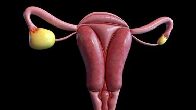
Two women with the same diagnosis on the same morning can get operations that share almost nothing. What decides it is who measured the disease first.
-
Debulking Surgery
Surgeons removed a median of 57 lymph nodes from women already cleared of visible disease. It bought no extra months, and tripled deaths within 60 days.