When a tumor presses on the spinal cord, an operation followed by radiotherapy kept 84 percent of patients walking in a randomized trial, against 57 percent with radiotherapy alone. This guide explains which spinal tumors need surgery, how benign tumors are removed, how metastases are treated with fixation and radiosurgery, and the stay.
About This Department
Orthopedics
A spinal tumor pressing on the cord is a race against time. Surgery wins back walking.
In a randomized trial of 101 patients with spinal cord compression from cancer, 84 percent of those operated on and then irradiated could walk afterward, against 57 percent with radiotherapy alone. Send the MRI of the whole spine, any CT and biopsy report, and a note on weakness, numbness and bladder control, and our spine surgery team will tell you whether an operation fits and how soon.
84%
Walking after surgery and radiotherapy, against 57% with radiotherapy alone
Spinal tumors grow in the bones of the spine, in the coverings of the spinal cord, or inside the cord itself. Metastases make up the majority, cancer spread from the breast, lung, prostate, kidney or thyroid, or myeloma of the bone marrow. A smaller group starts in the spine, from benign meningiomas and nerve sheath tumors to ependymomas, chordomas and sarcomas.
Location decides the operation.
This table scrolls sideways on a narrow screen. Swipe or drag to see every column.
Where spinal tumors grow
Location
Common tumors
Aim of surgery
In the vertebra and outside the dura (extradural)
Metastases, myeloma, chordoma, sarcoma
Free the cord, stabilize the spine, and remove the tumor in one piece for primary bone cancers
Inside the dura, outside the cord (intradural extramedullary)
Meningioma, schwannoma, neurofibroma
Complete removal, which cures most of these benign tumors
Inside the spinal cord (intramedullary)
Ependymoma, astrocytoma, hemangioblastoma
Maximum safe removal under nerve monitoring
Primary tumors and metastases
A metastasis tells the team the cancer has spread, and surgery then aims to protect the spinal cord, keep the spine stable and relieve pain, while radiotherapy and drug treatment control the cancer. The operation alone cures many primary tumors of the spine, especially benign ones.
The Spine Instability Neoplastic Score is a comprehensive classification system with content validity that can guide clinicians in identifying when patients with neoplastic disease of the spine may benefit from surgical consultation.
Spine Oncology Study Group, Spine, 2010
What to send for a free review
MRI of the whole spine with and without contrast, as image files, with the report. CT of the affected level if done.
Any biopsy report, the primary cancer diagnosis and treatment so far, and a short note of symptoms, with the date weakness or numbness began and whether bladder or bowel control has changed.
A spine surgeon reviews the file within the week. With weakness, the same day.
1
MRI of the whole spine
Shows the tumor and every level it touches.
2
CT and staging
CT shows how much bone the tumor has destroyed and maps the path of every screw, while a PET-CT or a CT of the chest, abdomen and pelvis finds the primary cancer and any other spread.
3
Tumor board
Spine surgeon, radiation oncologist and medical oncologist weigh the neurological, oncological, mechanical and general health picture.
Why a biopsy sometimes comes first
A tumor that may be a primary bone cancer, such as a chordoma or sarcoma, needs a biopsy planned by the surgeon who will operate, through a path that the operation can remove, because these tumors must come out in one piece. Myeloma and lymphoma respond to radiotherapy and chemotherapy, and a biopsy that confirms them can spare the patient an operation.
Tumors that need no operation
Myeloma, lymphoma and many breast and prostate metastases without cord compression or instability are treated with radiotherapy, stereotactic radiosurgery and drugs.
Steroids when weakness starts
New leg weakness, numbness rising up the body or loss of bladder control in a patient with cancer points to spinal cord compression. High-dose dexamethasone the same day reduces swelling around the cord while the MRI and surgical plan are arranged.
Tumors that radiation controls, and those it does not
Lymphoma, myeloma, breast, prostate and small cell lung cancers respond to conventional radiotherapy. Kidney, thyroid and colon cancers, melanoma and sarcoma resist it, and need high-dose stereotactic radiosurgery or surgery to control them.
4
Urgent pathway
Patients losing strength are assessed, scanned and operated on within 24 to 48 hours, because function lost for days returns less often than function lost for hours.
When surgery is urgent
Speed matters most for patients who can still walk. Surgery that frees the cord before paralysis sets in keeps most of them walking.
Who needs surgery for a spinal tumor?
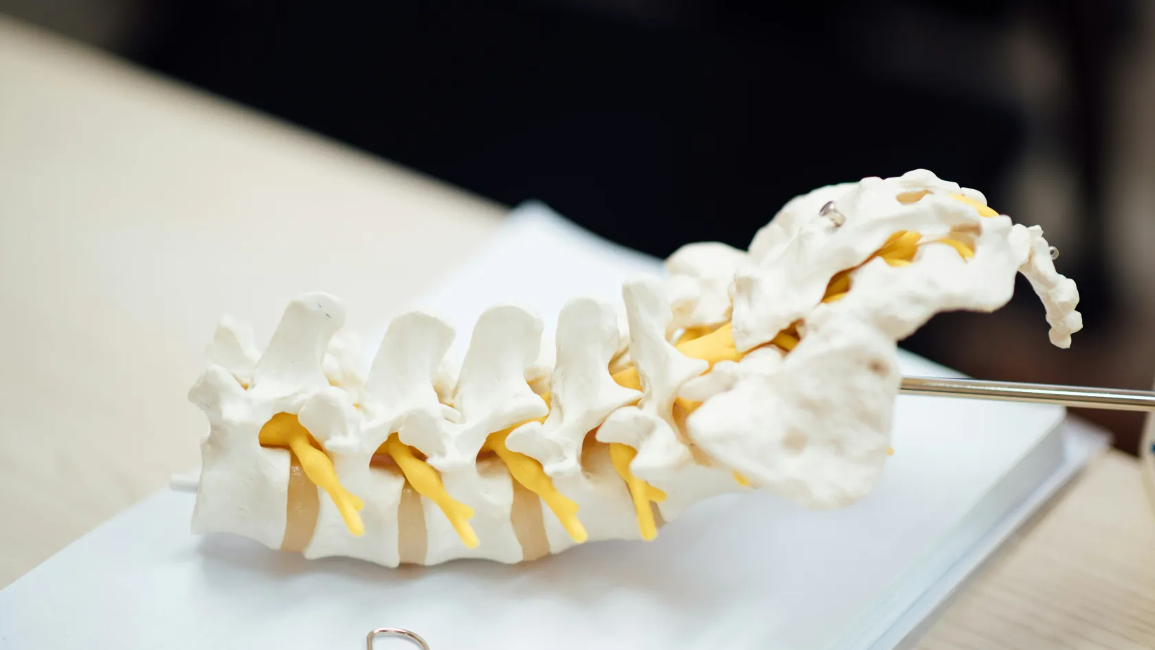
Four questions decide it, covering compression of the cord and any weakening, whether radiotherapy controls this tumor type, whether the spine stays stable or collapses and hurts on movement, and whether the patient's general health and outlook can carry an operation and its recovery. The Spine Instability Neoplastic Score, built by an international panel, grades stability from the level of the tumor, pain that worsens with movement, the type of bone damage, alignment, collapse and involvement of the joints at the back of the spine. A high score means the spine needs screws and rods whatever the tumor type, while a stable spine with a radiosensitive tumor and no compression goes straight to radiotherapy. Patients expected to live at least three months with the cancer, and fit enough for anesthesia, gain the most from an operation.
Removing tumors inside the spinal canal
Meningiomas and schwannomas grow inside the dura, the tough covering of the cord, and press on it slowly over years. The surgeon removes the back of one or two vertebrae (laminectomy), opens the dura under the microscope, and lifts the tumor away from the cord while electrodes monitor the signals running through it. In an Essen series of 131 patients with spinal meningioma, removal was complete in 97 percent, and 96.2 percent were improved or unchanged at follow-up, with permanent complications in 3 percent.
Tumors inside the cord itself, ependymomas above all, have a plane between tumor and cord that a skilled surgeon can follow. Complete removal of an ependymoma cures it in most cases, while astrocytomas blend into the cord and are only partly removable.
Surgery for spinal metastases
Surgery for a metastasis rarely removes every cancer cell. It aims at four things.
Freeing the spinal cord and nerve roots.
Stabilizing the spine with screws and rods, in many cases through small incisions.
Relieving pain from collapse and instability.
Creating a gap between tumor and cord so that high-dose radiosurgery can treat the rest safely.
The evidence for operating
A trial led from the University of Kentucky compared surgery followed by radiotherapy with radiotherapy alone in 101 patients with cord compression from cancer. It stopped early because surgery was clearly better. Patients who had the operation walked for a median of 122 days after treatment, against 13 days, and needed fewer steroids and painkillers.
Direct decompressive surgery plus postoperative radiotherapy is superior to treatment with radiotherapy alone for patients with spinal cord compression caused by metastatic cancer.
Randomized trial, 101 patients, The Lancet, 2005
Patients who could not walk before treatment
Of the 32 patients who entered the trial unable to walk, 10 of 16 in the surgery group walked again, against 3 of 16 with radiotherapy alone.
Separation surgery and radiosurgery
Memorial Sloan Kettering treated 186 patients by removing only the tumor pressing on the cord, fixing the spine and then giving high-dose stereotactic radiosurgery. With the high-dose three-session schedule, the tumor grew back at the treated level in 4.1 percent at one year, whatever the tumor type, so even radiation-resistant cancers came under control.
The ESCC scale provides a valid and reliable instrument that may be used to describe the degree of ESCC based on T2-weighted MR images.
Spine Oncology Study Group, Journal of Neurosurgery Spine, 2010
Cement for collapsed vertebrae
A vertebra weakened by cancer and painful without compression of the cord can be strengthened by injecting bone cement through a needle (vertebroplasty), or after restoring height with a small balloon (kyphoplasty). The procedure takes under an hour and patients go home the next day.
This table scrolls sideways on a narrow screen. Swipe or drag to see every column.
Epidural spinal cord compression on MRI
Grade
What the MRI shows
Usual approach
0
Tumor in bone only
Radiotherapy or radiosurgery
1
Tumor reaching the dura without touching the cord
Radiosurgery in most cases
2
Tumor touching and pressing the cord, fluid still visible
Surgery for resistant tumors, radiotherapy for sensitive ones
3
Cord compressed with no fluid around it
Surgery, then radiosurgery, for most tumor types
General anesthesia with nerve monitoring.
Screws placed above and below the tumor, through small incisions or open.
Tumor removed from around the cord to leave a gap of two to three millimeters.
Rods connect the screws, and the wound closes.
High-dose radiosurgery two to four weeks later.
Recovery, risks and the stay in Istanbul
Radical resection of spinal meningiomas can be performed with good functional results.
Essen series, 131 patients, European Spine Journal, 2008
Risks and how they are managed
Worsening weakness or numbness, temporary in many cases, occurs more often after operations inside the cord. A leak of spinal fluid needs flat bed rest or a repair. Wound infection, blood clots and screw problems are treated as they arise, and the nerve monitoring during surgery warns the surgeon before damage is done.
This table scrolls sideways on a narrow screen. Swipe or drag to see every column.
Hospital stay and the flight home
Operation
Nights in hospital
Cleared to fly
Removal of a meningioma or schwannoma
Four to six
Ten to fourteen days after surgery
Removal of a tumor inside the cord
Seven to ten, with rehabilitation
Two to three weeks
Decompression and fixation for a metastasis
Five to eight
Two weeks, with radiosurgery planned here or at home
Vertebroplasty or kyphoplasty
One
Two to three days
Patients stand and walk with a physiotherapist on the day after surgery.
Follow-up after you return home
Back home, MRI scans follow at three months and then every three to six months for metastases, and yearly for benign tumors. The coordinator stays on the same WhatsApp number, and the surgeon reviews each scan sent from home. Radiotherapy, radiosurgery and chemotherapy also run at Biruni Hospital, so treatment after surgery can continue with the same team.
Patients who received high-dose hypofractionated SRS demonstrated 1-year local progression rates of less than 5% (95% CI 0%-12.2%), which were superior to the results of low-dose hypofractionated SRS.
Memorial Sloan Kettering, 186 patients, Journal of Neurosurgery Spine, 2013
Call the team the same day
New weakness or numbness in the legs, loss of bladder or bowel control, clear fluid leaking from the wound, fever, a calf that swells and hurts, or sudden breathlessness.
What moves the cost
The operation weighs most. Removing a benign tumor through a small laminectomy differs widely from a multilevel fixation with screws and rods or an en bloc removal of a primary bone cancer, and the number of levels fixed, nerve monitoring, intensive care nights, rehabilitation and radiosurgery after surgery all change the figure. Published packages for spinal tumor surgery in Turkey include the operation, anesthesia, implants named in the plan, the ward nights, the interpreter and transfers, and leave out flights, insurance and the treatment of complications. A written estimate, free after the surgeon reads your MRI, names the operation and the implants it assumes.
Your companion, language and food
One family member stays in your room every night on the companion bed. The international patients office arranges hotel nights, airport and daily transport, and the visa invitation letter, sent ten days before you travel, and your coordinator works in English, Arabic, French, Russian, Serbian, Romanian or Spanish, with interpreters for other languages. Halal, vegetarian and diabetic meals, a prayer room and a female doctor on request wherever the rota allows complete the arrangements.
Spinal tumor removal FAQ
Is spinal tumor surgery dangerous?
Every spinal operation carries a risk to nerve function, kept low by microsurgery and continuous nerve monitoring. In 131 patients with spinal meningioma, permanent complications occurred in 3 percent.
Can a benign spinal tumor be cured?
Yes. Complete removal cures most meningiomas and schwannomas, and in one large series removal was complete in 97 percent.
Is surgery worth it for a spinal metastasis?
For cord compression, yes. In a randomized trial, 84 percent of patients walked after surgery and radiotherapy, against 57 percent with radiotherapy alone.
How long will I stay in Istanbul?
Ten days to three weeks depending on the operation, longer if radiosurgery follows here.
Can a family member stay with me?
Yes. One family member sleeps on the companion bed in your room every night.
References
Patchell RA, Tibbs PA, Regine WF, et al. Direct decompressive surgical resection in the treatment of spinal cord compression caused by metastatic cancer: a randomised trial. Lancet. 2005;366(9486):643-648.
Fisher CG, DiPaola CP, Ryken TC, et al. A novel classification system for spinal instability in neoplastic disease: an evidence-based approach and expert consensus from the Spine Oncology Study Group. Spine. 2010;35(22):E1221-E1229.
Laufer I, Iorgulescu JB, Chapman T, et al. Local disease control for spinal metastases following "separation surgery" and adjuvant hypofractionated or high-dose single-fraction stereotactic radiosurgery: outcome analysis in 186 patients. Journal of Neurosurgery Spine. 2013;18(3):207-214.
Sandalcioglu IE, Hunold A, Muller O, et al. Spinal meningiomas: critical review of 131 surgically treated patients. European Spine Journal. 2008;17(8):1035-1041.
Bilsky MH, Laufer I, Fourney DR, et al. Reliability analysis of the epidural spinal cord compression scale. Journal of Neurosurgery Spine. 2010;13(3):324-328.
Editor's note
Written by the Biruni Hospital medical editorial team. Reviewed by Assoc. Prof. Dr. Suat BATAR, Orthopedics and Traumatology.