
Soft Tissue Sarcoma Surgery
Nearly three in five tumour beds re-opened after an accidental removal still contain sarcoma. What decides the outcome is whether anybody thought the word before the first incision.
About This Department
Most of what decides how this goes is settled before anybody picks up a scalpel, by whether a single word crossed somebody's mind. A lump in a thigh comes out as a presumed lipoma, the wound heals nicely, and a fortnight later a pathology report arrives describing something nobody was expecting. The lump has gone. The tissue planes it was sitting in are now open and seeded, and every operation from that point onward is repair work carried out in a field somebody else already disturbed. Between a fifth and a half of all new patients walking into specialist sarcoma centres walk in having already had this happen.
It is close to entirely preventable, and what prevents it is a scan before a knife. Nothing else comes close.
Free consultation
Send the scan first, and the slides if a biopsy has already been done
An MRI of the affected part with contrast is what a sarcoma team actually reads, and a written report on its own leaves out most of what the team needs, so send the images themselves. Send the study on a disc or through a file transfer, along with any biopsy report, the exact wording of the diagnosis, and a note of whether the lump has already been operated on and when. Tell us how big it is, how deep it feels, how long it has been there and whether it has changed recently. If tissue has already been removed anywhere, say so plainly, because that single fact reshapes the entire plan. Where a diagnosis exists, our pathologists will want the original glass slides in front of them, and a summary of what somebody else saw does not serve. The review costs nothing and commits you to nothing.
The operation that decides the rest
Soft tissue sarcomas are rare enough that a general surgeon in a district hospital may see one every few years and a family doctor may never knowingly see one at all, which is exactly why they get removed by accident. The tumour behaves considerately at first. It sits in a thigh or a shoulder or a buttock, grows without pain, feels reasonably well defined under the fingers and looks for all the world like the lipoma that a hundred other lumps in that position turned out to be. So it is removed the way a lipoma is removed, which is to say shelled out along whatever plane offers the least resistance, and that plane runs straight through the pseudocapsule that sarcomas push ahead of themselves as they enlarge. That pseudocapsule is not a capsule. It is compressed normal tissue with tumour fingers running into it, and cutting along it leaves those fingers behind while smearing cells across every layer the dissection opened. Then the wound is closed, the haematoma spreads through the same layers over the following week, and the contaminated field is now considerably larger than the tumour ever was. All of it avoidable.
What follows is a re-excision, and re-excision after an unplanned removal is a harder operation than the correct one would have been. There is nothing left to aim at. The surgeon is not resecting a tumour with a visible edge and a measurable margin but excising a scar, a drain track, a biopsy site and every tissue layer the first operation entered, guessing at where contamination stopped from the operative note and the appearance of the field. More tissue comes out than would have come out originally. Reconstruction that would not have been necessary becomes necessary. Radiotherapy fields widen because the volume at risk widened, and a wider field buys you more stiffness later.
All of that is the price of a diagnosis nobody made in time. Nobody set out to cause it.
What is actually found when the bed is re-opened
A 2024 study followed 185 patients referred after an unplanned sarcoma resection. In 156 of them the tumour bed was re-excised, and residual disease was present in 93 of those specimens, which is 59.6 percent, with contaminated margins in a further 11. That is close to three in five people who had been told the lump was out and who still had sarcoma in them. Local recurrence afterwards ran at 8 percent in the re-excised group against 14 percent in the 29 who were not re-excised, a difference that did not reach statistical significance across the whole cohort. The detail underneath it matters more than the headline. Ten of the twelve recurrences in the re-excised group occurred in the subgroup where residual disease had been found, and that subgroup also had compromised overall survival. So the re-excision itself does not rescue everybody. It identifies who was already in trouble.
The honest reading of that evidence cuts in two directions at once and any unit that gives you only one of them is selling something. For a small, superficial, low grade lesion where the first operation was reasonably clean, an immediate radical re-excision is not automatically the right answer, and the authors of that study argued for a more conservative approach in exactly those cases. For a deep, large or high grade tumour shelled out along its pseudocapsule, the argument runs the other way with some force. What decides it is a specialist assessment with the original imaging, the original slides and the original operative note in front of the same group of people, which is the thing that did not happen the first time.
Which lumps earn a scan
Nobody is proposing an MRI for every lump, and most soft tissue lumps in adults are benign by an enormous margin. What sarcoma services ask for instead is a short list of features that shift a lump from the routine pile to the imaging pile, and the reason those features are worth memorising is that they are all things you can notice yourself, without a scan and without a specialist, in about fifteen seconds. Five features, no equipment.
This table scrolls sideways on a narrow screen. Swipe or drag to see every column.
| Feature | Why it matters, and what it should trigger |
|---|---|
| Bigger than a golf ball | Five centimetres is the conventional threshold and it is not arbitrary, since benign lumps rarely reach it and sarcomas routinely exceed it. A lump you can no longer cover with a thumb pad earns an MRI. Measure it against something and write the number down. |
| Deep to the fascia | Lumps that sit in the fat just under the skin move freely when you push the skin around. Lumps that sit underneath the sheet of fascia within the muscle compartment feel fixed and hard to define. Depth is the single most useful physical sign and it is the one most often skipped. |
| Getting bigger | Growth over weeks or months, in a lump of any size, outweighs almost everything else on this list. A stable lump that has been the same for a decade is a different animal from one that has changed since Easter. Rate of change is information that only you hold. |
| Painful, or recurring | Pain is unreliable in both directions, since plenty of sarcomas are painless and plenty of benign lesions hurt, but new pain in a growing mass counts. A lump that has come back at the site of a previous removal counts a great deal more, and it should go straight to a sarcoma service rather than back to whoever removed it. |
| Called a haematoma without an injury | Sarcomas bleed into themselves and the result can look convincingly like a bruise on a scan and under the fingers. A haematoma in someone who cannot recall a corresponding blow, or one that is not shrinking after several weeks, needs an MRI rather than patience. |
Any one of those is enough.
Magnetic resonance with contrast is the imaging that answers the question, covering the whole compartment rather than a window around the lump, and it takes half an hour. Ultrasound is a reasonable first look in experienced hands and a poor substitute in inexperienced ones, because a sarcoma that has bled into itself can look cystic and a well differentiated liposarcoma can look exactly like fat, which is what it is made of. If a scan is reported as a lipoma but the lump is deep, or large, or growing, the reasonable response is to ask a sarcoma radiologist before anybody books a removal. Scans are cheap. Second operations are not.
The needle and where it goes in
Once imaging has raised the question, the answer comes from a core needle biopsy performed under ultrasound or computed tomography guidance, and the placement of that needle is a surgical decision rather than a radiological convenience. Whatever track the needle takes is assumed to be contaminated and has to be excised along with the tumour at the definitive operation, which means the track must run through tissue the surgeon was going to remove anyway, through a single compartment, avoiding neurovascular bundles and avoiding any plane that would turn a limb sparing operation into an amputation. A needle placed by somebody who has not discussed the eventual operation can, in one careless pass, cost a patient a limb. This is why sarcoma units insist on doing their own biopsies and why they are so consistently unhappy when one arrives already done. Route matters as much as tissue.
Performance of the technique itself is good.
A Japanese cancer centre reviewed 284 image-guided core needle biopsies drawn from 1,899 consecutive musculoskeletal biopsy procedures and found 252 of them, or 88.7 percent, clinically effective. Sensitivity for detecting malignancy was 94.0 percent and specificity 95.3 percent, giving an accuracy of 94.4 percent for the malignant against benign question and 92.3 percent for naming the histological subtype. What varied was the route. Effectiveness correlated significantly with how complicated the approach had to be, and the trans-pedicular, trans-retroperitoneal and trans-sciatic foramen approaches tended to produce uninformative results, which is a polite way of saying that awkwardly placed lesions in the spine and pelvis are where this fails. Repeating the biopsy did not significantly improve matters. Getting it right the first time did. Placement is everything.
Practically, the biopsy is a twenty minute outpatient procedure under local anaesthetic. You lie on a scanner table, the skin is numbed, several cores of tissue roughly the width of a pencil lead are taken through a small nick, and you go home with a dressing. Soreness lasts a day or two. What takes longer is the laboratory, because a modern sarcoma diagnosis is rarely made by looking at the tissue alone. Immunohistochemistry and, for many subtypes, molecular testing for specific gene fusions or amplifications are what separate one sarcoma from another, and those tests decide whether chemotherapy is worth giving at all.
Expecting an answer in a week is realistic. Expecting one in a day is not. Molecular work takes time.
Who reads the slide
Soft tissue sarcoma covers more than seventy named subtypes, and a general pathologist may encounter a handful of them across an entire career, so the diagnosis you arrive with is not necessarily the diagnosis you leave with. This is not a criticism of anybody. It is a straightforward consequence of rarity, and it is the reason every serious sarcoma centre re-reads outside slides as standard practice whenever they arrive. Rarity does this.
How often the second reading changes something
A high volume sarcoma centre formally re-reviewed the outside pathology of 196 referred patients in 2025. Full concordance was reached in 44 percent, minor discordance in 12 percent and major discordance in 44 percent. Within that major group, 26 percent were a benign against malignant mismatch, 29 percent were a different histological subtype, 39 percent went from nondiagnostic to diagnostic and 6 percent were a major disagreement about grade. When cases referred from other high volume centres were excluded, major discordance rose to 48 percent. Roughly one in four of those major discordances was a case where the fundamental question of whether this was cancer at all had been answered differently.
A benign to malignant switch changes everything about the operation.
A subtype switch changes whether chemotherapy is offered, since a myxoid liposarcoma and a leiomyosarcoma and a synovial sarcoma respond to entirely different things, and it changes where surveillance scanning is aimed, because myxoid liposarcoma metastasises to soft tissue and the spine instead of obediently to the lungs. A grade switch changes whether radiotherapy is given at all. None of that requires new tissue or a new procedure. It requires the glass slides and the paraffin blocks, which your original hospital holds and will release on request, and which any unit treating you should be asking for without being prompted. Ask for them by name.
What the operation involves
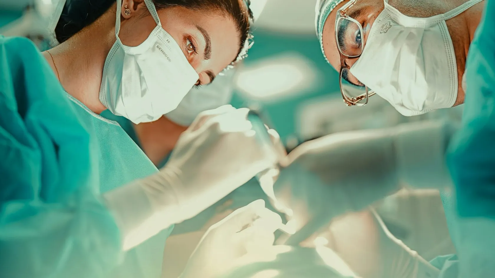
The operation is a wide local excision, which means the tumour is removed inside an unbroken envelope of normal tissue without ever being seen. Nothing is shelled out, nothing is entered, and if the surgeon has looked at the tumour during the operation something has gone wrong. In a limb this is usually a two to four hour procedure under general anaesthetic. Where a major nerve, vessel or bone is involved, or where the resulting defect will not close on itself, a reconstructive team works alongside the resecting team and the day becomes considerably longer. Two teams, one plan.
Planning against the scan
The MRI goes up on the screen in theatre and stays there. The incision is drawn to take in the previous biopsy track and any previous scar, and it runs along the length of the limb rather than across it, because a transverse incision on a limb forces a much larger operation if anything ever has to be revised.
Working outside the tumour
Dissection proceeds through normal tissue on every surface, taking the fascia above, muscle around and a cuff below. Where the tumour abuts a major nerve or artery, the sheath of that structure is stripped and taken with the specimen instead of sacrificing the structure itself, which is one of the main reasons amputation has become uncommon.
Marking the specimen and the bed
The specimen is orientated with sutures so that any involved margin can be traced back to a precise surface rather than a vague direction, and small metal clips are left in the tumour bed. Those clips are what the radiotherapy planning scan will aim at weeks later, and leaving them out makes accurate targeting far harder.
Filling and closing
Large dead spaces collect fluid and fluid collections become infections, so the defect is filled with a rotated local muscle flap or, where the loss is greater, with tissue transferred from elsewhere in the body. Drains stay in until output settles, commonly for several days, and closure is done without tension because a wound in an irradiated field is unforgiving.
Amputation is now the exception rather than the expectation.
In roughly nine out of ten limb sarcomas the tumour comes out without removing the limb, and the cases where it does not are ones where the tumour has encircled the main artery, nerve and bone together, where a previous operation has contaminated multiple compartments beyond salvage, or where the limb that would remain would be more of a burden than a prosthesis. That last consideration is a genuine one and it deserves an honest discussion instead of being treated as a defeat, because a salvaged limb that is painful, insensate and does not work is not a better outcome than a functional prosthesis. That conversation belongs beforehand.
What a clear margin means here
Margin in sarcoma surgery does not translate into a single number, and anybody who gives you one is simplifying. What counts is the quality of the tissue at the edge rather than its thickness. A millimetre of dense fascia is a better barrier than three centimetres of fat, because sarcomas spread along the path of least resistance and fascia resists while fat does not. So a report saying the margin was one millimetre at the fascial surface and four centimetres in the muscle can describe an excellent operation, and a report giving a comfortable-sounding margin all round in soft fatty tissue can describe a worse one. The surgeon's description of what structure lay at the closest point is the part worth reading. Quality beats thickness here.
This table scrolls sideways on a narrow screen. Swipe or drag to see every column.
| What it says | What it changes |
|---|---|
| R0, margins negative | No tumour at any inked surface. This is the aim and it is achieved in the large majority of planned operations. It does not by itself remove the case for radiotherapy in a high grade or deep tumour, since the argument there rests on grade and depth rather than on margin alone. |
| R1, microscopic disease at the edge | Tumour reaches an inked surface without being visible to the eye. The response depends on where. A planned close margin against a preserved nerve is managed with radiotherapy. An unplanned positive margin in tissue that could have been taken is usually managed by going back. |
| Grade, from one to three | Grade is built from how much the cells resemble their tissue of origin, how fast they divide and how much dead tissue sits inside the tumour. It drives the risk of spread more strongly than size does, and it is the main determinant of whether drug treatment is discussed at all. |
| Named subtype and molecular result | The specific name matters more than the word sarcoma. It sets which drugs have any prospect of working, how the tumour tends to spread and therefore where the follow-up scans are pointed, and how likely it is to come back locally at all. |
| Percentage of dead tumour after treatment | Where radiotherapy or chemotherapy was given before surgery, the proportion of the specimen that is no longer viable is recorded. It is a retrospective read on how the tumour responded and it informs what is offered afterwards, though it does not on its own dictate it. |
Radiotherapy, drugs and sequence
Radiotherapy for limb sarcoma can be given before or after the operation and the choice between them is a real trade.
Given beforehand it uses a smaller field and a lower dose, because the target is the tumour with a margin rather than the whole surgical bed, and it can firm up the pseudocapsule so that the dissection is cleaner. The cost is wound healing. Operating on irradiated tissue roughly doubles the rate of wound complications in the early months. Given afterwards the wound heals in untreated tissue, but the field is larger, the dose higher and the long term stiffness, swelling and fibrosis correspondingly worse. Most units now favour giving it first for large, deep, high grade limb tumours and accepting the wound problems, because the wound problems settle within a year and the fibrosis does not. That is the trade.
Chemotherapy is the genuinely contested question, and this is where the evidence rewards reading past the summary. A network meta-analysis published in 2021 pooled 25 randomised trials covering 3,453 patients with resectable soft tissue sarcoma and compared every sequence against surgery with or without radiotherapy. Adjuvant chemotherapy given after surgery improved overall survival. The hazard ratio was 0.86, confidence interval 0.75 to 0.97, at a p value of 0.017. Neoadjuvant chemotherapy combined with regional hyperthermia performed best of all, at a hazard ratio of 0.45, though its confidence interval touched 1.00 and the p value sat at 0.049. Neoadjuvant chemotherapy on its own did not improve survival, at a hazard ratio of 0.61 and a p value of 0.195. Nor did perioperative chemotherapy, at 0.66 and 0.317. Nor did histology-tailored chemotherapy, which came out at 1.08.
The finding nobody quotes in a brochure
In that same analysis the survival benefit of adjuvant chemotherapy was confined to male patients, with a hazard ratio of 0.78 and a p value of 0.004, while in female patients the hazard ratio was 1.08 with a p value of 0.410, meaning no detectable benefit at all. The authors were clear that this warrants further investigation before anybody changes practice, and a subgroup finding in a network meta-analysis is not the same class of evidence as the main result. It is nonetheless a reasonable thing to raise if chemotherapy is being proposed to you, and a team that has not heard of it is a team that has not read the literature it is quoting at you.
What that adds up to in practice is that chemotherapy in soft tissue sarcoma is a discussion rather than a protocol. The absolute gain from adjuvant treatment is modest, the toxicity is not, and the size of the gain depends heavily on subtype, grade, size and age. For a large high grade synovial sarcoma in a young patient the argument is strong. For a low grade tumour in an older patient with other health problems it is often weak enough that not giving it is the better medicine.
Anyone offering it as automatic, in either direction, is not giving you the actual state of the evidence.
When the sarcoma is in the abdomen
Retroperitoneal sarcomas, which grow in the space behind the abdominal organs, behave like a separate disease and are worth understanding separately. They are usually large before anything is noticed, because there is room back there for a tumour to reach the size of a rugby ball without producing more than a vague fullness. They have indistinct borders, they are constrained by the spine, the great vessels and the abdominal wall, and they are covered by a peritoneal layer thin enough that a true wide margin is often geometrically impossible. So local recurrence, which is a manageable problem in a limb, becomes the leading cause of death in low grade retroperitoneal sarcoma. High grade tumours in the same space are more likely to kill through distant spread instead.
Surgeons answered that with the extended compartmental resection, in which the first operation removes not just the tumour but all the fat on that side of the retroperitoneum together with whatever adjacent organs sit against it, most often a kidney and a segment of colon. It is a demanding operation and it is not universally the right one. Evidence indicates it improves local control in low grade sarcoma and in liposarcoma but not in leiomyosarcoma, for which complete removal of all visible tumour appears to be sufficient. Preoperative radiotherapy follows a similarly divided pattern, appearing beneficial in well differentiated liposarcoma and in grade one and two dedifferentiated liposarcoma, and probably in solitary fibrous tumour, without being indicated across the board.
The benefit of chemotherapy before surgery in high grade retroperitoneal disease remains unsettled while a dedicated randomised trial reports.
What all of that means for a patient is that the subtype has to be established before the abdomen is opened, since the same operation is correct for one histology and excessive for another. It also means that the first attempt is by a wide margin the best chance, more so here than anywhere else in sarcoma surgery, because a recurrent retroperitoneal sarcoma grows back into a field with no planes left in it. First attempt, best attempt.
Recovery and the wound
For a limb sarcoma removed without complex reconstruction, hospital stay is commonly three to six days and much of that is spent waiting for drain output to fall. Where a flap has been used it is longer, seven to twelve days, because the flap needs watching. Retroperitoneal surgery involving other organs runs to seven to fourteen days with a period of not eating at the start. Pain after limb surgery is moderate and manageable with tablets after the first day or two, and the thing most patients find harder than pain is the stiffness, which is a function of how much muscle came out and how much scar forms. Wound problems are the characteristic complication of this operation and pretending otherwise helps nobody. Across series of limb sparing wide local excisions, wound complications including infection, fluid collections and skin loss run at around a third of cases, while the serious ones needing readmission or a return to theatre stay under one in ten. Those figures rise where radiotherapy came first. They matter beyond the discomfort because a wound problem delays the start of adjuvant radiotherapy, and a delayed field is a worse field. Practically this means the drain stays until it is genuinely ready, the dressing is checked more often than seems necessary, and compression and elevation are worth taking seriously rather than treating as advice.
Physiotherapy starts within a day or two and continues for months. Function afterwards depends on what came out. Losing one muscle from a group of four leaves very little detectable deficit within a few months. Losing a whole compartment, or a major nerve, does not, and the honest conversation about that belongs in the consultation beforehand. Most people with a limb sarcoma removed and reconstructed return to desk work at four to six weeks, to driving once they can perform an emergency stop without hesitation, and to sport at three to six months if at all.
Swelling in the limb below the operation is common for six months and improves slowly. Compression helps.
Coming to Istanbul
How long you need to stay in Istanbul comes to two and a half to four weeks for a limb sarcoma and three to five for a retroperitoneal one, hotel nights included. The first several days go on assessment rather than treatment, which is deliberate. Imaging is repeated or reviewed, the original slides are re-read by our own pathologists, staging scans of the chest are completed, and the case is presented to a multidisciplinary meeting that includes the sarcoma surgeon, the reconstructive surgeon, the radiologist, the pathologist, the radiation oncologist and the medical oncologist before a plan is fixed. If a biopsy has not been done, or has been done unhelpfully, it is done here first and the wait for molecular results is built into the schedule. Where preoperative radiotherapy is part of the plan the arithmetic changes considerably, because five weeks of daily treatment followed by a gap of four to six weeks before surgery is not a single trip. Most patients in that position either have the radiotherapy at home and travel for the operation, which works well provided the planning scans and the field details travel too, or make two separate visits. We will say which of those applies before you book anything, and the answer comes out of the multidisciplinary discussion, and a coordinator cannot give it to you. We will say so early.
Flying home is reasonable once the wound is dry, the drains are out, the pathology has been discussed with you in person and any further treatment has been arranged in writing with whoever will deliver it. For most limb cases that lands at ten to fourteen days after surgery. Clearance for a long flight also depends on clot risk, which is elevated after cancer surgery and pelvic surgery in particular, so compression stockings, an anticoagulant for a defined period and moving around the cabin are not optional extras. Interpreting is arranged in advance in English, Arabic, Russian, French and German, and a written summary in your own language goes home with you along with the imaging on a disc, the operative note and the full pathology report.
What moves the cost
No single figure covers soft tissue sarcoma surgery, and any quotation given before the subtype, the grade, the site and the extent are known is a guess wearing a suit. The variables are not administrative. They are the operation itself. A superficial low grade lesion in a thigh removed with a straightforward closure and a lesion of the same size wrapped around the femoral vessels requiring vascular reconstruction and a free flap are different operations with different theatre times, different teams and different intensive care requirements, and they sit at opposite ends of any sensible range.
Eight things move the figure, and every one of them is a clinical variable.
- Whether the tumour is superficial or deep, and whether it involves a major nerve, a major vessel or bone.
- Whether reconstruction is needed and of what kind, since a rotated local muscle flap is a different proposition from a free tissue transfer with microvascular anastomosis.
- Whether other organs come out, which is routine in retroperitoneal surgery and rare in a limb.
- How long the operation runs, and whether two consultant teams are operating together for part of it.
- Whether an intensive care bed is required overnight.
- Whether the pathology needs molecular testing, which a good many subtypes do.
- Whether radiotherapy or chemotherapy forms part of the plan, and whether it is delivered here or at home.
- Whether this is a first operation or a re-excision after an unplanned removal elsewhere, which runs longer, takes more tissue and is likelier to need reconstruction.
Any written quotation should say which of those it includes and which it does not, and the exclusions are where people get caught. Ask whether the figure covers the pathology in full including immunohistochemistry and molecular testing, whether it covers a second operation if the margin comes back involved, whether it covers intensive care if it turns out to be needed rather than only if it was predicted, whether it covers the treatment of a wound complication, and how many follow-up appointments and scans sit inside it. Ask what happens to the price if the plan changes after the multidisciplinary meeting, because for sarcoma it quite often does. A number that cannot survive those five questions was never a real number. Ask all five.
Follow-up once you are home
Surveillance after sarcoma surgery is intensive at first and relaxes slowly, typically every three to four months for the first two to three years, then every six months to year five, then annually. Two things are being watched. The site itself, by examination and by MRI where the anatomy is complicated enough that fingers cannot tell scar from recurrence, and the chest by computed tomography, because the lungs are where most soft tissue sarcomas go if they go anywhere. Myxoid liposarcoma is the notable exception and needs imaging of the abdomen and spine as well, which is one more reason the subtype on the report has to be right. All of this can be delivered by your own oncologist at home, and it should be. What we send you away with is what makes that possible, which is the operative note describing exactly what was taken and what was preserved, the full pathology report with the subtype, grade, margins and molecular results, the position of the clips left in the bed, the radiotherapy details if any were given, and a written surveillance schedule with the specific scans and intervals named. Our team stays reachable for questions from you or from your doctor, and if something shows up on a scan two years from now we would rather look at it than hear about it later.
If you have already been operated on somewhere else
Where a lump was removed and the report came back as a sarcoma, the useful next step is a review by a sarcoma team with the original imaging, the operative note and the glass slides in front of them, before anybody decides whether to go back in. The single most valuable document is usually the scan performed before that first operation, because it shows what the tumour looked like while it was still intact. Bring it, along with anything else you hold, and expect the discussion to include the option of watching rather than operating where that is reasonable.
Frequently asked questions about soft tissue sarcoma surgery
My lump was removed as a lipoma and turned out to be a sarcoma. Do I need another operation?
Should I get the pathology looked at again?
Will I lose my arm or leg?
How reliable is a needle biopsy, and could it spread the tumour?
Do I need chemotherapy after surgery?
Is a sarcoma in the abdomen treated the same way?
How bad are the wound problems?
How long should I plan to be in Istanbul?
References
- Fromm J, Klein A, Mentrup F, et al. Unplanned resections of soft tissue sarcomas, necessity of re-resection? Cancers. 2024;16(10):1851.
- Eckardt MA, Siena NM, Copeland AR, et al. Dedicated review of sarcoma pathology is necessary for corroborative diagnosis in nearly one half of referred patients. Surgery. 2025;188:109610.
- Toki S, Sone M, Yoshida A, et al. Image-guided core needle biopsy for musculoskeletal lesions. Journal of Orthopaedic Science. 2022;27(2):448-455.
- Haussmann J, Matuschek C, Bölke E, et al. Comparison of different systemic therapeutic regimes in resectable soft-tissue sarcoma, results of a network meta-analysis. Cancers. 2021;13(22):5631.
- de Bree E, Michelakis D, Heretis I, et al. Retroperitoneal soft tissue sarcoma, emerging therapeutic strategies. Cancers. 2023;15(22):5469.
Editor's note
Written by the Biruni Hospital medical editorial team. Reviewed by Prof. Dr. Mustafa Teoman YANMAZ, Medical Oncology.
Medically reviewed by
Prof. Dr. Mustafa Teoman YANMAZ
Medical Oncology
Related Treatments
View All-
Bilateral Mastectomy
Whether you carry a BRCA mutation, have cancer in one breast or in both, bilateral mastectomy removes both breasts in one operation, and how much it helps depends on which of those three paths brought you to it.
-
Breast Conserving - Lumpectomy Surgery
Keeping the breast has never been the less thorough choice, and the largest recent studies now point the other way. This page explains who can have breast conserving surgery, what the margin report decides, and how long a woman traveling for it needs to stay.
-
Cytoreductive - Tumor Reduction Surgery
Patients cleared of every visible deposit lived 30 months in one gastric series. Those left with anything at all lived 7.3 months, however little remained.
-
Debulking - Ovarian Cancer Tumor Reduction
Two women with the same diagnosis on the same morning can get operations that share almost nothing. What decides it is who measured the disease first.
-
Debulking Surgery
Surgeons removed a median of 57 lymph nodes from women already cleared of visible disease. It bought no extra months, and tripled deaths within 60 days.