Most tumors at the base of the skull are benign, and many come out through the nose with an endoscope, leaving no scar on the face or scalp. This page explains which route suits which tumor, why a sodium check around day seven sets the length of the stay, and where radiotherapy fits after chordoma surgery.
About This Department
Otorhinolaryngology
Many tumors under the brain come out through the nose. No scalp incision at all.
Skull base surgery removes tumors and repairs defects in the floor of the skull, where the brain sits on the bones behind the eyes, nose and ears, and for a large share of these tumors the route now runs through the nostrils with an endoscope. Across 4,088 patients in a 2025 meta-analysis, a flap of the patient's own nasal lining kept spinal fluid leaks after endoscopic surgery to 3.4 percent. This page explains which tumors suit which route, what the operation and the hormone checks afterward involve, why the stay in Istanbul runs to two weeks, and what to send for a free review of your MRI.
3.4%
Spinal fluid leak with a nasoseptal flap, 4,088 patients
Over 90%
Complete removal of anterior midline meningiomas, either route
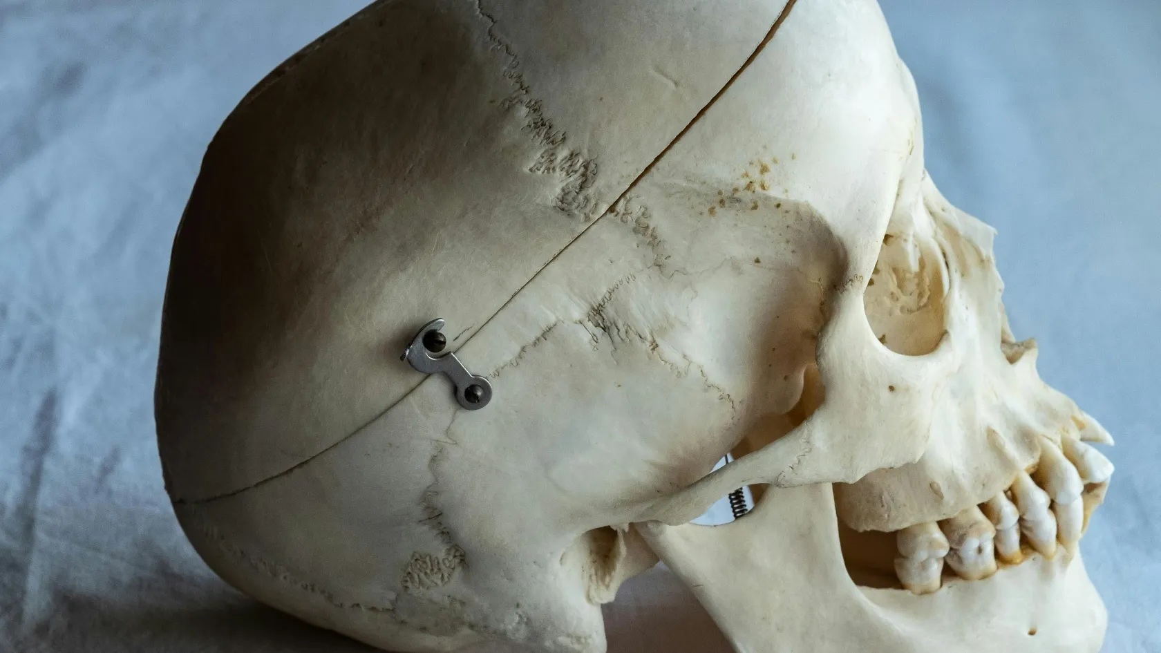
Above it sit the frontal and temporal lobes and the brainstem, below it lie the eye sockets, the sinuses, the back of the nose and the upper neck, and through small openings in it pass the optic nerves, the nerves for smell, eye movement, facial feeling, hearing and swallowing, and the carotid arteries on their way to the brain. Tumors here grow in a crowded space. Every one of those structures is one a patient cannot afford to lose, and that crowding, more than the tumor itself, is what makes the surgery specialized. Pituitary adenomas are the commonest reason for skull base surgery, and nearly all of them are benign. Meningiomas come next, arising from the membrane that covers the brain. Craniopharyngiomas, Rathke cleft cysts, chordomas, chondrosarcomas, acoustic neuromas, tumors growing up from the sinuses, and leaks of spinal fluid through a thin or broken floor make up most of the rest.
Most are not cancer.
Through the nose or through the skull
Two families of approach reach the skull base. In the endoscopic endonasal approach, an ear, nose and throat surgeon and a neurosurgeon work together through both nostrils, with a thin camera on a rod, opening the back wall of the sinus to reach the tumor from below without touching the brain or leaving a scar on the face or scalp. In the transcranial approach, a window of bone is lifted from the side or front of the skull and the tumor is reached from above or the side under the operating microscope, with the brain gently moved aside. Neither route wins every time. The tumor's position relative to the optic nerves and the carotid arteries decides, and a skull base team that offers both will choose the route the anatomy favors, which is a reason to send the MRI before choosing a hospital. A few tumors need both routes.
This table scrolls sideways on a narrow screen. Swipe or drag to see every column.
How the two routes compare
Endoscopic endonasal
Transcranial
Best for
Midline tumors below and between the optic nerves, such as pituitary tumors, chordomas and tuberculum sellae meningiomas
Tumors that spread sideways past the carotid arteries or optic nerves, and large olfactory groove meningiomas
Visible scar
None
A scalp incision hidden behind the hairline
Main trade-off
A higher risk of spinal fluid leak, and weeks of nasal crusting
The brain is moved aside to reach the tumor, and recovery is slower
Usual hospital stay
Three to five nights
Five to seven nights
Pituitary tumors
Pituitary adenomas cause trouble in two ways. Some press on the optic nerves above them and narrow the outer field of vision on both sides, which many people first notice when driving. Others make too much of a hormone, prolactin, growth hormone or cortisol, and cause infertility, enlarged hands and jaw, or Cushing syndrome, and of these the prolactin-secreting tumors are the exception to the surgical rule, since tablets shrink most of them without an operation.
Surgery first
A tumor pressing on the optic nerves, an adenoma making growth hormone or cortisol, or a nonfunctioning tumor that is growing.
Tablets or watching first
A prolactin-secreting tumor, which cabergoline shrinks in most patients, or a small nonfunctioning tumor found by chance that causes no symptoms.
For the rest, removing the whole tumor matters. A 2023 patient-level meta-analysis of 3,941 people with nonfunctioning adenomas found that those left with residual tumor had clearly worse progression-free survival than those whose tumor was removed completely, and that radiotherapy after an incomplete removal cut the risk of regrowth sharply. Endoscope or microscope made no difference to long-term control. A 2026 systematic review of 769 patients found the endoscopic route linked with better hormonal results, either remission of an overactive tumor or preserved normal pituitary function, with a risk ratio of 1.26. Hormone tests come before surgery and again in the first days after it, when cortisol, thyroid hormone, sodium and urine output tell the endocrine team whether the remaining gland is working and whether steroid tablets are needed.
Meningiomas near the eyes and smell nerves
Meningiomas along the front of the skull base, over the smell nerves or just above the pituitary, offer the clearest example of the route depending on the goal, and in a 2026 systematic review of 35 studies and 1,866 patients complete removal exceeded 90 percent with either approach.
Tuberculum sellae and planum
The endoscopic route gave better vision recovery for meningiomas of the tuberculum sellae and planum, the shelf of bone just in front of the pituitary, especially when tumor had grown into the optic canal.
Olfactory groove
The transcranial route preserved the sense of smell in more patients with olfactory groove meningiomas, because the endonasal route has to pass through the area where smell begins.
For tumors pressing on the optic chiasm, a 2026 meta-analysis of 582 patients found vision improved in more patients after the endoscopic route, with a risk ratio of 1.32, and complete removal rates were similar. The spinal fluid leak rate was higher after the endoscopic route.
Small meningiomas that cause no symptoms can simply be watched with yearly scans.
Chordoma and the radiotherapy that follows
Chordomas are rare. They grow in the clivus, the sloping bone behind the nose that supports the brainstem. Growth is slow and spread is uncommon, but they come back if any is left, so the operation aims for the largest safe removal and radiotherapy follows in most cases. A 2026 meta-analysis of 18 studies and 1,197 patients treated by the endonasal route found complete removal in 48.3 percent, with near-total removal in a further 29.2 percent, recurrence in roughly one patient in three over a mean follow-up of 55 months, a spinal fluid leak rate of 12.4 percent, a new cranial nerve problem in 7.5 percent and major bleeding in 0.3 percent. A separate 2025 meta-analysis comparing the endonasal and transcranial routes found similar survival, with cranial nerve function worsening in fewer than half as many patients after the endonasal route. After surgery, high-dose radiotherapy controls what remains. A 2024 meta-analysis of 30 studies found five-year progression-free survival of 67.8 percent after particle therapy, protons or carbon ions, against 40.2 percent after conventional photon radiotherapy, though brain tissue damage was reported more frequently after particle treatment.
Radiotherapy on the same site
Radiotherapy and chemotherapy are both delivered at Biruni Hospital, on the same site as surgery, using an Elekta Versa HD linear accelerator for photon and stereotactic treatment. Where a chordoma or chondrosarcoma would be better served by particle therapy, the written plan says so, so that it can be sought at a particle center, and a patient who prefers to have photon radiotherapy at home leaves with a written handover to their own radiation oncologist.
Sealing the skull floor
Removing a tumor through the nose leaves an opening between the nose and the fluid around the brain, and closing it well matters as much as the removal.
Repair uses layers.
An inlay graft. A piece of fat from the abdomen or thigh, a graft of tough tissue from the thigh, or a synthetic membrane fills the opening from the inside.
The nasoseptal flap. A strip of the lining of the septum, still attached to its own artery, is laid over the graft so it keeps a blood supply and heals into place.
Support while it heals. Soft splints or a balloon hold the layers against the bone for several days, and in high-flow cases a lumbar drain lowers the fluid pressure for two to three days.
A 2025 meta-analysis of 20 studies and 4,088 patients found spinal fluid leaks in 3.4 percent of those repaired with this flap against 5.6 percent with other methods, which halves the odds. Some teams add injectable bone cement or a lumbar drain for a few days in high-flow cases.
Because the flap is planned from the start, the ear, nose and throat surgeon raises it at the beginning of the operation before the tumor work begins.
Risks, with their rates
Spinal fluid leak
Spinal fluid leak is the complication specific to the endonasal route. It shows as a clear, salty drip from one nostril, worse when leaning forward, in the first days or weeks, and most leaks are repaired with a short second endoscopic procedure. An untreated leak carries a risk of meningitis. For that reason the instructions not to blow the nose or strain matter. Leak rates depend heavily on the tumor. Small pituitary tumors carry the lowest risk, while olfactory groove meningiomas removed through the nose leaked in 16 to 30 percent of patients in a 2026 meta-analysis against 2 to 7 percent after transcranial removal.
This table scrolls sideways on a narrow screen. Swipe or drag to see every column.
Published spinal fluid leak rates by tumor and route
Tumor and route
Leak rate
Evidence
Any endonasal tumor, repaired with a nasoseptal flap
3.4%
20 studies, 4,088 patients
Any endonasal tumor, other repair methods
5.6%
Same meta-analysis
Clival chordoma, endonasal
12.4%
18 studies, 1,197 patients
Olfactory groove meningioma, endonasal
16 to 30%
8 studies, 382 patients
Olfactory groove meningioma, transcranial
2 to 7%
Same meta-analysis
Hormones, nerves and vessels
Hormone changes follow pituitary surgery in a minority. Diabetes insipidus, passing large volumes of dilute urine because the gland stops releasing the hormone that holds water, appears in the first days and settles in most patients within weeks. A drop in blood sodium can follow around day seven to ten, and lasting loss of one or more pituitary hormones is treated with daily tablets. New cranial nerve problems, such as double vision or facial numbness, occur in single-digit percentages after the more extensive operations. Damage to the carotid artery is rare.
Surgeons plan around it with angiographic imaging and navigation.
Crusting in the nose lasts four to eight weeks and needs regular saline rinses and one or two cleaning visits.
The first two weeks, and planning the trip
Days one and two
Patients wake in intensive care or a high-dependency bed. Soft splints sit in the nose, and it feels like a heavy cold. Fluid balance and sodium are checked several times a day after pituitary surgery. Walking starts on the first day.
Days three to five
Discharge to the hotel follows after an uncomplicated endonasal operation once sodium and cortisol are stable, while after a transcranial operation the stay runs to five to seven nights, with an MRI before discharge.
Days seven to ten
A blood test for sodium around day seven takes minutes. It catches the late drop that follows pituitary surgery in some patients, and the ear, nose and throat surgeon cleans the nose and checks the flap on the same day, and that visit and the sodium result are the main reasons to stay in Istanbul for two weeks.
No nose blowing, heavy lifting, straws or straining until the surgeon clears it, at four to six weeks.
Planning the trip
Published packages from Turkish hospitals and medical travel agencies plan two weeks in Istanbul for endonasal skull base surgery and two to three weeks for a transcranial operation, and flying is cleared ten to fourteen days after endonasal surgery once the nose has been checked and there is no sign of a leak. A chordoma treated with radiotherapy here adds six to eight weeks for the radiation course, which can start three to six weeks after surgery, so many patients go home in between. The patient room has a companion bed, so one person stays overnight for the whole admission, and the international patients office arranges the hotel for the nights either side along with airport and hospital transfers. A coordinator assigned from the first message stays with you through discharge, and the international patients team works in English, Arabic, French, Russian, Serbian, Romanian and Spanish, and interpreting in other languages is arranged on request, so the consent conversation about risks to vision and hormones takes place in your own language. A request for a female physician goes to the department and is met wherever the rota allows, while halal, vegetarian and diabetic meals come from the hospital kitchen, a prayer room is on site, and an invitation letter naming the hospital and the surgeon goes out ten days before travel for the visa application.
Bring every MRI and CT disc, the hormone results, any visual field test, and a list of current medications.
Reading a quote
Tumor type and route set most of the cost.
Removing a small pituitary adenoma endonasally in two to three hours sits at one end of the range, and a chordoma reaching from the clivus into the neck, removed over five or six hours with nerve monitoring and a lumbar drain, sits far from it. Intensive care nights, navigation and intraoperative monitoring, the repair materials, a second procedure for a leak, hormone replacement started in hospital and the pathology report each change the total, and radiotherapy, where needed, is a separate course.
Packages for skull base surgery in this market include the surgeons and anesthesia, the operating room, a set number of intensive care and ward nights, the MRI after surgery, the pathology, an interpreter, transfers and hotel nights, and they leave out flights, travel insurance, extra hospital nights, repair of a delayed leak, radiotherapy and long-term hormone medication.
Ask any hospital which route its figure assumes, whether two surgeons are included, whether the day seven sodium test and nasal cleaning visit are inside it, and what happens financially if a leak needs repair. A real figure comes after a skull base team has read your MRI, and that review is free.
Scans and hormones in the years after
Once you are back home, the follow-up has two strands. An MRI at three months sets the baseline, followed by yearly scans for most benign tumors and scans every six months for chordoma. Hormone blood tests at six weeks and three months show whether the pituitary is working. An endocrinologist at home adjusts any replacement. Each scan and result goes by WhatsApp to the same coordinator, and the surgeon who operated reviews it against the earlier images at no charge, so the person who knows the anatomy best stays involved for years after the operation.
Call the same day for any of these.
A clear, salty drip from one nostril, worse when leaning forward
Fever with headache or a stiff neck
Sudden worsening of vision
Passing large amounts of urine with severe thirst, or new confusion
Your local emergency department does the first assessment, and the surgeon speaks to that doctor directly with the operation note to hand.
Skull base surgery FAQ
Is skull base surgery through the nose safe?
In experienced hands, yes. The main specific risk is a spinal fluid leak, which occurred in 3.4 percent of patients repaired with a nasoseptal flap across 4,088 patients in a 2025 meta-analysis, and most leaks are fixed with a short second procedure.
How long is recovery after endoscopic skull base surgery?
Three to five nights in hospital, a return to desk work in two to four weeks, and nasal crusting that settles over four to eight weeks. Heavy lifting and straining wait four to six weeks.
How long do I need to stay in Turkey?
About two weeks after endonasal surgery and two to three weeks after a transcranial operation. The stay covers the sodium check around day seven, the nasal cleaning visit and clearance to fly.
Will I need hormone tablets afterward?
Most people after pituitary surgery do not, or need them only for a few weeks. Those whose gland does not recover take daily replacement, adjusted by an endocrinologist at home on the basis of blood tests at six weeks and three months.
Can a family member stay with me?
Yes. The patient room has a companion bed for the whole admission, and the international patients office arranges the hotel and transport for both of you.
What should I send for a free review?
The MRI with contrast as images and its report, any CT of the skull base, hormone results, a visual field test if you have one, and a short note on symptoms. The team replies with the likely diagnosis, which route would be used, how long the stay would be and whether radiotherapy is likely, at no charge and with no obligation to travel.
References
Lino-Filho AM, Fernandes MNF, Teixeira OAPM, et al. Pedicled nasoseptal flap after endoscopic endonasal resection of skull base tumors: a comparative meta-analysis with other modalities of reconstruction. J Neurol Surg B Skull Base. 2025;87(4):469-476.
Fong KY, Lim MJR, Fu S, et al. Postsurgical outcomes of nonfunctioning pituitary adenomas: a patient-level meta-analysis. Pituitary. 2023;26(4):461-473.
Sulaimanov U, Uslu I, Alomari O, et al. Anterior midline skull base meningiomas: a systematic review of resection rates, functional outcomes, and perioperative complications following contemporary endoscopic endonasal versus transcranial approaches. J Clin Med. 2026;15(12):4676.
Arend RB, Lima NL, Ieggli PH, et al. Endoscopic endonasal approach for resection of clival chordomas: a systematic review and meta-analysis. Oper Neurosurg. 2026. doi:10.1227/ons.0000000000002147
Pahlavi IR, Usman H, Azhari NTP. Endoscopic endonasal approach vs microsurgical transcranial approach in intracranial chordoma surgery: a systematic review and meta-analysis. Neurosurg Rev. 2025;49(1):38.
Saito T, Mizumoto M, Oshiro Y, et al. Systematic review and meta-analysis of particle beam therapy versus photon radiotherapy for skull base chordoma: TRP-Chordoma 2024. Cancers (Basel). 2024;16(14):2569.
Hafiz B, Alsharif T, Okal F, et al. Comparative efficacy of endoscopic endonasal versus transcranial approaches for treating olfactory groove meningiomas: a systematic review and meta-analysis. World Neurosurg. 2026;211:125015.
Editor's note
Written by the Biruni Hospital medical editorial team. Reviewed by Assoc. Prof. Dr. Melih ÜÇER, Neurosurgery.