
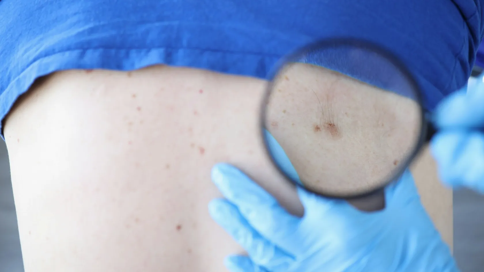
Skin Cancer Surgery
Cutting out a skin cancer is easy. Knowing you got all of it depends on how much of the edge somebody actually examined, and that varies far more than patients are ever told.
About This Department
Cutting out a skin cancer is easy. Knowing you got all of it is the entire problem, and it is decided not by how much tissue was taken but by how much of the edge somebody actually examined down a microscope. With an ordinary excision the pathologist slices the specimen like a loaf and looks at the faces of those slices, which samples the true margin rather than inspecting it. With margin-controlled techniques the whole edge is examined while you are still in the building. That single difference in method, invisible to the patient and rarely mentioned in a consultation, shows up plainly in how often the cancer comes back. Ask about it. That is a yes or no question with a yes or no answer, and any unit that hesitates over it has told you something useful without meaning to.
Free consultation
Send clear photographs with something for scale beside the lesion
Put a ruler, or a coin, next to the lesion and turns a photograph from an impression into a measurement, and size on the face changes the plan more than almost anything else. Send photographs in daylight from two distances, any biopsy report with the exact diagnosis and, for melanoma, the Breslow thickness in millimetres, plus a note of previous skin cancers, previous radiotherapy to the area and any medication that suppresses your immune system. Say if the lesion sits on an eyelid, a nose, an ear or a lip, because those four locations change both the technique and who should perform it. The review costs nothing and carries no obligation. A photograph with a ruler beside it answers more questions in ten seconds than a paragraph of description does in ten minutes, and the position on the face matters as much to the plan as the diagnosis does.
Three diseases, one phrase
Skin cancer surgery covers three diseases. They share a technique and almost nothing else. The commonest of the three grows locally, destroys whatever it grows into and essentially never spreads to the rest of the body, which means the entire risk is what it does to the face it is sitting on. The second can spread, does so in a minority of cases, and is judged on features a pathologist reads from the specimen. The third is melanoma, which is the reason people are frightened of the phrase, and it behaves differently enough that half this page belongs to it. Getting told which of the three you have changes everything that follows, and a great many people arrive at a consultation having read about the wrong one. Establish which you have. Everything below depends on it, and the difference between the first of the three and the third is the difference between an inconvenience and a serious illness.
This table scrolls sideways on a narrow screen. Swipe or drag to see every column.
| Type | How it behaves, and what surgery is trying to achieve |
|---|---|
| Basal cell carcinoma | Slow, local and destructive, spreading through the body in only a handful of documented cases ever. Left alone on a nose or an eyelid it will eventually eat through cartilage and bone, which is the whole argument for treating it. Surgery aims at complete local clearance with as little tissue removed as the margin allows. |
| Squamous cell carcinoma | Also mostly local, and capable of spreading to lymph nodes in a minority. Thickness, poor differentiation, perineural invasion, ear and lip location and a suppressed immune system all raise that risk, and all of them come from the pathology report rather than from looking at it. |
| Melanoma | Genuinely dangerous, and staged principally on how deep it goes measured in millimetres. Surgery is wider, may include mapping the first lymph node it drains to, and the margins used have been getting narrower rather than wider as the trials have reported. |
| Rarer types | Merkel cell carcinoma, dermatofibrosarcoma and others behave individually and belong in a specialist discussion. If a report names something unfamiliar, that is a reason to ask for a multidisciplinary opinion rather than a reason to panic. |
What actually gets looked at
How the specimen is examined
Picture the piece of skin that has been removed as a small disc with the tumour in the middle. The margin that matters is the entire outside surface of that disc, all the way round and underneath. In a standard excision the specimen goes into a pot of preservative, arrives at a laboratory some days later and is cut into parallel slices, and the pathologist examines the flat faces of those slices. Anything sitting between two slices is not seen. That method samples the margin instead of inspecting it, and it works perfectly well most of the time because most tumours are round and predictable, and fails precisely where tumours send out a finger in one direction that happens to run between two cuts. Margin-controlled surgery inverts the geometry. The specimen is cut and oriented so that the whole outside surface lies flat in one plane, examined immediately while the patient waits, and any positive area is mapped back to its exact position so that only that spot is re-excised.
What that difference does to recurrence
Recurrence rates for basal cell carcinomas were pooled around the eye, where the consequences of getting it wrong are highest and the tissue available is least. Recurrence after margin-controlled micrographic surgery was 2.9 percent across an average of nearly four years of follow-up, and after excision under frozen section control 1.9 percent across almost six years. After standard wide excision with the specimen processed conventionally it was 5.9 percent. Meta-regression found the standard excision rate significantly higher than either margin-controlled method, while the two margin-controlled methods did not differ significantly from each other.
What that means for you
Two conclusions follow, and the second is the useful one. Examining the margin during the operation roughly halves recurrence in the hardest location on the body, which is a large effect for a change in laboratory method, not in surgical skill. And it does not matter greatly which margin-controlled method is used, since micrographic surgery and frozen section control performed similarly. The question is whether anybody looked at the edge before you went home, and that question has a yes or no answer that any unit can give you in advance.
When the margin is not clear
How often it happens
Sometimes the tumour reaches the edge of what was taken, which is called an incomplete excision and is not rare. What happens next divides sharply depending on whether anybody had planned for the possibility, and a study looking specifically at that question found a difference nobody would guess from the size of the tumours involved.
Planned for, or discovered
Researchers at a tertiary centre reviewed 127 cases in which micrographic surgery had stopped with cancer still at the margin, and separated them into cases where a multidisciplinary plan had been assembled beforehand and cases where the positive margin was handled as it arose. Where it had been anticipated, 98.5 percent of patients went on to further surgery, against 72.6 percent where it had not. The delay to that further surgery averaged 3.9 days when planned and 13.2 days when not. Final clearance of the margin was achieved in 84.6 percent of the anticipated group and 59.7 percent of the unanticipated one. Every one of those differences was statistically significant. The tumours involved were no bigger in one group than the other. What differed was whether anybody had made a plan for the possibility before the operation started, which costs nothing and appears to change what happens to a quarter of the people it applies to.
The number worth carrying
More than a quarter of patients whose incomplete excision came as a surprise never had further surgery at all. Carry that finding away, and it is not really about surgery. It comes down to whether somebody had thought in advance about what would happen if the first attempt did not clear the tumour, and had arranged the next step before it was needed. Ask what the plan is if the margin comes back involved. A unit that has an answer ready is telling you something about how the rest of your care will run.
What the day involves
Almost all of this happens while you are awake. Local anaesthetic, and you talking to the surgeon throughout.
Marking the plan
Outlining comes first, marking the visible edge of the tumour, frequently under magnification and sometimes with a dermatoscope, then the intended margin is drawn around it. On the face the shape is planned along the lines that skin naturally folds into, since a scar following one of those lines becomes very hard to see and a scar crossing one never does.
Local anaesthetic
Stinging from the injection is the only genuinely unpleasant part of the whole procedure and it lasts under a minute. After that you feel pressure and movement but no pain, and telling the surgeon if you feel anything sharp is expected rather than a complaint.
Excision and orientation
Out comes the tissue, and it is then marked, generally with a stitch at a nominated position, so that anything found at the margin can be traced back to a point on your face and not to a vague direction. Skipping that step is why some reports say the margin is involved without saying where.
Waiting, then closing
Where the margin is being checked during the operation you sit in a waiting area with a dressing on for around an hour per round, and most tumours need one or two rounds. Only once the edge is clear is the defect closed, which is the correct order and the reason these appointments take a morning, not twenty minutes.
When you would be asleep
General anaesthetic is reserved for very large tumours, for children, for lesions in awkward places such as inside the nose or on the eyelid margin, and for people who genuinely cannot tolerate lying still. Nobody should feel obliged to be brave about it, and asking for sedation is entirely reasonable.
Melanoma, and the shrinking margin
Two operations, not one
Melanoma surgery happens in two stages and the second is the one that matters. First the lesion is removed with a narrow margin purely to make the diagnosis and measure how deep it goes, a figure called the Breslow thickness that drives everything afterwards. Then a wider excision is performed around the scar, taking a margin determined by that thickness. Forty years ago those margins were enormous, with five centimetres of normal skin taken in every direction on the theory that melanoma cells crept invisibly outward, which meant grafts and disfigurement for tumours that in many cases were never going to spread. Every trial run since has pushed the numbers down, and the most recent evidence has pushed them down again. Wider is not safer here. Wider is just wider, and it takes more skin from a face that has to go on being a face afterwards.
Narrow against wide, in thick melanomas
Three randomised trials were gathered in a meta-analysis totalling 2,304 patients, restricted deliberately to melanomas thicker than two millimetres, the group in which wide margins had been most confidently defended. Ten year all-cause mortality showed no significant difference between narrow margins of one to two centimetres and wide margins of three to four, with a risk difference of 3.3 percent and a p value of 0.202. Overall survival showed no significant difference either, at a hazard ratio of 1.09. Heterogeneity between the trials was low. The reviewers support the non-inferiority of narrow margins while noting that one centimetre may be inadequate on the evidence from individual studies, which places the sensible working figure at two centimetres for a thick melanoma rather than anywhere near the four that used to be standard.
What that means on the operating table
The practical consequence for a patient is considerable. A two centimetre margin around a thick melanoma on a shoulder closes directly in most cases, and four centimetres frequently cannot, and the difference between those two operations is the difference between stitches and a skin graft. Nobody should now be offered the older wider margins as a matter of routine, and being offered them is a reason to ask which guideline is being followed and when it was written.
The node biopsy and what follows it
What the node biopsy is for
For melanomas above a certain thickness, a small amount of radioactive tracer and a blue dye are injected around the original site so that the first lymph node the area drains into can be located and removed. The purpose is information, not treatment, since finding cancer in that node moves you into a different stage and opens the door to drug treatment that has genuinely changed outcomes. What used to follow automatically was an operation to remove every remaining node in that group, on the reasonable-sounding theory that if one node contained cancer, others might too. Two large randomised trials tested that assumption and the answer reorganised the field. Both trials pointed the same way. That is rarer in surgery than anybody would like, and it is the reason the change in practice happened quickly rather than over a decade of argument.
What clearing the rest of the nodes achieved
One review summarising the two major randomised trials in this area records that early completion lymph node dissection, compared with simply observing the remaining nodes after a positive sentinel biopsy, produced no significant difference in survival between the two groups. Control of disease in that region was better after dissection. The reviewer concludes that the role and value of early completion dissection for patients with microscopic nodal disease is now very limited worldwide, and notes that the arrival of adjuvant drug therapy has further reduced the reasons for performing it.
What a positive node now means
So a positive sentinel node no longer commits you to an operation that clears an armpit or a groin, with the lymphoedema and stiffness that came with it. It commits you to closer surveillance and a conversation about drug treatment, which is a substantially better trade. The sentinel biopsy itself remains worth having in the right patients, because the information it produces determines whether that conversation happens at all, and it remains a genuine operation with a small risk of its own, not automatically right for a thin melanoma.
Closing the hole
How the defect gets closed is a separate decision from how the cancer gets removed, and confusing the two leads to bad outcomes in both. The cancer clearance comes first and is never compromised to make the closure easier, which sounds obvious and is exactly what goes wrong when somebody plans a flap before knowing how much tissue will actually be missing. Four options exist and the choice gets made once the defect is sitting there to be looked at.
This table scrolls sideways on a narrow screen. Swipe or drag to see every column.
| Method | Where it suits, and what it leaves |
|---|---|
| Direct closure | The edges are brought together into a line. Suits small defects where the surrounding skin has enough slack, and it leaves the least visible result of the four because a line hides in a crease and a patch never does. |
| Local flap | Skin from immediately beside the defect is moved across on its own blood supply, bringing matched colour, thickness and texture with it. The workhorse of facial reconstruction, and the reason flaps look better than grafts on a face. |
| Skin graft | Skin taken from behind an ear or above a collarbone and laid into the defect. Reliable, quicker, and always visible as a patch to some degree because donor skin never quite matches. It also creates a second wound that nobody warned you about. |
| Left to heal | Deliberately leaving the wound open to heal on its own, which sounds like neglect and produces surprisingly good results in concave places such as the inner corner of the eye or the bowl of the ear. |
Two things to settle first
Two points belong in the conversation before the operation, not after it. Where the margin is being checked during the procedure, the closure happens the same day but only once the edge is clear, so the shape of it cannot be finalised in advance. And where a graft is likely, ask where the donor skin will come from, because that is a second wound nobody mentioned and it usually ends up being the one that itches.
Why one is rarely the last
Why the field matters more than the lesion
Skin that produced one cancer produced it because of decades of accumulated sun damage across a wide area, and that damage does not stop at the edge of the lesion that was removed. Follow-up after skin cancer is therefore about the rest of your skin at least as much as about the site that was treated, which is a different proposition from follow-up after most cancers and takes people by surprise.
What happened to 1,077 people after their first one
Investigators followed 1,077 patients in a population cohort from their first basal cell carcinoma, linked to a national pathology registry, to build a model predicting who develops more. A third of patients with a first such cancer went on to develop further ones. A second occurred in 293 patients, a third in 122, a fourth in 58 and a fifth in 36, and the median intervals shortened as the count rose, from 3.0 years to the second, then 2.1, 1.7 and 1.8 years. The strongest predictors were the number of previous diagnosis dates and having had more than one tumour at the original diagnosis, both of which are known on the day you are first diagnosed rather than discovered later.
Reading the intervals
Read the shortening intervals rather than the headline. Somebody on their third skin cancer is likely to meet their fourth within about eighteen months, so surveillance intervals tighten as the count goes up instead of relaxing, and why the man who has had four is examined more frequently than the man who has had one. None of that means the disease is becoming more dangerous. It means the skin is behaving consistently, and consistent behaviour is something a follow-up schedule can be built around.
Recovery and the scar
The fortnight itself
Physically there is very little to recover from, and what people actually experience is a fortnight of minor inconvenience followed by a year of watching a scar change.
- Expect the anaesthetic to wear off after a few hours and simple painkillers to be enough. Anything worse than that on a face wound is unusual and worth reporting.
- Bruising around an eye after surgery anywhere on the upper face is normal, gravitational and can look dramatic for a week.
- Stitches come out at five to seven days on the face and ten to fourteen elsewhere, and leaving facial stitches longer is what produces the railway-track marks people associate with old scars.
- A graft looks alarming for the first fortnight, going through purple and crusted stages before settling, and judging it in that window is pointless.
- Keep the area out of direct sun for a year and use sunscreen on it daily, because a fresh scar pigments permanently if it burns.
The scar, month by month
The scar itself follows a course that alarms anybody who was not warned. It looks neat at two weeks, then reddens, firms and thickens between about six weeks and three months, then fades and flattens across the rest of the first year. That middle phase is the normal biology of healing and not a sign of a problem, and the number of people who seek a revision at three months and would not have wanted one at twelve is considerable enough that most surgeons decline to operate again inside a year on principle. Judge the result at a year, and if it still needs improving at that point, revision is straightforward and can be planned properly. Twelve months. Not three, and not six.
How long you stay
Five to eight days for a straightforward excision with margin control, and ten to fourteen where melanoma surgery includes a sentinel node biopsy or where a staged reconstruction is planned. Assessment takes one to two days, covering examination of the whole skin surface and not only the lesion you came about, photography, review or repeat of the biopsy, and imaging where the diagnosis calls for it. That whole-skin examination is not a formality. It finds a second lesion frequently enough to justify the twenty minutes, and finding one now is considerably easier than finding it in eighteen months from another country.
What the days are actually for
Almost all of this is day surgery, so hospital stay is measured in hours except for general anaesthetic cases and larger reconstructions. The days that follow cover a wound check at forty-eight hours where a graft or flap was used, suture removal at five to seven days, and the pathology discussion that establishes whether the margin was clear and whether anything further is needed. Staying for the pathology is the argument for the longer figure, because being told the margin is involved after you have flown home turns a straightforward second procedure into a considerably harder problem.
When you are cleared to fly
Two to three days after simple excision and once any graft has been checked at the first dressing change, so five to seven days in most cases. A window seat and a cabin blind down are worth having if the wound is on your face, since aircraft windows transmit more ultraviolet than most people expect and a fresh scar is exactly what should not be exposed to it.
What drives the cost
Of every cancer operation described on this site, this is the least expensive, and the variation within it comes almost entirely from what happens to the specimen rather than from the operation. Six things move the figure.
- Whether the margin is examined during the operation, which occupies a laboratory and a technician for the duration and is the single largest driver.
- How many rounds of margin checking are needed, since each one repeats that laboratory work.
- How the defect is closed, with direct closure, a local flap, a graft and a staged reconstruction all sitting at different levels.
- Local or general anaesthetic, and whether theatre time is needed at all.
- Sentinel node biopsy, which requires nuclear medicine, a dye and a separate procedure.
- The number of lesions treated, since people frequently arrive with one and leave having had three dealt with.
The open-ended item
One item is genuinely open-ended and it sits outside the operation entirely. Given that a third of people with one basal cell carcinoma develop another, and that the intervals shorten with each one, the real long-term cost of this diagnosis is a lifetime of skin surveillance and periodic further procedures, all of it happening wherever you live. Packages here ordinarily cover the transfers, the assessment, the operating fees, the stated stay, an interpreter, accommodation and the appointments before you fly, while outside them sit the flights, insurance, extra nights, any further surgery for an involved margin, and that ongoing surveillance.
Five questions to ask before you accept a figure
Is the margin being examined during the operation, and is that inside the quoted figure. What further rounds of margin checking would cost if more than one is needed. Is the reconstruction quoted, and what changes between a direct closure and a flap or graft. What a re-excision would cost if the margin comes back involved. And is the whole-skin examination and treatment of any additional lesions included or charged separately.
Nothing here means anything until somebody has seen a photograph with a ruler beside it. That review costs nothing.
Once you are home
What follow-up consists of
Follow-up after skin cancer means examination, not scanning, which makes it the easiest of any cancer on this site to arrange at home and the easiest to let slide. For basal cell carcinoma it is a skin check at intervals that tighten with the number you have had. For squamous cell carcinoma it includes feeling the draining lymph nodes as well. For melanoma it is more structured, running for years and including examination of the scar, the surrounding skin and the node basins, with imaging added according to stage.
Reading your own report
This table scrolls sideways on a narrow screen. Swipe or drag to see every column.
| What it says | Why it changes the plan |
|---|---|
| Breslow thickness | How deep the melanoma reaches, measured in millimetres from the surface. This single number drives the width of the second excision, whether a node biopsy is offered and the whole staging. Ask for it as a figure rather than as a description. |
| Ulceration | Whether the surface of the melanoma had broken down. Present or absent, and its presence moves the stage upward independently of thickness, which is why a thin ulcerated lesion is not treated like a thin one that was intact. |
| Margin status | How far the tumour sat from the edge of what was removed, ideally stated in millimetres rather than as clear or involved. A margin described only as clear tells you less than one described as three millimetres. |
| Perineural invasion | Tumour tracking along a nerve, which matters most in squamous cell carcinoma and on the face. Its presence changes the surgical plan and sometimes adds radiotherapy, and it is easy to miss in a report that runs to several pages. |
| Differentiation and subtype | How closely the cells resemble normal tissue, and which pattern the tumour follows. Infiltrative and morphoeic basal cell patterns spread further under intact skin than they appear to, which is precisely where sampling a margin rather than examining it goes wrong. |
What to take with you
Four documents should travel home with you, in English, since whoever picks up that surveillance was not in the room.
- The pathology report in full, naming the exact diagnosis and, for melanoma, the Breslow thickness, ulceration status and mitotic activity.
- A clear statement of the margin status, in millimetres, and whether any margin-controlled technique was used.
- A diagram or photograph showing exactly where each lesion was, which matters more than people expect once several scars accumulate.
- The surveillance schedule with real intervals, and for melanoma the stage on which it is based.
When to make contact
Get in touch here for a wound that opens, spreading redness, a graft that turns black rather than pink, or any new lump appearing near the scar or in the neighbouring lymph nodes. Send a photograph in daylight rather than describing the colour, because colour described in words is close to useless across a message and a picture settles the question immediately. Daylight. Not a bathroom bulb.
The only thing that reduces the count
Sun protection from here on is not a lifestyle suggestion, it is the treatment for the field of damaged skin that produced this in the first place, and it is the only intervention on this page that reduces the number of operations you will have across the next twenty years. Hats and shade do more than any cream. Creams still help.
Frequently asked questions about skin cancer surgery
Does it matter how the margin is checked?
What happens if the cancer reaches the edge of what was removed?
How much normal skin has to come out around a melanoma?
If the sentinel node has melanoma in it, must the rest be removed?
Will I get another skin cancer?
How long do I need to stay, and when can I fly?
References
- Phan K, Oh LJ, Goyal S, Rutherford T, Yazdabadi A. Recurrence rates following surgical excision of periocular basal cell carcinomas, systematic review and meta-analysis. Journal of Dermatological Treatment. 2020;31(6):597-601.
- Lin SK, Deitermann AM, Lubeck M, et al. Anticipated versus unanticipated incomplete Mohs micrographic surgery for keratinocyte carcinomas, impact on treatment delays and final margin status. Dermatologic Surgery. 2023;49(12):1066-1071.
- Floriano LS, Picon RV, Dagostim C, Chedid MF. Surgical excision margins in skin melanomas with Breslow thickness greater than 2 mm, a systematic review and meta-analysis. Current Pharmaceutical Design. 2026;32(4):301-305.
- Nakamura Y. The role and necessity of sentinel lymph node biopsy for invasive melanoma. Frontiers in Medicine. 2019;6:231.
- Smedinga H, Verkouteren JAC, Steyerberg EW, Hofman A, Nijsten T, Vergouwe Y. Occurrence of metachronous basal cell carcinomas, a prognostic model. The British Journal of Dermatology. 2017;177(4):1113-1121.
Editor's note
Written by the Biruni Hospital medical editorial team. Reviewed by Prof. Dr. Özcan YILDIZ, Medical Oncology.
Medically reviewed by

Prof. Dr. Özcan YILDIZ
Medical Oncology
Related Treatments
View All-
Bilateral Mastectomy
Whether you carry a BRCA mutation, have cancer in one breast or in both, bilateral mastectomy removes both breasts in one operation, and how much it helps depends on which of those three paths brought you to it.
-
Breast Conserving - Lumpectomy Surgery
Keeping the breast has never been the less thorough choice, and the largest recent studies now point the other way. This page explains who can have breast conserving surgery, what the margin report decides, and how long a woman traveling for it needs to stay.
-
Cytoreductive - Tumor Reduction Surgery
Patients cleared of every visible deposit lived 30 months in one gastric series. Those left with anything at all lived 7.3 months, however little remained.
-
Debulking - Ovarian Cancer Tumor Reduction
Two women with the same diagnosis on the same morning can get operations that share almost nothing. What decides it is who measured the disease first.
-
Debulking Surgery
Surgeons removed a median of 57 lymph nodes from women already cleared of visible disease. It bought no extra months, and tripled deaths within 60 days.