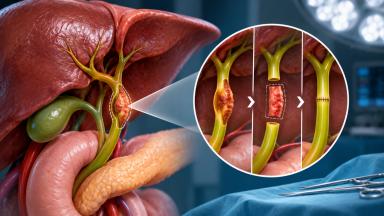
Sentinel Lymph Node Biopsy For Melanoma
The thickness of a melanoma, measured in millimeters, decides whether the nearest lymph nodes need checking. This guide explains how the sentinel node is found with a tracer and a probe, who needs the biopsy, what a positive node means since MSLT-II, and a stay of seven to ten days in Istanbul.
About This Department
One small lymph node decides the stage of a melanoma. Finding it takes a short operation.
Melanoma that spreads goes first to the nearest lymph node basin, and within that basin to one or two sentinel nodes. Sentinel lymph node biopsy finds and removes those nodes through a short incision, usually in the same session as the wider excision of the scar. The result sets the stage, decides whether immunotherapy is offered, and spares most patients the full lymph node dissection that once followed every positive node.
A mole was removed and the report says melanoma. It also gives a number in millimeters.
That number, the Breslow thickness, decides almost everything on this page.
Plan the sentinel node biopsy and the wider excision together, ideally within a few weeks of the diagnosis. A wide excision done first, on its own, can change the lymph flow and make the sentinel node harder to find.
What a sentinel node is
Lymph from every patch of skin drains along fixed channels to a group of lymph nodes, in the armpit, the groin or the neck. The first node or two to receive that lymph are the sentinel nodes. If melanoma cells have left the skin, these nodes are where they settle first, so a pathologist who examines them closely, slice by slice and with special stains, can say whether the melanoma has spread at a stage when no scan and no examination can yet detect it. A clear sentinel node makes spread to the rest of that basin very unlikely. A positive one changes the stage from II to III and opens the door to treatment after surgery. Before this test existed, surgeons either removed every node in the basin to check it, with the swelling and stiffness that follow, or waited for a lump to appear, and the sentinel node biopsy replaced both with one small operation that answers the question early.
How the biopsy runs
Forty-five to ninety minutes in theater. Home the same day or the next morning.
Who should have one
A 2018 guideline from the American Society of Clinical Oncology and the Society of Surgical Oncology ties the decision to thickness and ulceration.
This table scrolls sideways on a narrow screen. Swipe or drag to see every column.
| Category | Thickness | Sentinel node biopsy |
|---|---|---|
| T1a | Under 0.8 mm without ulceration | Not recommended routinely |
| T1b | 0.8 to 1.0 mm, or under 0.8 mm with ulceration | May be considered after discussion |
| T2 and T3 | Over 1.0 to 4.0 mm | Recommended |
| T4 | Over 4.0 mm | May be recommended after discussion |
The wider excision
The first excision removed the melanoma. The second removes a rim of skin around the scar, because cells can sit a short distance from the visible tumor.
This table scrolls sideways on a narrow screen. Swipe or drag to see every column.
| Breslow thickness | Margin of healthy skin |
|---|---|
| Up to 1.0 mm | 1 cm |
| 1.01 to 2.0 mm | 1 to 2 cm |
| Over 2.0 mm | 2 cm |
On the face, ears, hands and feet, the margin is balanced against function and appearance, and closing the wound can need a local skin flap or a graft.
What the result tells you
MSLT-I, whose final report appeared in the New England Journal of Medicine in 2014, randomized 2,001 patients to sentinel node biopsy or to watching the nodes. In melanomas of intermediate thickness, 1.2 to 3.5 mm, ten-year melanoma-specific survival was 85.1 percent with a clear sentinel node and 62.1 percent with a positive one. Node status split survival at every thickness studied. Thick melanomas over 3.5 mm showed the same pattern, with ten-year melanoma-specific survival of 48.0 percent with nodal spread and 64.6 percent without it, so the result carries weight wherever the biopsy is offered. Patients in the biopsy group also stayed free of recurrence longer, with ten-year disease-free survival of 71.3 percent against 64.7 percent with observation, and among patients whose intermediate-thickness melanoma had already reached the nodes, biopsy-based management lowered the risk of death from melanoma. Across the whole trial, however, survival did not differ between the two groups, so the biopsy is first of all a staging test.
If the node is positive
For years a positive sentinel node meant removing every remaining node in that basin. The MSLT-II trial, published in 2017, tested that in 1,934 patients and found three-year melanoma-specific survival of 86 percent whether the basin was cleared or simply watched with regular ultrasound. Full dissection controlled the nodes better, but lymphedema, a lasting swelling of the arm or leg, affected 24.1 percent after dissection against 6.3 percent with observation, and watching the basin with ultrasound has since become standard practice. Fewer swollen limbs. Same survival.
Treatment after surgery then depends on the stage. Stage III melanoma is discussed for adjuvant immunotherapy or targeted therapy. Some node-negative melanomas qualify too, and the decision weighs the chance of the cancer coming back against a year of treatment and its side effects, which in that trial included grade 3 or 4 treatment-related events in 16 percent of patients. In KEYNOTE-716, published in the Lancet in 2022, a year of pembrolizumab after surgery for thick, node-negative stage IIB or IIC melanoma reduced first recurrence or death to 11 percent from 17 percent on placebo at the first analysis. Biruni Hospital's medical oncology team gives these treatments in Istanbul or hands a written plan to your oncologist at home.
Recovery week by week
- Day one. Walking, normal food, simple painkillers. Blue dye can tint the urine and the skin near the injection for a while.
- Week one. Dressings stay dry, and a small drain, if used, comes out.
- Week two. Stitches out and the pathology report discussed.
- Weeks three to four. Full arm or leg movement, swimming and the gym, once the wounds have closed and the surgeon has checked the node site for a fluid collection.
Risks
A seroma, a pocket of fluid under the node scar, is the commonest problem, and it is drained with a needle in the clinic. It settles. Wound infection, numbness near the scar and an allergic reaction to the blue dye are uncommon. Lymphedema after sentinel biopsy alone is far less frequent than after a full dissection. No test is perfect. A node can be missed, so follow-up continues once you are back home, with clinical checks and ultrasound of the basin at intervals your oncologist sets from the stage, the thickness and whether the sentinel node held melanoma cells.
Fever or a spreading red wound needs a call the same day.
Days in Istanbul
One day for the clinic visit and tests, the lymph map and surgery on the next one or two, then hotel days until the stitches come out and the final node report is ready. Day surgery or one night on the ward. Fitness to fly is confirmed at the stitch removal visit.
Coming to Istanbul
Your coordinator, assigned from your first message, works in English, Arabic, French, Russian, Serbian, Romanian or Spanish, with interpreters arranged for other languages. Ten days before travel the international patients office sends the appointment confirmation and a visa invitation letter, then books the airport transfer, every trip between hotel and hospital, and a hotel for you and a companion. One office handles all of it. Halal, vegetarian and diabetic meals come from the hospital kitchen, and there is a prayer room on site.
The original pathology slides or blocks let our pathologists confirm the thickness and ulceration before surgery. A photo of where the mole was helps the surgeon place the excision.
What moves the cost
Quotes differ. Cost follows the number of node basins, the size and site of the wider excision, a flap or graft to close it, and whether the night is spent on the ward. A review of your slides and any staging scans add to the total. Ask for a written quote that names the lymph map, both operations, the pathology and the follow-up visit, and check whether flights, hotel nights after surgery and any immunotherapy that the final report might lead to sit inside or outside it, because these are the items that most often separate two quotes for what looks like the same treatment. A melanoma on the back that drains to both armpits, for example, means two node operations under one anesthetic, while a thin melanoma on the forearm with a single basin is the simplest case, with one small node scar and a same-day discharge from the ward. Then compare.
Sentinel node biopsy FAQ
Is the radioactive tracer safe?
Do I need the biopsy for a thin melanoma?
Will all my lymph nodes be removed if the node is positive?
How long do I stay in Istanbul?
References
- Morton DL, Thompson JF, Cochran AJ, et al. Final trial report of sentinel-node biopsy versus nodal observation in melanoma. N Engl J Med. 2014;370(7):599-609.
- Faries MB, Thompson JF, Cochran AJ, et al. Completion Dissection or Observation for Sentinel-Node Metastasis in Melanoma. N Engl J Med. 2017;376(23):2211-2222.
- Wong SL, Faries MB, Kennedy EB, et al. Sentinel Lymph Node Biopsy and Management of Regional Lymph Nodes in Melanoma. American Society of Clinical Oncology and Society of Surgical Oncology Clinical Practice Guideline Update. Ann Surg Oncol. 2018;25(2):356-377.
- Luke JJ, Rutkowski P, Queirolo P, et al. Pembrolizumab versus placebo as adjuvant therapy in completely resected stage IIB or IIC melanoma (KEYNOTE-716). A randomised, double-blind, phase 3 trial. Lancet. 2022;399(10336):1718-1729.
Editor's note
Written by the Biruni Hospital medical editorial team. Reviewed by Prof. Dr. Özcan YILDIZ, Medical Oncology.
Medically reviewed by

Prof. Dr. Özcan YILDIZ
Medical Oncology
Last updated: