Retina surgery repairs detachments, closes macular holes, removes scar membranes and clears diabetic bleeding, most often by vitrectomy under local anesthesia. In a randomized trial, 98.7 percent of detached retinas were attached at one year. A gas bubble after surgery means no flying for two to eight weeks, so we plan the stay around it.
About This Department
Ophthalmology
Retina surgery reattaches, seals and clears the layer that lets you see. If a gas bubble is used, you cannot fly until it is gone.
Retina surgery treats problems of the thin light-sensitive layer at the back of the eye, above all a retinal detachment, a macular hole, a scar membrane over the macula, or bleeding from diabetic eye disease. Surgeons do most of these operations through three tiny ports in the white of the eye under local anesthesia, and most patients go home the same day. Most repairs succeed. In a randomized trial of 176 detachments, 98.7 percent of retinas were attached at one year after one or more operations. For a patient from abroad, one detail shapes the whole trip. Many repairs end with a gas bubble in the eye, and flying with that bubble can blind the eye, so the stay has to be planned around it.
98.7%
Retinas attached at 1 year after one or more operations, Ophthalmology 2019
9 times
Higher odds of macular hole closure with membrane peeling, Cochrane 2013
2 to 8 weeks
Typical time a gas bubble takes to clear, with no flying until then
Free
Retina surgeon review of your scans
Free consultation
Send your eye examination report, the OCT scan images of the macula, any ultrasound or wide-field retina photographs, and a note of when the symptoms began. A retina surgeon tells you what the problem is, how soon it needs treatment, which operation fits, and how long you would need to stay. If the problem cannot wait for travel, we say so plainly. The review is free and binds you to nothing.
This page on retina surgery is written for patients who have been told they need an operation on the retina, or who fear they might. Read the timing section first. It explains the conditions, the warning signs that cannot wait, the operations and what the trials show, the gas bubble and the positioning that follow many repairs, and how the flying ban shapes a visit to Istanbul.
What retina surgery treats
Four problems account for most retinal operations.
The retina in one paragraph
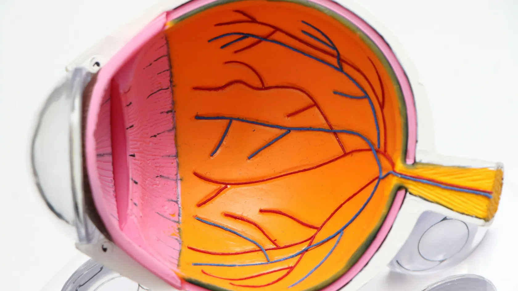
The retina lines the back of the eye like the film in a camera. Its center, the macula, gives sharp reading and face vision. The inside of the eye is filled with a clear gel, the vitreous, which with age shrinks and pulls away from the retina. Most retinal operations start with removing that gel, a vitrectomy, so the surgeon can work on the retina directly.
1
Retinal detachment
A tear lets fluid seep under the retina and lift it off like wallpaper. Without surgery it spreads and the eye goes blind.
2
Macular hole
A small full-thickness hole at the center of the macula, causing a central blur or gap when reading.
3
Epiretinal membrane
A thin scar film on the macula that wrinkles it, bending straight lines and blurring detail.
4
Diabetic eye disease and bleeding
Bleeding into the vitreous that will not clear, or scar tissue pulling on the retina in advanced diabetic retinopathy.
Warning signs, and how soon each problem needs treatment
Timing decides the result more than anything else in retinal surgery. A detachment caught while the macula is still attached can leave reading vision intact. A week later, once the macula has lifted, the same detachment leaves permanent blur in many eyes however well the operation goes. So the table below puts some problems out of reach of a planned trip. For a patient abroad with a fresh detachment, the honest advice is to have it repaired at home and to come to Istanbul for a later operation, a second opinion or a problem that can wait.
When to act
The symptoms
Signs of a tear or detachment
A sudden shower of new floaters, flashes of light at the edge of vision, or a shadow or curtain spreading across part of the field of view. Any of these needs an eye examination the same day or the next, wherever you are.
How soon
This table scrolls sideways on a narrow screen. Swipe or drag to see every column.
How soon common retinal problems need surgery
Problem
Timing
What this means for travel
Detachment with the macula still attached
Within one to two days
Treat where you are, do not wait to travel
Detachment with the macula already off
Within days
Travel only if surgery can happen within the week
Macular hole
Weeks
Time to plan a visit, results are better if done within months
Epiretinal membrane
Months
Planned surgery, when the distortion troubles you
Diabetic bleeding or traction
Weeks to months, unless the macula is threatened
Planned, with diabetes control checked first
The operations
Three operations, chosen by the problem and the eye.
Vitrectomy
Through three tiny ports
Three ports thinner than a pin are placed in the white of the eye. The surgeon removes the vitreous gel, releases any pull on the retina, peels membranes, seals tears with laser, and fills the eye with gas, air or silicone oil to hold the retina in place while it heals. Vitrectomy treats almost every retinal condition on this page.
Scleral buckle and pneumatic retinopexy
Two alternatives exist for some detachments. A scleral buckle stitches a soft silicone band to the outside of the eye, indenting the wall toward the tear, and suits younger patients who still have their natural lens. Pneumatic retinopexy injects a small gas bubble in the clinic, then seals the tear with freezing or laser while the patient holds a set head position for several days, and suits a single tear in the upper part of the retina.
Repairing a detachment, the evidence
No single operation suits every detachment.
Two randomized trials
Pneumatic retinopexy against vitrectomy
The Canadian PIVOT trial randomized 176 patients with simpler detachments. At one year, vision was better after pneumatic retinopexy, 79.9 against 75.0 letters, and straight lines were less distorted. The retina stayed attached after the first procedure in 80.8 percent with pneumatic retinopexy against 93.2 percent with vitrectomy, and after further surgery in 98.7 and 98.6 percent. Cataract surgery was needed within a year in 65 percent of vitrectomy eyes with a natural lens against 16 percent. Hillier and colleagues, Ophthalmology, 2019.
Scleral buckle against vitrectomy
It depends on the lens
A European trial randomized 681 patients with medium-complexity detachments. In eyes with their natural lens, vision improved more after a buckle, and cataract progressed more after vitrectomy. In eyes that had had cataract surgery, vitrectomy reattached the retina at the first attempt in 72.0 percent against 53.4 percent with a buckle, and needed fewer further operations. Heimann and colleagues, Ophthalmology, 2007.
Closing a macular hole
Most macular holes close with one vitrectomy. Technique decides how many.
Peeling the inner membrane
Cochrane review of four trials
Peeling the thin internal limiting membrane around the hole during vitrectomy raised the odds of closing the hole at the first operation more than ninefold, odds ratio 9.27, and cut the need for further surgery, odds ratio 0.11, without more complications. Vision at six months was similar either way, but fewer patients needed a second operation. Spiteri Cornish and colleagues, Cochrane Database of Systematic Reviews, 2013.
Large holes
The inverted flap
For holes wider than 400 micrometers, surgeons can fold a flap of that membrane over the hole instead of removing it. A Cochrane review of four small trials with 285 eyes found closure 10 percent more likely with the flap and better vision from three months, on low to moderate certainty evidence. Ghoraba and colleagues, Cochrane Database of Systematic Reviews, 2023.
The day of surgery
On the day
What happens
1
Before
Drops to widen the pupil, a numbing injection beside the eye and, if you wish, light sedation, while children and some anxious patients have general anesthesia instead and stay a night.
2
During
Thirty minutes to two hours depending on the problem. You feel pressure but no pain, and you see light and shadow.
3
After
A pad and shield over the eye, a check, and the positioning instructions. Most patients go back to the hotel the same day.
Afterward
Hold the head position the surgeon prescribes, face down or on one side, for the hours per day and the number of days stated.
Use the drops exactly as written, usually for four weeks.
Keep water and dust out of the eye and do not rub it.
Wear the identity band that says a gas bubble is in the eye, if one was used.
Come to the check the next day and at one week.
Gas, oil and positioning
At the end of a vitrectomy the eye is filled with something that presses the retina flat while the laser or freezing marks around a tear turn into a firm seal, a process that takes one to two weeks. The choice of filling decides how long vision stays blurred, what position you hold, and above all whether and when you can fly.
What holds the retina while it heals
A gas bubble
No flying, no mountains, no nitrous oxide while gas is in the eye. A gas bubble expands as air pressure falls, in an aircraft cabin or at high altitude, and the pressure rise inside the eye can destroy the optic nerve within hours. Some gases take about two weeks to clear, others six to eight. Any anesthetist treating you in that time must know about the bubble, since nitrous oxide makes it expand too. Vision is blurred while the bubble is present, and a line appears as it shrinks.
Silicone oil
1
When oil is chosen
For complex or repeated detachments, or when a patient must fly soon, since oil does not expand and flying stays safe.
2
Living with oil
Vision is blurry through oil. It stays in for two to six months.
3
Removal
A second, shorter operation removes the oil, often combined with cataract surgery.
Risks and results
What can go wrong
Cataract
Vitrectomy speeds up cataract in eyes with a natural lens, within a year for the majority of patients over fifty. The cataract operation gets planned in advance or combined.
Redetachment and other risks
A retina can detach again. In the European trial this happened to roughly one eye in four among those with a natural lens, whichever operation was used. Raised eye pressure, bleeding and scar tissue regrowth are the other risks, along with infection inside the eye, which is rare but serious. Vision recovers over weeks to months, and how much it recovers depends above all on whether the macula was detached and for how long, which is the strongest argument for early surgery.
Length of stay and flying
The length of stay depends on what fills the eye at the end of surgery. With air or no tamponade, as after many membrane peels, five to seven days covers the consultation, surgery and the checks before flying. With a gas bubble, you cannot fly until it has cleared, two to eight weeks, so patients either stay in Istanbul for that time, travel home overland, or, if flying soon is essential, ask whether silicone oil suits their case. The surgeon tells you before the operation which is planned. Patients holding a face-down position benefit from a hotel room chosen for it, and the coordinator can arrange rental of a positioning chair.
Before you travel
Four steps help before you book.
Send the OCT images and eye report so the surgeon can confirm the plan and the likely tamponade.
Check whether you take blood thinners, and ask your doctor whether they can be paused.
If you have diabetes, bring recent blood sugar results.
Plan the return, air or overland, once the surgeon has said whether gas is likely.
1
The cost
Cost depends on the operation, the tamponade, whether cataract surgery is combined, and the length of stay, which gas can extend. Published packages in Istanbul cover the consultation, tests, surgery, drops, the checks, an interpreter and transfers, and leave out the hotel and flights. The review of your scans is free.
2
The arrangements
The coordinator writes in English, Arabic, French, Russian, Serbian, Romanian or Spanish, or arranges an interpreter, sends an appointment confirmation and an invitation letter naming the hospital and the treating doctor ten days before travel, and books the hotel and transfers. Two family members may join consultations, halal, vegetarian and diabetic meals are available, a prayer room is on site, and you may request a female doctor at first contact.
Follow-up at home
After you fly home
Checks
1
One month
An eye doctor at home checks the retina and pressure, using the surgical report.
2
Three months
An OCT scan of the macula, and planning for cataract surgery or oil removal if needed.
Warning signs
See an eye doctor the same day
For increasing pain, worsening redness, a sudden drop in vision, new floaters or flashes, or a shadow spreading across the vision. Once you are back home, the coordinator stays reachable on the same WhatsApp number and passes questions to the operating surgeon.
Retina Surgery FAQ
What is retina surgery?
Operations on the light-sensitive layer at the back of the eye, most often vitrectomy, to repair a detachment, close a macular hole, remove a scar membrane or clear bleeding. Scleral buckle and pneumatic retinopexy are alternatives for some detachments.
How successful is retinal detachment surgery?
In the PIVOT trial the retina was attached at one year in 98.7 percent after pneumatic retinopexy and 98.6 percent after vitrectomy, including those who needed a second operation. The first operation succeeded in 80.8 and 93.2 percent.
Can I fly after retina surgery?
Not while a gas bubble is in the eye, which takes two to eight weeks to clear, because the bubble expands in the cabin and can damage the optic nerve. With air, silicone oil or no bubble, flying is allowed after the surgeon's check.
Why do I need to keep my head face down?
The bubble floats upward, and face-down or side positioning places it against the hole or tear so the retina seals. The surgeon prescribes the position and the number of days.
Does retina surgery cause cataract?
Vitrectomy speeds up cataract in eyes with a natural lens. In the PIVOT trial, 65 percent of such eyes needed cataract surgery within a year after vitrectomy.
How long do I need to stay in Istanbul?
Five to seven days if no gas bubble is used. With gas, until it clears, two to eight weeks, unless you travel home overland. The surgeon tells you in advance which applies.
References
Hillier RJ, Felfeli T, Berger AR, et al. The Pneumatic Retinopexy versus Vitrectomy for the Management of Primary Rhegmatogenous Retinal Detachment Outcomes Randomized Trial (PIVOT). Ophthalmology. 2019;126(4):531-539.
Heimann H, Bartz-Schmidt KU, Bornfeld N, Weiss C, Hilgers RD, Foerster MH. Scleral buckling versus primary vitrectomy in rhegmatogenous retinal detachment, a prospective randomized multicenter clinical study. Ophthalmology. 2007;114(12):2142-54.
Spiteri Cornish K, Lois N, Scott N, et al. Vitrectomy with internal limiting membrane (ILM) peeling versus vitrectomy with no peeling for idiopathic full-thickness macular hole (FTMH). Cochrane Database Syst Rev. 2013;2013(6):CD009306.
Ghoraba H, Rittiphairoj T, Akhavanrezayat A, et al. Pars plana vitrectomy with internal limiting membrane flap versus pars plana vitrectomy with conventional internal limiting membrane peeling for large macular hole. Cochrane Database Syst Rev. 2023;8(8):CD015031.
Editor's note
Written by the Biruni Hospital medical editorial team. Reviewed by Assistant Professor Selin ŞİMŞEK ALKAN, Ophthalmology.