
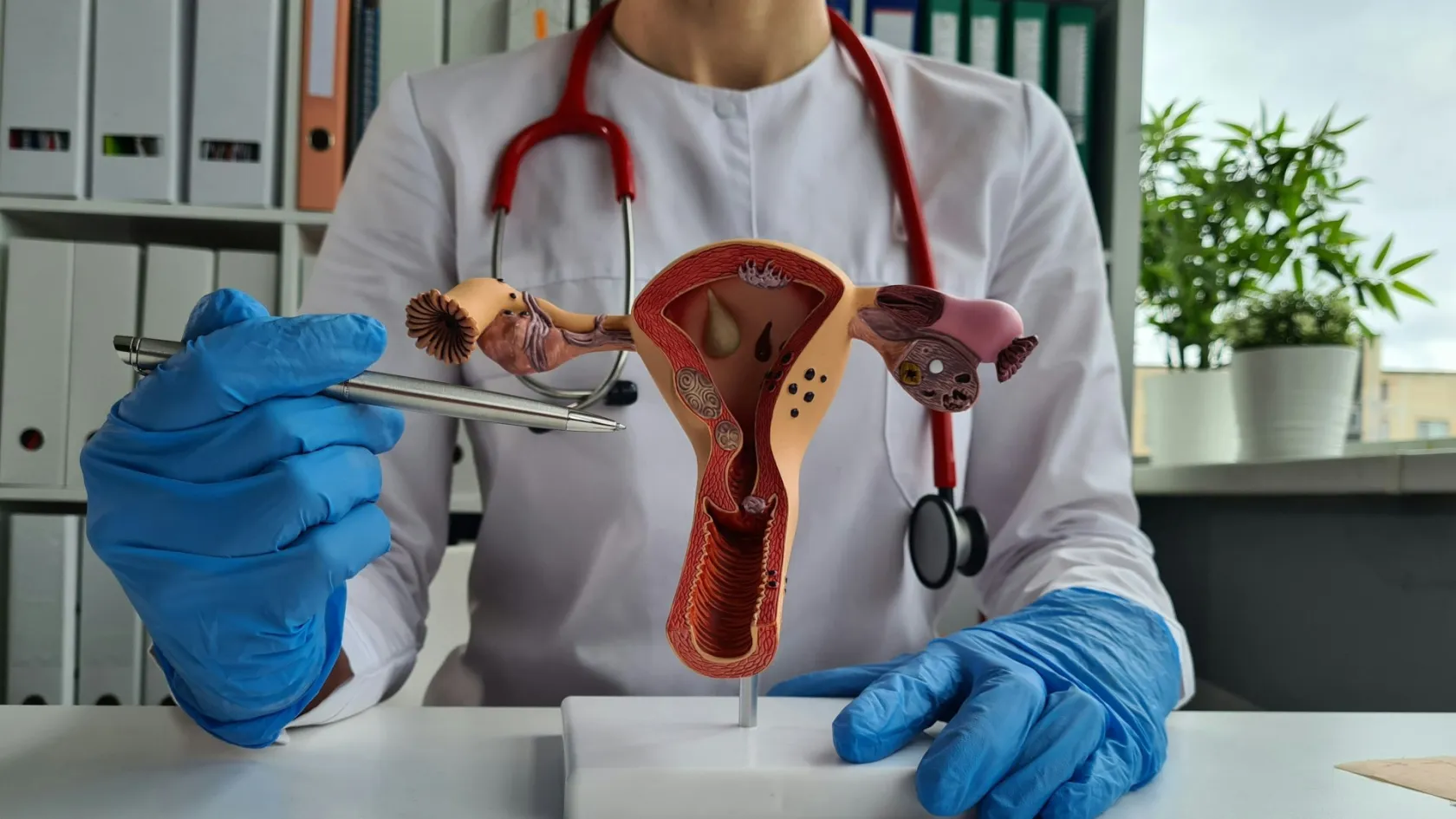
Radical Hysterectomy for Cervical Cancer
Radical is a graded scale from A to D, and the letter you are given decides whether you will be emptying your bladder normally in six months.
About This Department
Radical is not one thing.
It is a graded scale running from A to D, each step taking more of the tissue on either side of the cervix, and the letter you are given decides whether you will be emptying your bladder normally in six months. A consent form that says radical hysterectomy without naming a type has left out the part that matters most to how you will live afterwards. That omission is common.
Ask which letter.
Free consultation
Two measurements decide this operation, tumour size and parametrial spread
Send the cervical biopsy or cone specimen report with the cell type written in it, and the MRI of the pelvis on a disc rather than only the written report, since the size of the tumour and whether it has reached the tissue beside the cervix are measured off those images and together they set the type of operation. Add the HPV and smear results, any earlier scan, and a note of any previous cone biopsy or loop treatment with its date. Tell us your age, whether you still have periods, whether you hope to have children, and whether you have had a caesarean or any other pelvic surgery. If a cone biopsy has already removed the visible tumour, say so, because that changes what the MRI can still show and how the operation is planned.
Radical is a scale, not a word
In 2008 two surgeons, Querleu and Morrow, proposed a classification that the field adopted quickly because it solved a real problem. Until then, surgeons in different countries described the same operation with different words and different operations with the same word, which made comparing results across hospitals close to meaningless. The classification defines four types by anatomical landmarks, meaning by where exactly the cut is made and never by how radical it feels, and a 2024 review revisiting it proposes further refinement including a selective nerve-sparing version of the most extensive type.
This table scrolls sideways on a narrow screen. Swipe or drag to see every column.
| Type | How far the dissection goes, and who it suits |
|---|---|
| Type A | The least extensive. The cut is made between the cervix and the ureter without following the tissue out to the pelvic sidewall, and only a few millimetres of surrounding tissue and vagina come with it. Used for the smallest tumours, and functionally close to an ordinary hysterectomy. |
| Type B | The ureter is freed and the tissue beside the cervix is divided at the level of the ureteric tunnel, taking roughly a centimetre of vagina. A middle position, and the type most often used where the tumour is small but not tiny. |
| Type C1 | The full dissection out to the pelvic sidewall and internal iliac vessels, taking one to two centimetres of vagina, but with the autonomic nerves identified and preserved. This is the nerve-sparing version and it is the type most patients with early cervical cancer should be discussing. |
| Type C2 and Type D | C2 is the same extent as C1 with the nerves sacrificed rather than preserved. D goes further still, out to the pelvic sidewall vessels and muscle, and is reserved for the most advanced situations. Both carry the highest rates of bladder and bowel dysfunction. |
Two things follow from having a scale at all. The first is that the extent should be matched to the tumour, and one of the most cited recent papers on this subject is titled around abandoning the idea that one operation fits every patient. The second is that when a hospital quotes you for a radical hysterectomy without naming a type, they may be quoting for a C2 and delivering a B, or the reverse, and neither of you would know. Ask for the letter in writing.
What the nerves in there do
The tissue on either side of the cervix is not packing material.
Running through it are the autonomic nerves that tell the bladder when it is full and make it contract to empty, the nerves supplying the lower bowel, and nerves involved in genital sensation and lubrication. A conventional radical hysterectomy divides them, since they sit inside the tissue being removed. A nerve-sparing operation identifies them, separates them from the tissue and leaves them intact, which takes longer and requires a surgeon who has learned the anatomy properly. It is a taught skill.
What preserving them actually buys
A meta-analysis pooled thirteen randomised controlled trials covering 1,002 women having laparoscopic radical hysterectomy with or without nerve preservation. Nerve-sparing operations took significantly longer. Time to the catheter coming out was significantly shorter with nerve sparing, at a standardised mean difference of minus 1.24, and time to bowel function returning was shorter as well, at minus 1.27. Crucially for anybody worried that preserving nerves means removing less cancer, there was no significant difference in the length of tissue resected beside the uterus, at a standardised mean difference of minus 0.10, nor in the length of vagina removed, nor in blood loss, nor in complications during the operation. The authors noted that sample sizes remain limited and that the technique should still be applied with care.
That last point about the length of tissue removed is the one to hold onto. Nerve sparing is not a smaller cancer operation with a nicer name. The same amount of tissue beside the cervix comes out, and the same length of vagina, and the difference lies entirely in whether the surgeon takes the extra time to dissect the nerves away from it first.
What is being spared is your bladder. The tumour is treated exactly the same. Hold onto that distinction.
The outcome you most want to avoid
Cervical cancer can be treated by surgery or by chemoradiation, and for early disease the two produce similar cure rates.
Trouble arises when a woman receives both. That happens when the pathology after surgery reveals risk factors nobody predicted beforehand, at which point radiotherapy is added to an already operated pelvis, and the combined damage is greater than either treatment would have caused alone.
A cross-sectional study of 427 cervical cancer survivors aged sixty or under, all at least six months past treatment, compared three groups. Two hundred and forty-one had nerve-sparing radical hysterectomy, sixty had the conventional operation and one hundred and twenty-six had chemoradiation. Urinary symptoms were reported by 27.4 percent after nerve-sparing surgery, 40.0 percent after conventional surgery and 18.6 percent after chemoradiation, a significant difference. Sexual function scores were highest after nerve-sparing surgery at 23.8, then conventional surgery at 23.3, then chemoradiation at 21.6. Nerve-sparing surgery also scored better on constipation, on leg swelling and on sexual activity. Read the urinary figure again, because it complicates the surgical story in a way a hospital selling surgery has no incentive to mention. Chemoradiation produced fewer urinary symptoms than either operation. Surgery scored better on sexual function and on leg swelling, so the picture is genuinely mixed rather than one-sided, and that is the honest summary. Neither route wins outright.
The study's subgroup analysis carries the practical lesson.
Women who had nerve-sparing surgery alone had fewer complications than women who had nerve-sparing surgery followed by radiotherapy. So the worst functional outcome in this disease is not surgery and it is not radiotherapy. It is both, one after the other, in the same pelvis. Which means the single most useful question before consenting is what the chance is, given your imaging and biopsy, that radiotherapy will be recommended after the operation. Where that chance is high, going straight to chemoradiation and avoiding the operation entirely is a legitimate and sometimes better plan, and a unit that will not discuss it is not giving you the full set of options. Ask for all three.
How they decide beforehand
Everything above depends on knowing the size of the tumour and whether it has reached the tissue beside the cervix, and both are measured before the operation by imaging. How well that works was examined in a post-hoc analysis of a large international study, SENTIX, which ran at 47 sites across 18 countries and enrolled 690 women with early cervical cancer between 2016 and 2020.
Every woman had either pelvic magnetic resonance imaging or an expert ultrasound before surgery, and the researchers compared what the scan said against what the pathologist found. Tumour size was out by a centimetre or more in 13.1 percent of the ultrasound group and 16.5 percent of the MRI group, with no significant difference between the two methods. For predicting that the tissue beside the cervix was clear, the negative predictive value was 97.0 percent for ultrasound and 95.3 percent for MRI. For predicting that the lymph nodes were clear it was 94.0 percent and 94.1 percent. The two were comparable on every measure tested. Expert hands matter more than the machine. Two conclusions come out of that and both are useful to a patient. The first is that expert ultrasound is as good as MRI for this specific job, which matters if you are in a place where MRI is hard to obtain, and the authors argued explicitly that ultrasound deserves a place in routine preoperative staging. The second is quieter and more important. In roughly one woman in seven, the tumour turns out to be a centimetre or more different in size from what the scan showed, and since tumour size is one of the thresholds deciding which type of operation you get, the plan made in clinic is a well-informed estimate rather than a certainty. A surgeon who tells you the plan may change once the final pathology arrives is being accurate. Plans are estimates until the specimen is read.
The operation, step by step
Allow three or four hours for a nerve-sparing type C1 radical hysterectomy.
The route, meaning open or keyhole, is a separate discussion covered on our page about hysterectomy for cancer generally, and it does not change the sequence below.
Opening the spaces
The pelvis contains natural planes between structures that can be opened bloodlessly, and the operation begins by developing them on both sides. Doing this properly exposes the anatomy the rest of the procedure depends on, and rushing it is where difficulty later comes from.
Nodes first, and a decision point
The sentinel nodes or the pelvic nodes are removed and, in many units, examined while you are still asleep. If cancer is found in them the surgeon may stop, because a woman who needs chemoradiation anyway is better served by not also having the hysterectomy. Knowing that this may happen makes it a plan rather than a shock.
Unroofing the ureter
The tube carrying urine from the kidney runs through a tunnel in the tissue being removed and has to be lifted out of it under direct vision. This step defines the difference between type B and type C, it is where injury occurs when it occurs, and it is unhurried in good hands.
Finding and sparing the nerves
In a C1 the nerve bundles running to the bladder are identified where they lie in the deep part of the tissue and are separated from what is being removed. This is the step that distinguishes C1 from C2, it adds time to the operation, and it is the reason the catheter comes out sooner.
Taking the vaginal cuff and closing
One to two centimetres of upper vagina comes out with the specimen and the remaining vagina is closed. That closure is why nothing enters the vagina for six weeks, and the length removed is worth asking about, since it is what shortens the vagina and it varies with the type.
Expect a catheter at the end.
In a nerve-sparing operation it usually comes out within a few days, and after a conventional radical hysterectomy it often stays a week or two while the bladder relearns its job. Blood loss is moderate and transfusion is uncommon. The risks worth naming are injury to the ureter or bladder, which is uncommon and usually recognised and repaired during the operation, a fistula between bladder and vagina, which is rarer still, clots, and infection. All are uncommon.
Moving the ovaries out of range
Cervical cancer rarely spreads to the ovaries, so a young woman having this operation can usually keep hers.
Radiotherapy is what threatens them, since ovaries sitting in the pelvis receive a dose that ends their function permanently. The answer is a small additional procedure done during the hysterectomy called ovarian transposition, in which the ovaries are detached from their normal position, lifted up out of the pelvis and fixed high in the abdomen with a marker clip, keeping their blood supply intact. It adds minutes, not hours.
A meta-analysis gathered 29 publications covering 1,160 women with cervical cancer who had this done, and the numbers vary sharply depending on what treatment followed. In women who had surgery alone, 91 percent kept working ovaries. With surgery plus internal radiotherapy the figure was 93 percent. Where external beam radiotherapy was given, it fell to 61 percent. Ovarian cysts formed in a proportion of women in every group and were the commonest side effect. Metastases to the transposed ovaries were close to non-existent, at 99 percent of women free of them in the surgery groups and none at all reported in the external beam group. Three practical points follow. Transposition works best when radiotherapy is avoided altogether, which is the same argument made further up this page about not needing both treatments. It still preserves function in three women out of five even when external beam radiotherapy is given. For a woman in her thirties that is a great deal better than nothing. And the marker clips matter, since they tell the radiotherapy planner exactly where the ovaries now are, so that the beams can be arranged to avoid them. Clips are not optional.
The bladder, and the first weeks
Bladder function is the defining recovery issue after this operation and it is the one patients are least prepared for.
Nerves that were disturbed, or divided, normally signal fullness and drive the muscle that empties. Without them a woman does not feel the urge until the bladder is very full, finds it hard to start the stream, and does not empty completely, which is what leads to repeated infections. Nobody warns you about this enough.
Management of it is unglamorous and it works.
This table scrolls sideways on a narrow screen. Swipe or drag to see every column.
| Measure | What it involves, and why it helps |
|---|---|
| Timed voiding | Going to the toilet by the clock, usually every two to three hours, rather than waiting for an urge that may never arrive. It prevents the bladder from overfilling and stretching, which is what makes emptying harder still. |
| Double voiding | Waiting a minute after you think you have finished, then trying again, sometimes leaning forward. Costs nothing, takes a minute, and clears a surprising amount of the urine that would otherwise sit there breeding infection. |
| Measuring the residual | A quick bladder ultrasound after you have emptied, done before the catheter is removed for good. It turns the question of whether you are emptying from a guess into a number, and it is the step that decides what happens next. |
| Self-catheterisation | Where emptying stays incomplete, passing a fine catheter yourself a few times a day. It sounds far worse than it is. Taught in an afternoon, it beats recurrent infections or a permanent catheter. Most women who need it need it temporarily. |
Three other things follow the same nerves and the same pattern, and all three are treatable. Say so out loud in clinic.
- Constipation, common in the early months, responding to the ordinary measures of fluid, fibre and a stool softener started before it becomes a problem rather than after.
- Vaginal dryness and reduced lubrication, which have a nerve component as well as a hormonal one, which is why they occur even when the ovaries were kept, and which respond to moisturisers, lubricants, dilators and pelvic floor physiotherapy.
- Leg swelling, which relates to the lymph node dissection and not to the hysterectomy itself, and which responds far better when reported in the first weeks than in the first year.
Recovery and living with it
Hospital stay is three to five days after a keyhole radical hysterectomy and five to seven after an open one. Return to desk work takes four to six weeks, and to full activity six to eight. Nothing enters the vagina for six weeks. Lifting is restricted over the same period and driving resumes once an emergency stop is possible without hesitating. Six weeks is the number.
Longer term, the arc deserves more attention than it gets.
Bladder function improves over months rather than weeks, and the trajectory in the first fortnight is a poor guide to where you will end up. Sexual activity resumes after six weeks and takes longer than that to feel normal, particularly where the vagina was shortened, and dilator use started early makes a measurable difference. If both ovaries were removed or irradiated, surgical menopause arrives abruptly and hormone replacement is appropriate in this cancer, which is worth knowing since women are sometimes refused it by clinicians extrapolating from breast cancer. The two are different diseases.
Fertility ends with this operation.
Where childbearing is still hoped for and the tumour is small enough, an alternative operation that removes the cervix and leaves the body of the womb exists and is discussed on our hysterectomy page. That conversation belongs before surgery is booked, since it cannot be had afterwards. Raise it at the first appointment.
Coming to Istanbul
How long you need to stay in Istanbul is two and a half to three and a half weeks, hotel nights included. The first days go on measurement and discussion rather than surgery. The outside biopsy is re-read by our own pathologists, the MRI is reviewed or repeated and an expert ultrasound is performed alongside it, and the case goes to a gynaecological oncology multidisciplinary meeting where the type of operation is decided and written down as a letter before you consent to it. In writing, as a letter.
That meeting also settles the question this page keeps returning to, which is how likely radiotherapy is to be needed afterwards. Where the imaging and biopsy suggest it is probable, we will say so and set out the option of chemoradiation without surgery, since avoiding the combination is worth more to your long term function than any surgical technique can add. Pathology after surgery takes seven to ten days and it decides whether anything else is needed, so waiting for it is why the stay is longer than the physical recovery requires. Flying home is reasonable at around two weeks, once the catheter is out or you have been taught to manage it, the wound is healed and the pathology has been discussed with you face to face. Clot risk is raised after pelvic cancer surgery, so anticoagulant injections continue after discharge and compression stockings matter on the flight. Interpreting is arranged in advance in English, Arabic, Russian, French and German, and you go home with the operative note naming the Querleu-Morrow type performed, whether the nerves were spared and whether the ovaries were transposed. Check all three lines.
What moves the cost
Because radical covers a graded scale, a figure attached to the phrase without a type behind it describes an operation nobody has yet specified. What moves it is clinical.
- Which Querleu-Morrow type is planned, since a type A and a type C1 differ substantially in operating time.
- Whether nerve sparing is performed, which the evidence shows takes significantly longer.
- The route, since robotic instruments carry costs that keyhole and open surgery do not.
- Whether sentinel node mapping is used, needing a tracer and often a fluorescence camera, or a full pelvic node dissection is performed.
- Whether nodes are examined while you are asleep, which requires a pathologist on standby.
- Whether ovarian transposition is added, with the marker clips that go with it.
- Whether an expert ultrasound and an MRI are both performed here or one has already been done to an acceptable standard.
- Whether radiotherapy or chemoradiation forms part of the plan and where it will be delivered.
Ask any written quotation five questions. Does it name the Querleu-Morrow type. Does it include nerve sparing explicitly. Does it cover a ureteric injury or a fistula if one occurs. Does it include the full pathology and the multidisciplinary review. And what happens to the figure if the nodes are positive during surgery and the hysterectomy is abandoned in favour of chemoradiation, which is a real possibility and the right decision when it happens. A quotation that answers those five is one you can plan around. Ask all five.
Follow-up once you are home
Surveillance is clinical examination every three to four months for two years, then six-monthly to year five, with a vaginal vault examination each time and imaging when a symptom or a finding raises a question. Most recurrences announce themselves through bleeding, discharge, pelvic or back pain, or leg swelling on one side, so reporting those promptly is more useful than any routine scan.
Bladder symptoms that are worsening rather than improving after the first few months deserve assessment rather than patience. Getting worse is not normal.
Four documents make the handover work, and you should leave with all of them.
- The operative note naming the Querleu-Morrow type, whether nerves were spared, the length of vagina removed and whether the ovaries were transposed and clipped.
- The pathology report with tumour size, depth of invasion, lymphovascular space involvement, margin status and node results.
- A written statement of whether radiotherapy or chemoradiation is recommended, with the reasoning.
- A bladder plan naming what to do if emptying stays incomplete, and a menopause and sexual health plan where the ovaries were removed or irradiated.
Our team stays reachable for your own doctor's questions afterwards, and where a symptom turns on exactly what was done in theatre we would rather answer it than have it guessed at. Send it over.
Frequently asked questions about radical hysterectomy
What do the types A, B, C and D mean?
Does nerve-sparing surgery remove less cancer?
Should I have surgery or chemoradiation?
How accurate is the scan that decides my operation?
Can I keep my ovaries?
How long will I have a catheter?
Why might the surgeon stop before removing the womb?
How long should I plan to be in Istanbul?
References
- Kostov S, Kornovski Y, Watrowski R, et al. Revisiting Querleu-Morrow radical hysterectomy, how to apply the anatomy of parametrium and pelvic autonomic nerves to cervical cancer surgery? Cancers. 2024;16(15):2729.
- Ma L, Li Q, Guo Y, Tan X, Wang M, Qi Q. Laparoscopic nerve-sparing radical hysterectomy for the treatment of cervical cancer, a meta-analysis of randomized controlled trials. World Journal of Surgical Oncology. 2021;19(1):301.
- Li W, Wang X, Wu M, Tan X. Quality of life in cervical cancer survivors after nerve-sparing radical hysterectomy, conventional radical hysterectomy or concurrent chemoradiotherapy. Frontiers in Oncology. 2025;15:1654730.
- Cibula D, Köhler C, Jarkovský J, et al. Magnetic resonance imaging and ultrasound examination in preoperative pelvic staging of early-stage cervical cancer, post-hoc analysis of SENTIX study. Ultrasound in Obstetrics and Gynecology. 2025;65(4):495-502.
- Laios A, Otify M, Papadopoulou A, Gallos ID, Ind T. Outcomes of ovarian transposition in cervical cancer, an updated meta-analysis. BMC Women's Health. 2022;22(1):305.
Editor's note
Written by the Biruni Hospital medical editorial team. Reviewed by Prof. Dr. Özcan YILDIZ, Medical Oncology.
Medically reviewed by

Prof. Dr. Özcan YILDIZ
Medical Oncology
Related Treatments
View All-
Bilateral Mastectomy
Whether you carry a BRCA mutation, have cancer in one breast or in both, bilateral mastectomy removes both breasts in one operation, and how much it helps depends on which of those three paths brought you to it.
-

Breast Conserving - Lumpectomy Surgery
Keeping the breast has never been the less thorough choice, and the largest recent studies now point the other way. This page explains who can have breast conserving surgery, what the margin report decides, and how long a woman traveling for it needs to stay.
-

Cytoreductive - Tumor Reduction Surgery
Patients cleared of every visible deposit lived 30 months in one gastric series. Those left with anything at all lived 7.3 months, however little remained.
-
Debulking - Ovarian Cancer Tumor Reduction
Two women with the same diagnosis on the same morning can get operations that share almost nothing. What decides it is who measured the disease first.
-

Debulking Surgery
Surgeons removed a median of 57 lymph nodes from women already cleared of visible disease. It bought no extra months, and tripled deaths within 60 days.