
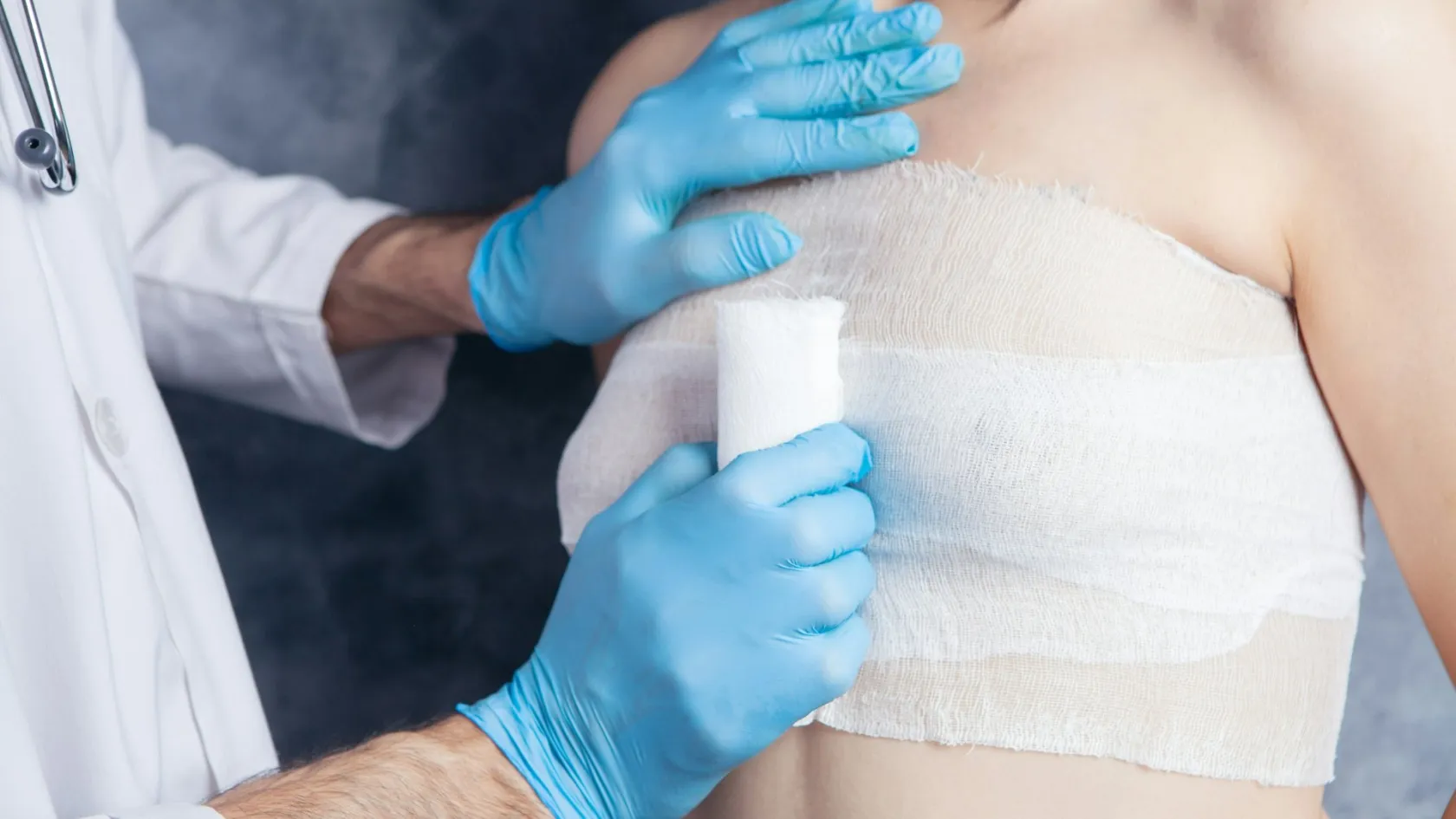
Oncoplastic Breast Surgery
The word oncoplastic sounds like cosmetic surgery bolted on to a cancer operation. It works the other way round. The reshaping is what lets the surgeon cut wider than a plain excision would survive.
About This Department
Oncoplastic breast surgery gets described as plastic surgery added on to a cancer operation, and that description has it backwards. The reshaping does not arrive as a finishing touch once the tumour is out. Reshaping is what allows the surgeon to take a wider piece of breast than that breast could otherwise absorb without collapsing into a dent, and that is how the technique lowers positive margin rates and lowers the number of women who end up losing the breast entirely. This page explains how that works, which version of it applies to which breast, and what it costs you in healing time.
Free consultation
Tell us the tumour size and your own breast size in the same message
Whether an oncoplastic repair is needed, and which one, comes down to the ratio between the two, plus where in the breast the tumour sits. Send the mammogram and ultrasound files themselves, and not only the report, any MRI, the core biopsy pathology, and your bra size. What comes back is an opinion on whether the breast can be conserved with reshaping, whether the other breast would need adjusting to match, and whether a mastectomy with reconstruction would give you a better result than either. The review costs nothing and carries no obligation.
The ratio that decides everything
Twenty five millimetres of tumour is a small operation in a large breast and a disfiguring one in a small breast. Surgeons therefore plan conservation around the proportion of the breast that has to leave rather than around the measurement on the report, and that proportion is the number worth asking about at your own consultation.
Where a plain excision starts to show
Take out less than roughly a fifth of the breast and the cavity looks after itself, filling with fluid in the first weeks, replacing that fluid gradually with scar tissue, and settling back close to the shape the breast had before. An ordinary lumpectomy for a screen-detected cancer therefore needs nothing done to it. Go past that fraction and the mechanism fails. There is not enough surrounding tissue to close the space, the skin sinks into the cavity, the nipple pulls towards the scar, and radiotherapy then contracts everything around a defect that is already there.
The threshold, and why it moves
Published practice puts the working figure at about 20 percent of breast volume, above which a plain excision starts to leave a visible defect. That figure moves, because position matters as much as proportion. The lower inner quadrant, where the breast has least tissue to spare and the skin is thinnest, tolerates loss badly and shows a defect well below 20 percent. The upper outer quadrant, which holds the most tissue in almost every breast, tolerates the same volume without any obvious change. So a surgeon planning a repair is reading two numbers together, and neither of them is the size of the tumour on its own.
Position decides as much as proportion.
Your own breast size is the other half of that ratio, and it is the half patients rarely think to mention.
Why a deformity is easier to prevent than to repair
Radiotherapy sets the result. Once the breast has been irradiated the tissue becomes firmer and less elastic, small blood vessels are reduced, and any operation done afterwards heals more slowly and carries a higher rate of wound problems and fat necrosis. Correcting a defect at that stage means operating on scarred, radiated tissue to move more scarred, radiated tissue into a hole, and the results are visibly poorer than the same reshaping done at the original operation. That asymmetry between prevention and repair is the whole clinical argument for planning the reconstruction before the excision rather than waiting to see how it turns out.
So the decision has to be made in the clinic, before the operating list, by a surgeon who has looked at the imaging alongside the breast in front of them. A patient who is told only that the tumour will be removed and the breast will be closed has not had that conversation, and it is a reasonable thing to ask for directly.
Level one and level two
Oncoplastic operations are graded by how much rearranging they require, and the grading matters to you because it predicts the scar, the theatre time, the healing, and who needs to be in the room. Level one covers modest volumes and modest rearrangement. Level two covers the operations that reshape the breast properly, move the nipple on its own blood supply, and remove skin as well as glandular tissue.
This table scrolls sideways on a narrow screen. Swipe or drag to see every column.
| Level | Volume removed | What the operation involves |
|---|---|---|
| Level one | Up to about a fifth | The breast tissue around the cavity is freed from the skin and the chest wall and slid together to close the space. No skin is removed, and the nipple stays exactly where it started, which is what keeps the scar short and the recovery close to that of an ordinary lumpectomy. Any breast surgeon trained in the technique can do it. |
| Level two | Roughly a fifth to a half | The breast is formally reshaped. Skin is removed, the remaining gland is built into a new mound, and the nipple travels on a pedicle of tissue that keeps its blood supply. The scars follow reduction patterns rather than sitting over the tumour. |
| Beyond level two | More than about half | Conservation stops being the better answer. A mastectomy with immediate reconstruction produces a more predictable breast than a conserved one that has lost most of its volume and then been irradiated. |
Nobody will hand you a percentage in the clinic, because it is an estimate and not a measurement. Ask instead which level the surgeon is planning, where the scars will run, and whether the nipple is being moved. Those three answers tell you what operation you are actually having.
Moving tissue, or bringing it in
Every oncoplastic repair answers the same question. Either the breast holds spare tissue to move into the gap, or the tissue has to come in from outside it.
Volume displacement, for a breast with tissue to spare
In a medium or large breast the remaining gland is rearranged to fill the cavity, and the operation borrows its patterns from breast reduction. A therapeutic mammoplasty is the commonest version, using the same wise pattern or vertical scar a cosmetic reduction would use, with the tumour taken as part of the tissue that a reduction would have removed anyway. Smaller variants exist for smaller defects. A round block technique works through an incision around the areola for tumours near the centre. A racquet mammoplasty adds a straight limb running out towards the tumour. A Grisotti flap rebuilds the centre of the breast when the tumour sits directly behind the nipple and the nipple has to go with it. The advantage that patients notice has nothing to do with the cancer. Their breast ends up smaller and lighter, and women who have carried heavy breasts for decades frequently report that the shoulder and neck pain they had lived with improved after the operation.
Volume replacement, for a breast with nothing to move
A small breast has no spare tissue, so rearranging it only makes it smaller and tighter. Tissue gets imported instead, most commonly on a chest wall perforator flap. The surgeon raises a strip of fat and skin from the side of the chest or the fold beneath the breast, keeps it alive on a small artery coming through the muscle, and swings it into the cavity through a tunnel. The lateral intercostal artery perforator flap and the thoracodorsal artery perforator flap are the two named most often. Nothing is taken from the other breast, and the muscle underneath is left intact, which is what separates these flaps from the older latissimus dorsi operation and why the recovery is shorter than that reputation suggests. The scar sits in the side fold, roughly where a bra band runs. Women who arrive expecting a visible reconstruction scar across the front of the breast are surprised by how little of it shows.
Which one you are offered is decided by your own breast and by where the tumour sits in it. A large-breasted woman with an upper outer tumour is a displacement case. A slim woman with a B cup and the same tumour is a replacement case, and being offered a reduction pattern in that situation is a reason to ask a second question.
Does taking more actually help
Underneath oncoplastic surgery sits a testable claim. If reshaping lets the surgeon cut wider, then fewer margins should come back involved, fewer women should be called back for a second operation, and fewer should end up losing the breast. Pooled data say all three happen.
What 52 studies and 46,835 women showed
An updated meta-analysis published in 2024 compared oncoplastic conservation against conventional conservation and found the oncoplastic group doing better on every primary measure. Re-excision fell by roughly a third, local recurrence by nearly 40 percent, positive surgical margins by around a quarter and conversion to mastectomy by a third, while total complications came out fewer as well and aesthetic outcomes were rated better by both the patients and the assessors doing the scoring. A separate pooled analysis of 31 studies covering 115,011 patients reported the same direction on margins, re-excision and local recurrence.
Set against that, a meta-analysis of 18 studies and 18,103 patients found no significant difference in recurrence, and its re-excision advantage disappeared once the authors adjusted for publication bias. That correction deserves to be taken seriously. None of this evidence comes from a randomised trial, the surgeons who choose the technique are also the ones reporting how well it went, and units that do oncoplastic work tend to be units that do a great deal of breast surgery of every kind.
Read narrowly, the finding is still clearly positive. Oncoplastic surgery lets a surgeon remove more tissue without paying for it in recurrence or in disfigurement, and for a woman whose only other option was a mastectomy that is a substantial gain.
It does not make a cancer less likely to come back. Clear margins are clear margins, however they were achieved.
One group deserves a specific warning. Invasive lobular carcinoma grows in single-file strands instead of forming a discrete lump, so it is chronically underestimated on imaging and its margins are the hardest in breast surgery to clear. A 2025 systematic review pooling eight studies found positive margins in 31 percent of lobular cancers treated with oncoplastic conservation, three and a half times the rate for the commoner ductal type. The same review found something more useful, though. Within lobular cancers, the larger tumours had roughly half the positive margin rate with an oncoplastic approach compared with a standard excision. So the technique is the right choice for lobular disease, carrying a frankly higher chance of needing a second operation, and knowing that number in advance is better than meeting it afterwards.
One practical thing follows from all of this, and it decides more than the technique does. It is who stands at the table.
Oncoplastic work is done either by a single surgeon trained in both cancer surgery and breast reconstruction, or by a breast surgeon and a plastic surgeon operating together on the same list. Both arrangements produce good results and neither is inherently superior, but the two-surgeon model needs the two of them to have planned the case in the same room beforehand, because a plastic surgeon meeting the patient for the first time under anaesthetic is reconstructing a defect somebody else designed. The question to ask is not how many operations the unit does. Ask whether the person reshaping the breast saw you in clinic before the day, and whether the reshaping was drawn on your skin while you were standing up. Marking a breast on a lying patient loses the effect of gravity, and gravity is most of what a breast looks like.
Who it suits and who it does not
Women who gain most are the ones who were about to be told that the breast could not be saved. Too large for a plain excision. Sitting under the nipple. In a breast with nothing to spare, or spread across a whole quadrant instead of sitting as one lump. Each of those was a mastectomy twenty years ago and is frequently a conserved breast now.
This table scrolls sideways on a narrow screen. Swipe or drag to see every column.
| Your situation | Where it usually lands |
|---|---|
| Large tumour, large breast | The best case for a therapeutic mammoplasty. The tumour comes out inside tissue a reduction would have removed anyway, and the breast is lighter afterwards. |
| Small breast, small tumour | Often a chest wall perforator flap, because there is no spare gland to move. A plain excision here is what produces the dents that get regretted. |
| Tumour behind the nipple | Conservation stays possible using a central technique, though the nipple normally goes with the specimen and can be rebuilt later or tattooed. |
| Invasive lobular cancer | Suitable, and a case for a frank warning beforehand. Around 31 percent of lobular cancers come back with an involved margin, so plan for the possibility of a second operation. |
| Breast already irradiated | Rarely suitable. Radiated tissue heals badly and a second course of radiotherapy is rarely possible, so mastectomy is the safer route. |
| Current smoker | The operation gets modified or postponed instead of refused. Nicotine narrows the small vessels these flaps and pedicles depend on, and skin necrosis rates climb sharply. |
Smoking is the one item on that list you can change, and stopping four weeks before the operation measurably reduces wound complications. Diabetes, a high body mass index, and previous surgery on the same breast all push in the same direction without being absolute barriers, and a surgeon commonly responds by choosing a level one repair over a level two rather than by refusing to conserve.
Another group could have this operation and should not want it. A woman carrying a BRCA mutation, a woman with disease in more than one quadrant, and a woman who simply does not want to spend the next five years having mammograms of a reconstructed breast are all making a reasonable choice when they choose mastectomy, and no surgeon should be arguing them out of it.
The other breast
Reshaping leaves a smaller breast, and radiotherapy shrinks and firms it a little more over the following year. Leave the other side alone and the two stop matching, and the difference is more noticeable in a woman who started with a larger bust because there is more to be asymmetric about.
Three routes exist. Leave the other breast alone, reduce it during the cancer operation, or reduce it as a separate procedure once the treated side has finished settling. For a woman having a therapeutic mammoplasty on a heavy breast, doing both sides in the same anaesthetic is the right answer, and it carries a second benefit that gets mentioned too rarely. A smaller, lighter breast takes radiotherapy better. The dose distributes more evenly through less tissue, the skin under the fold stays drier, and the acute skin reaction during treatment tends to be milder.
The argument for waiting is precision, because a breast that has been irradiated has not reached its final shape until about a year afterwards, and matching it before then means matching a moving target.
Tissue removed from the unaffected breast goes to the laboratory in the same way the cancer specimen does and is examined with the same care, because a small number of women turn out to have an unsuspected cancer or a high risk lesion sitting in the breast that nobody had been worried about. That result changes the plan, so it is one of the reasons the full pathology report takes seven to ten days and not two, and the wait covers two breasts and not one.
What goes wrong
Pooling 2,711 oncoplastic reduction procedures in 2,680 women, a systematic review put the overall complication rate at 20 percent, the positive margin rate at 11 percent, the re-excision rate at 6 percent, and the proportion who went on to mastectomy at 3 percent. Most of that 20 percent is wound healing of the slow, tedious kind. Dressings in the clinic settle nearly all of it.
Specific problems begin with delayed healing at the point where the scars meet, which is where the blood supply is thinnest, seroma, infection, fat necrosis producing a firm lump that has to be distinguished from recurrence on later imaging, altered or reduced nipple sensation, which is common and frequently permanent, and partial or complete nipple loss, which is uncommon and is the complication smoking makes far more likely.
One complication matters more than the rest, and it is not on the list above.
An unhealed wound delays radiotherapy and chemotherapy, and that is the real cost of a bigger operation on a cancer breast. Series looking specifically at this report delays to adjuvant treatment in a meaningful minority of women after oncoplastic breast reduction, in one analysis 22.5 percent of those managed with standard dressings.
What 436 patients showed about the longer term
A Swiss series covering operations between 2011 and 2018 reported the uncomfortable half of this honestly. Oncoplastic conservation carried more than twice the long-term morbidity of conventional conservation, at 25.5 against 11.3 events per 100 patient years, driven mainly by chronic pain at 13.3 against 6.6 and lymphoedema at 4.1 against 0.4. The same series found no difference between the two operations in positive margins or in recurrence. That is the fair summing up of the trade. You are buying a conserved breast and a better shape, and paying for it in healing time and in a higher chance of persistent discomfort, while cancer control stays where it was.
Recovery and the shape at a year
Most women are out of bed the same evening and off strong painkillers within a few days, and the discomfort is a heavy, bruised, tight sensation more than a sharp pain. A support bra worn day and night for four to six weeks does more for comfort than any medication, because it takes the weight off the healing tissue and stops the reshaped gland dragging on its own suture lines every time you stand up. Drains appear more here than after a plain lumpectomy, particularly when the other breast has been reduced at the same time, since two fresh cavities produce more fluid than one and a collection sitting under a reshaped gland distorts the result while it is being judged. They come out before you leave, or at the first wound check, and the timing is decided by how much is draining in twenty four hours rather than by the calendar.
- Wear the support bra continuously, including overnight, and do not switch to an underwired bra until you are told the scars have settled.
- Keep the arm moving gently from the first day, with the shoulder exercises you are given, so the shoulder does not stiffen while the chest is sore.
- Leave the dressings alone and ask before showering, since the answer depends on which dressing was used rather than on the operation.
- Report a fever, a spreading redness, an opening wound or a sudden increase in swelling on the day you notice it, without waiting for the next appointment.
- Avoid lifting anything heavy and avoid the gym for six weeks, then rebuild gradually.
Ordinary life resumes at around two weeks for desk work and around six for exercise. The shape takes far longer than that. Swelling and firmness disguise the result for the first three months, radiotherapy then adds its own swelling and skin change through the treatment weeks and for a while afterwards, and the breast does not reach its settled form until somewhere between nine and eighteen months. Scars follow the same slow course. Red and raised at three months, flat and pale at a year or more. Judge nothing before then, and that includes the size, the scars and the symmetry. Patients unhappy at six weeks are frequently content at a year, and the surgeons who see this every week will not discuss revision until the tissue has stopped changing, which is a discipline worth borrowing rather than resenting, because the commonest reason a woman ends up having a third operation on her breast is that somebody agreed to correct something at four months that would have corrected itself by twelve.
How long you stay
Plan on two to three weeks in the country. That is longer than a plain lumpectomy needs, and the difference is deliberate.
Assessment takes two to three days, covering examination, imaging repeated or reviewed here, a review of your pathology by a pathologist in this hospital, and a joint discussion between the surgeon, the oncologist and the radiologist before a date is set. The operation itself means one or two nights on the ward instead of a day case, because a level two repair takes longer, carries a drain more of the time, and benefits from being watched overnight. Wound checks follow at around a week, and again before you travel.
Full pathology on both specimens comes back at seven to ten days, and it settles whether the margins are clear, whether a second operation is needed, and what the radiotherapy and drug treatment will be. Have that conversation in a room and not over a video call.
It is the appointment the whole trip is built around.
Flying is normally cleared at about two weeks with a healed wound, no fever and an agreed plan for clot prevention, and leaving earlier is a worse idea after this operation than after a simple excision. The wound problems that matter here appear in the second week, so a woman who flies home on day eight is most likely to discover a healing complication in a country where nobody has seen the operation. Ask for the operative note, the photographs if any were taken, the pathology and the discharge summary before you leave, in a language your own doctors read.
What drives the cost
Seven things move the total for this operation, and the first two are the ones patients never anticipate.
- Whether the repair is level one or level two, since a formal reshaping is a substantially longer operation.
- Whether the other breast is adjusted in the same anaesthetic, which turns one procedure into two and produces a second specimen for the laboratory.
- Whether a perforator flap is raised, which adds theatre time and sometimes a second surgeon.
- Whether the armpit is staged at the same sitting, which adds the tracer, the probe and more theatre time.
- The pathology, covering both breasts where both were operated on, plus receptor and HER2 testing and any genomic recurrence score.
- Whether a drain and extra nights are needed.
- The radiotherapy, which is the largest single item of the seven and gets quoted separately, or left out of a quotation altogether.
Any quotation that looks unusually low is normally a quotation for the operating theatre alone.
Your own history moves it as well, through body mass index, diabetes, smoking, blood thinning medication and any previous operation on the same breast, because each of those raises the chance of a wound complication and therefore of extra care that nobody budgeted for. None of it appears on a headline figure.
Packages published in this market cover the airport transfers, the tests before admission, the surgeon and anaesthetist, the nights on the ward, the interpreter, a set number of hotel nights and the follow up appointments before you fly. They exclude flights, travel insurance, any additional night, intensive care beyond what was planned, and the management of a complication. Radiotherapy sits outside more often than inside.
So ask the five questions that turn a headline into a quotation. Which level of repair does this figure assume. Does it include surgery on the other breast, and if the surgeon later advises against doing both at once, what changes. Is the pathology on both specimens inside the figure. What happens financially if a second operation is needed for an involved margin. And what proportion of the radiotherapy, if any, sits inside the quotation.
No figure means anything until a surgeon has looked at your imaging and said what would actually be done. That review costs nothing. Ask for it before you compare prices.
Once you are home
Surveillance means a mammogram of both breasts every year alongside a clinical review. It continues indefinitely, well past the five years people expect, because a new cancer in either breast does not become impossible at any particular anniversary.
Expect it to go on for the rest of your life.
A reshaped breast photographs differently, and this is the practical problem an international patient runs into more than any other. Rearranged gland produces architectural distortion, the pedicle leaves a band of tissue where no band was before, fat that lost its blood supply forms oil cysts and calcifying nodules, and every one of those looks abnormal to a radiologist who has not been told what operation was done. That produces a recall, then an ultrasound, then sometimes a biopsy, in a woman who is entirely well. The way to avoid that sequence is to make sure your first mammogram after treatment happens somewhere that holds the operative note describing what was moved and where, and to keep a copy of that note yourself, so that any unit reporting your films in five years time can be handed the one document that turns an alarming picture into an expected one, saving you a fortnight of waiting for a result that was never going to be bad.
Contact the team here if the breast becomes red, hot or newly swollen, if a wound opens, if a new firm lump appears, or if the arm on the operated side starts to swell. Between check-ups, a photograph sent over WhatsApp answers most worries in a few hours.
Frequently asked questions about oncoplastic breast surgery
Is oncoplastic surgery cosmetic surgery?
Will my breasts match afterwards?
Could the bigger operation delay my radiotherapy or chemotherapy?
How long do I need to stay, and when can I fly home?
What happens if the margins come back involved after a reshaping?
References
- Tian R, Zheng Y, Liu R, Jiang C, Zheng H. Efficacy and safety of oncoplastic breast-conserving surgery versus conventional breast-conserving surgery, an updated meta-analysis. The Breast. 2024;77:103784.
- Kosasih S, Tayeh S, Mokbel K, Kasem A. Is oncoplastic breast conserving surgery oncologically safe? A meta-analysis of 18,103 patients. American Journal of Surgery. 2020;220(2):385-392.
- Tekdogan B, Martineau J, Scampa M, Kalbermatten DF, Oranges CM. Oncoplastic reduction mammoplasty, systematic review and proportional meta-analysis of surgical outcomes. Journal of Plastic, Reconstructive and Aesthetic Surgery. 2024;89:86-96.
- Oberhauser I, Zeindler J, Ritter M, et al. Impact of oncoplastic breast surgery on rate of complications, time to adjuvant treatment, and risk of recurrence. Breast Care. 2021;16(5):452-460.
- Switalla KM, Falade IO, Quirarte A, et al. Positive margin rates after breast-conserving surgery by histologic subtype, a systematic review and meta-analysis evaluating the impact of oncoplastic surgery. Annals of Surgical Oncology. 2025;32(7):4899-4909.
Editor's note
Written by the Biruni Hospital medical editorial team. Reviewed by Prof. Dr. Özcan YILDIZ, Medical Oncology.
Medically reviewed by

Prof. Dr. Özcan YILDIZ
Medical Oncology
Related Treatments
View All-
Bilateral Mastectomy
Whether you carry a BRCA mutation, have cancer in one breast or in both, bilateral mastectomy removes both breasts in one operation, and how much it helps depends on which of those three paths brought you to it.
-

Breast Conserving - Lumpectomy Surgery
Keeping the breast has never been the less thorough choice, and the largest recent studies now point the other way. This page explains who can have breast conserving surgery, what the margin report decides, and how long a woman traveling for it needs to stay.
-

Cytoreductive - Tumor Reduction Surgery
Patients cleared of every visible deposit lived 30 months in one gastric series. Those left with anything at all lived 7.3 months, however little remained.
-
Debulking - Ovarian Cancer Tumor Reduction
Two women with the same diagnosis on the same morning can get operations that share almost nothing. What decides it is who measured the disease first.
-
Debulking Surgery
Surgeons removed a median of 57 lymph nodes from women already cleared of visible disease. It bought no extra months, and tripled deaths within 60 days.