
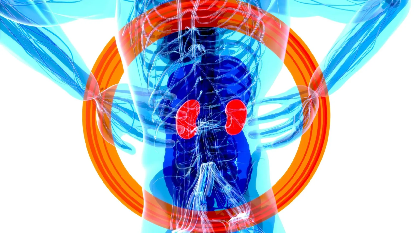
Nephrectomy - Kidney Removal Surgery
One patient in eight is left with a permanently changed shape to their side, and it has nothing to do with the kidney. It comes from the position on the table and the nerve that ran through the incision.
About This Department
Taking out a kidney is among the more dependable operations in surgery. What people complain about eighteen months later frequently has nothing to do with the kidney at all. It comes from the position they were held in on the operating table, from where the incision went and from which nerve happened to run through it, and almost nobody is warned about any of that beforehand. This page gives those things the space the kidney usually gets.
Free consultation
Tell us why the kidney is coming out
This operation is performed for at least five quite different reasons, and the reason changes the approach, the incision and how long you stay. Send the imaging itself along with any function study, the reason given to you for surgery, your creatinine and filtration rate, and details of previous abdominal operations, since scarring from an old operation frequently decides which route is used. Add your height and weight, because body mass is the single clearest predictor of the wound complications described further down. What comes back is an opinion on the approach, the likely length of stay and the specific risks that apply to your build and your history. The review costs nothing and carries no obligation.
Not one operation
Nephrectomy means removing a kidney, and that single word covers procedures that differ in almost every respect. Who the patient is, how much comes out, what route the surgeon takes and how much risk anyone is willing to accept all change with the reason for doing it. A kidney taken from a healthy volunteer to save somebody else is not the same undertaking as a kidney destroyed by twenty years of stones, and neither resembles a cancer operation.
This table scrolls sideways on a narrow screen. Swipe or drag to see every column.
| Reason | What it means for how the operation is done |
|---|---|
| Cancer | The kidney comes out inside its envelope of fat, untouched, so that nothing is cut across. The specimen must reach the pathologist whole, which limits how it can be removed from the body and rules out breaking it up. |
| A kidney that no longer works | Destroyed by long-standing obstruction, repeated stones or chronic infection, and taken out because it causes pain, infection or high blood pressure. Frequently the hardest of all these operations technically, because chronic inflammation glues everything together. |
| Living donation | A healthy person undergoes surgery for somebody else's benefit, which changes the acceptable risk entirely. The kidney must also come out in perfect condition and quickly, since every minute without blood supply matters to the recipient. |
| Injury | Uncommon now, since most injured kidneys are managed without surgery, and reserved for bleeding that cannot be stopped any other way. Done as an emergency, through the midline, alongside whatever else is damaged. |
| Severe infection | A kidney filled with pus or destroyed by an aggressive infection, removed to control sepsis. Urgent, and performed on somebody who is already unwell, which is what makes it risky rather than the surgery itself. |
What this page covers
Everything below applies most directly to planned surgery for cancer or for a kidney that has stopped working. Donation gets its own section, because it deserves one.
What the day looks like
Nothing by mouth from midnight, admission in the morning, and an anaesthetic that takes about twenty minutes to set up, and then comes the part patients never picture.
Being positioned
You are turned onto your side, the table is flexed to open the gap between the lowest rib and the hip, and you are strapped in place with the upper arm supported. This opens up the space the surgeon needs, and it is also where a large share of the trouble described in the next section begins.
Getting in
Three or four ports, carbon dioxide to create working space, and either the bowel swept aside to reach the kidney from the front or a balloon used to open a pocket behind the abdominal cavity. Where an open incision is needed, it runs below the ribs along the line of the flank.
The hilum
The artery, the vein and the ureter are identified where they enter the kidney. The artery is divided first so the kidney empties of blood rather than swelling, then the vein, then the ureter. This is the critical minute of the operation and it is over quickly when the anatomy is straightforward.
Getting it out
The kidney goes into a bag and leaves through a slightly widened port site or a small incision low down, which is the least discussed and most underestimated part of a keyhole operation. A specimen the size of a fist needs an opening it can pass through, and that opening is where hernias later appear.
Two to three hours, most of it before and after the minute that actually removes the organ.
The position, and what it costs
Here is the part that gets left out of consent conversations.
Two nerves run across the flank between the lowest rib and the hip, supplying both sensation to the skin and, crucially, the tone of the muscles of the side wall. A flank incision crosses their territory. They can be cut, stretched by a retractor, caught in a stitch or squeezed by scar tissue afterwards, and when a nerve stops supplying muscle, the muscle thins and the side of the abdomen sags outward. That sagging is called a flank bulge, and it is not a hernia. Nothing has come through a hole. The wall has simply stopped holding itself in, and to the person looking in a mirror the distinction is academic. Surgeons care about it because the repairs differ. Patients care about it because their trousers do not fit. The muscle has not been cut. It has simply stopped receiving the signal that told it to hold, and a muscle that receives no signal thins over months in a way that is visible from across a room.
How often it happens, in 184 patients
Swedish researchers followed 184 consecutive patients who had a kidney operation through a flank incision, examining them and their scans at three, twelve and twenty-four months. A visible bulge was noted in 36 patients at examination, of whom 20 still had one at last follow-up, meaning 12 percent. On imaging the figures ran higher, at 50 initially and 35 persisting, or 19 percent. Actual incisional hernia occurred in 5 patients clinically and 10 on imaging. Those who developed a hernia had a higher body mass index, at 30 against 26. The authors describe bulging as a common sequela and call for study of the pain, weakness and embarrassment it causes, noting that the rate of true incisional hernia after a flank incision is comparable to hernia rates after other kinds of abdominal surgery, which is to say the bulge and not the hernia is the distinctive problem here. Hernias get repaired. Bulges get lived with.
One patient in eight is left with a visible change in the shape of their side. That number is not small, and it is almost never quoted.
What actually predicts it
Four independent risk factors
Researchers prospectively followed 100 consecutive adults having kidney surgery through flank approaches recorded a flank bulge in 14 percent and a lumbar hernia in 10 percent at six months. Thirteen factors looked significant at first pass, and four survived multivariate analysis. These were a body mass index of 26.3 or above, the use of a self-retaining retractor during the operation, failure to identify and preserve the neurovascular bundle, and abdominal distension afterwards. Every patient whose wound was closed in a single mass layer, who developed a wound infection or who became constipated went on to develop a bulge or a hernia, which is a striking observation in a series of this size and one the authors present as an association rather than as proof of cause. Constipation is not usually thought of as a surgical risk factor, and in this operation it appears to behave like one, because a distended abdomen pushes outward on a wall that has temporarily lost its bracing.
Read that list again and notice where the items sit. Body mass is yours to work on before surgery. The retractor and the nerve belong entirely to the surgeon. Abdominal distension and constipation belong to the ward and to you together, which is the real reason nurses are so insistent about walking and about bowels after this particular operation. Three questions follow naturally. Does this operation need an open flank incision at all, or can it be done through ports. If an incision is needed, will the nerves be identified and preserved deliberately. And is a self-retaining retractor going to be used, given what that single instrument did to the risk in this series. None of those questions is impertinent. All three are answerable in a sentence by anybody who has thought about them. Put them at the consultation, and put them again in writing if the answers come back vague.
In from the front or the back
Kidneys sit behind the abdominal cavity rather than inside it, which gives keyhole surgery two genuinely different routes to them, since the surgeon can either go through the abdomen and sweep the bowel aside, or stay behind it entirely and work in a pocket created between the kidney and the back muscles.
This table scrolls sideways on a narrow screen. Swipe or drag to see every column.
| Route | What it offers, and what it costs |
|---|---|
| Through the abdomen | A large familiar working space with landmarks every surgeon trained on, at the price of moving the bowel out of the way and waiting for it to wake up afterwards. The default in most units, and the harder option where previous surgery has left adhesions. |
| Behind the abdomen | A pocket opened with a balloon between the kidney and the back muscles, reaching the artery almost immediately and never touching the bowel. A cramped space that takes longer to learn, and the better option after multiple previous abdominal operations. |
| Hand assisted | One hand inside through a small incision while the rest is done with instruments, giving back the sense of touch. Useful for very large kidneys and for surgeons transitioning from open technique, and it commits you to an incision that ports alone would not require. |
| Open flank | A single incision below the ribs with the patient on their side. Necessary for very large tumours, disease reaching the great vein and some heavily scarred kidneys, and it is the approach that carries the bulge and hernia risk described above. |
The first two of those have been compared properly.
Front against back, in fourteen studies
Fourteen studies were pooled in a systematic review, five of them randomised, comparing the two routes for keyhole removal of kidney tumours larger than seven centimetres. Working from behind was associated with a shorter operation, by a mean of 26.6 minutes, less blood loss, by 20.6 mL, and a faster return of bowel function, by 0.65 days. Length of stay, transfusion, conversion to open surgery, complications during and after the operation, positive margins and both local and distant recurrence showed no difference at all, while the reviewers note substantial heterogeneity between the studies and call for long-term randomised work before anybody treats this as settled. Half an hour of theatre time is a genuine gain and a poor reason to change hospitals, which is the sensible reading of a difference that size.
Half an hour of theatre time and a day of bowel function count for something and settle nothing. What that evidence really establishes is that both routes are legitimate, which means the real answer to which is better is whichever one your surgeon does more of.
One practical point does favour the back route in a specific group. Somebody who has had several previous abdominal operations may have adhesions that make the front route slow and hazardous, and going in from behind avoids all of it.
The gland above the kidney
Sitting like a cap on top of each kidney is a small triangular gland, producing adrenaline, cortisol and the hormone that manages salt and water. For decades surgeons removed it automatically along with the kidney in cancer operations, on the theory that it might harbour disease.
It rarely does.
What is done now
Modern practice leaves the adrenal gland alone unless a scan shows it is involved, the tumour sits at the upper pole and touches it, or the disease is advanced. Removing one gland unnecessarily causes no immediate problem, since the other side compensates, but it matters later if that remaining gland ever fails or needs removing itself, and it matters in the meantime because there is no benefit to set against the loss.
Confirm the plan beforehand. A note in an operative record saying the adrenal was removed, with no explanation of why, is a question worth putting before rather than after.
When the patient is healthy
Living donors gain nothing medically from the operation being performed on them, which makes them unique among surgical patients. That single fact changes the standard applied to everything, and it explains why this version of nephrectomy has been tested far more rigorously than the version done for disease.
Four techniques, thirteen trials, 1,280 donors
A Cochrane review gathered thirteen randomised trials comparing open, keyhole, hand-assisted and robotic removal in living kidney donors. Against open surgery, keyhole removal reduced painkiller requirements and shortened hospital stay, while taking longer to perform and leaving the kidney without blood supply for longer. Blood loss, complications and reoperations showed no overall difference. Hand-assisted and keyhole surgery were equivalent on painkillers, stay, operating time, blood loss and complications. Conversion to open surgery occurred in about 1 percent of keyhole cases. Graft outcomes for the recipient, including early graft loss, delayed function, rejection and kidney function at a year, did not differ between keyhole and open donation, which is the finding that made keyhole donation acceptable in the first place, since a donor benefit purchased at the recipient s expense would not have been.
What the donor is trading
Two things in that summary deserve attention. Keyhole donation trades a longer operation and a longer interval without blood flow for less pain and a shorter stay, which is a trade made on behalf of the donor and paid for, in principle, by the recipient. The reassuring part is that graft outcomes came out the same, so the recipient is not, in fact, paying anything.
Reviewers also found the operation getting faster and safer over time, with procedure duration falling by around seven minutes for every year of accumulated experience across the studies, which is an unusual thing to be able to measure and a reassuring one to read.
Law and strict independent assessment govern donor surgery in every country that permits it, and Turkey is no exception. Anyone contacting a hospital about donation should expect an evaluation process that includes people whose specific job is to make sure nobody is being pressured or paid.
How short this can be
Kidney surgery once meant a week in hospital and six weeks off work, and keyhole technique combined with structured recovery protocols has compressed that steadily, considerably further than most patients realise.
Going home the same day, across 20,548 patients
A systematic review pooled eleven studies covering 20,548 patients having keyhole kidney surgery, of whom 1,419, or 7 percent, went home in under a day. Total complication rates did not differ significantly between the same-day and standard-stay groups, and neither did readmission within thirty days, while serious complications came out less common in the same-day group, which reflects careful selection rather than any benefit of leaving early. The reviewers conclude that in appropriately selected patients this is safe and feasible, and call for better data on cost, on satisfaction and on precisely which patients belong in that selected group. Seven percent sounds small. It also describes a group that did not exist fifteen years ago.
What that means if you have travelled
None of which means an international patient should aim for it. The relevant point is different and it is about direction. If the fittest patients are safely going home the same day, then two or three nights for someone who has travelled represents caution rather than necessity, and a unit proposing a week should be able to say what it is for.
Recovery, week by week
Most people find this easier than they had prepared for, and the things that end up bothering them are rarely the things they had worried about beforehand, which is worth remembering during the fortnight before the operation.
- Day one brings shoulder tip pain from the gas, which is referred pain rather than anything wrong with the shoulder, and it clears within a few days.
- Walking starts the same evening, and it is the main thing preventing the abdominal distension that predicts wound problems later.
- Bowels take two to four days to wake up after a front approach and rather less after a back one. Constipation counts for more here than it usually does, given what it was associated with in the risk data above.
- Numbness across the skin below the incision is expected, often permanent in patches, and unrelated to how well the operation went.
- Desk work at two weeks, driving when you can perform an emergency stop without hesitating, heavy lifting at six weeks and not a day earlier.
That six week rule is the one people break, and it is the one that matters most, because the specimen extraction site and the flank musculature are both still knitting at four weeks whatever your energy levels suggest.
The change that shows up late
Where a flank bulge develops, it generally appears between three and twelve months rather than immediately. If your side looks different at four months, that is the reason, and it should be photographed and reported, not written off as weight gain.
How long you stay
Ten to twelve days for keyhole surgery and fourteen to eighteen for an open operation, with both figures driven by the wound and by wanting a blood test that shows the remaining kidney settling, and not by anything dramatic.
Assessment takes two days. Imaging gets reviewed here, kidney function measured, a split function study is added where the other kidney is in any doubt, and the anaesthetic assessment happens. For a kidney being removed because it no longer works, that split study is not optional, since the whole justification rests on how little the kidney contributes. Hospital stay runs two to three nights for keyhole surgery and four to six for open. Then a week nearby, covering wound review, removal of any stitches, a blood test at around day seven and the pathology discussion where there was a tumour. That week earns its place. It covers the window in which a wound problem declares itself, and dealing with one here is considerably simpler than dealing with one four thousand kilometres away.
When you are cleared to fly
Around ten days after keyhole surgery and two weeks after an open operation, once the wounds are dry and the kidney blood tests have stopped moving. Get up and walk every hour on the flight, since a long operation followed by a long period sitting still is precisely the combination that produces clots, and both halves of it apply to you in the same week.
What drives the cost
Nephrectomy sits at the more predictable end of surgical pricing, which makes the few genuine variables easier to identify than they are for most of the operations described elsewhere on this site. Six things move the figure.
- Keyhole against open, and within keyhole, robotic against laparoscopic, since console time and single-use robotic instruments dominate everything else here.
- Why the kidney is coming out, because a kidney destroyed by chronic infection takes considerably longer to remove than a tumour-bearing one.
- Whether previous abdominal surgery forces a longer or different approach.
- The split function scan, where one is needed to justify removal at all.
- Length of stay, and the accommodation across the week that follows discharge.
- The pathology on the whole specimen, which is more involved for a tumour than for a scarred kidney.
The one that arrives years later
Repairing a flank bulge or a lumbar hernia is a real operation with mesh, and it is not covered by anything associated with the original surgery. Given rates of 10 to 19 percent in the published series, this is not a remote possibility and it deserves a place in your thinking, alongside the observation that avoiding it is largely a matter of technique and of your own weight beforehand. Packages here ordinarily cover the transfers, the assessment, the operating fees, the stated hospital nights, an interpreter, accommodation and the appointments before you fly. Outside them sit the flights, insurance, extra nights, the treatment of a complication, and any later repair. Raise that last item specifically, since a bulge repair years afterwards is the cost most people never think to enquire about and the one this page has spent most of its length explaining.
Five questions to ask before you accept a figure
Does the figure assume keyhole surgery, and what changes if the operation has to be converted to open. Is the split function scan included where one is needed. How many hospital nights are covered and what an extra one costs. Is the accommodation for the week after discharge inside the figure or outside it. And what happens financially if the wound becomes infected, given how strongly that predicts the later problems described above.
Nothing here means anything until a urologist has seen your images and knows why the kidney is being removed. That review costs nothing.
Once you are home
What happens next depends entirely on why the kidney came out, in that a cancer operation brings a surveillance schedule of scans and blood tests running for years. A kidney removed for infection or obstruction brings a single check that the remaining kidney has settled, and then nothing.
Four documents should travel home with you, in English, because whoever picks up your care afterwards was not in theatre.
- The operative note, naming the approach used, whether the adrenal gland was taken, and whether the nerves were identified where an open incision was made.
- The pathology report, which matters even for a kidney removed for benign disease, because it occasionally finds something nobody expected.
- Your filtration rate before the operation and at discharge, which is the baseline every future doctor will measure against.
- The follow-up plan, with actual dates, and a clear statement of whether any surveillance is needed at all.
Get in touch here for fever, a wound that opens or discharges, pain that is increasing rather than decreasing after the first week, or blood test results that are drifting in the wrong direction. Also get in touch about a change in the shape of your side, even months later, because that is the complication this page exists to make visible and it is far easier to assess with a photograph from you than to guess at over a message. Carry a note saying you have one kidney. Every doctor you meet for the rest of your life needs that information before prescribing anything, and you will not always be in a position to tell them. Put it on your phone. Put it in your wallet as well, because phones run out of battery at exactly the wrong moment. Contrast for a scan, doses of common antibiotics and several classes of painkiller all depend on how much kidney a person has, and a doctor making those decisions at three in the morning has no way of knowing unless something tells them.
Frequently asked questions about nephrectomy
What is a flank bulge, and how likely is it?
Can anything reduce that risk?
Is it better to go in through the front or the back?
Will the adrenal gland be removed too?
Is donating a kidney different from having one removed for disease?
How long do I need to stay, and when can I fly?
References
- Inkiläinen A, Styrke J, Ljungberg B, Strigård K. Occurrence of abdominal bulging and hernia after open partial nephrectomy, a retrospective cohort study. Scandinavian Journal of Urology. 2018;52(1):54-58.
- Osman T, Emam A, Farouk A, ElSaeed K, Tawfeek AM, AbuHalima A. Risk factors for the development of flank hernias and bulges following surgical flank approaches to the kidney in adults. Arab Journal of Urology. 2018;16(4):453-459.
- Wang L, Li KP, Liu Y, Yin S, Zhu PY. Perioperative and oncologic outcomes of transperitoneal versus retroperitoneal laparoscopic radical nephrectomy for large-volume renal carcinoma. World Journal of Surgical Oncology. 2023;21(1):86.
- Kourounis G, Tingle SJ, Hoather TJ, et al. Robotic versus laparoscopic versus open nephrectomy for live kidney donors. Cochrane Database of Systematic Reviews. 2024;5(5):CD006124.
- Paynter A, Uy M, Millan B, Le Nguyen D, Bansal R, Shayegan B. The safety and feasibility of ambulatory minimally invasive partial nephrectomy, a systematic review and meta-analysis. Journal of Endourology. 2024;38(11):1112-1120.
Editor's note
Written by the Biruni Hospital medical editorial team. Reviewed by Prof. Dr. Mustafa Teoman YANMAZ, Medical Oncology.
Medically reviewed by

Prof. Dr. Mustafa Teoman YANMAZ
Medical Oncology
Related Treatments
View All-
Bilateral Mastectomy
Whether you carry a BRCA mutation, have cancer in one breast or in both, bilateral mastectomy removes both breasts in one operation, and how much it helps depends on which of those three paths brought you to it.
-

Breast Conserving - Lumpectomy Surgery
Keeping the breast has never been the less thorough choice, and the largest recent studies now point the other way. This page explains who can have breast conserving surgery, what the margin report decides, and how long a woman traveling for it needs to stay.
-

Cytoreductive - Tumor Reduction Surgery
Patients cleared of every visible deposit lived 30 months in one gastric series. Those left with anything at all lived 7.3 months, however little remained.
-
Debulking - Ovarian Cancer Tumor Reduction
Two women with the same diagnosis on the same morning can get operations that share almost nothing. What decides it is who measured the disease first.
-
Debulking Surgery
Surgeons removed a median of 57 lymph nodes from women already cleared of visible disease. It bought no extra months, and tripled deaths within 60 days.