Mandibulectomy
Patients facing jaw bone cancer surgery usually ask two things first, whether they will eat normally again and what happens to the leg that donates the new jaw. This guide answers both with published figures, explains marginal and segmental mandibulectomy, and sets out the weeks in Istanbul.
About This Department
The diseased jaw comes out and a new one goes in. Same operation, built from your own leg bone.
A 2024 systematic review of 36 studies on jaws rebuilt with the fibula found that dental implants placed in a fibula that had not received radiotherapy survived in 93 percent of cases, which means fixed teeth back in the new jaw for many patients after jaw bone cancer surgery.
Two questions come first from almost every patient told they need part of the lower jaw removed. Will I eat normally again? And what happens to the leg that gives the bone? This page answers both with published figures, then explains how surgeons decide between removing the rim of the jaw and removing a full section, how the new jaw is planned on a computer before the operation, what the teeth look like a year later, and how the weeks in Istanbul run from the first message to the follow-up scan at home.
Mandibulectomy, or jaw bone cancer surgery, in plain terms
Mandibulectomy removes part of the mandible, the lower jaw, because a tumor has grown against it or into it.
Patients and family members searching from abroad tend to call it jaw bone cancer surgery, and the two names describe the same operation.
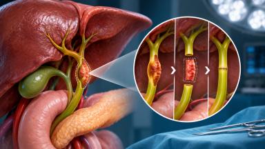
Surgeons describe it by how much bone goes. A marginal mandibulectomy takes the upper rim of the jaw, the ridge that holds the teeth, and leaves the lower border in one continuous piece, so the jaw keeps its shape and its hinge and needs no bone rebuilding. A segmental mandibulectomy cuts through the full height of the jaw and removes a whole section, which breaks the arch in two. Without reconstruction the remaining pieces drift toward each other, the chin falls back, and chewing and closing the lips both suffer, so a segmental resection almost always goes together with a new piece of living bone set into the gap during the same anesthetic. A hemimandibulectomy removes one side of the jaw from the chin back to the joint, and it follows the same rebuilding logic.
Which tumors reach the lower jaw
Squamous cell carcinoma of the gum, the floor of the mouth or the area behind the last molar accounts for most mandibulectomies, and these cancers start in the lining of the mouth and press against the bone from outside, first wearing down the hard outer shell, the cortex, and later entering the soft marrow inside. Gum cancer reaches bone soonest, because only a thin layer of tissue separates the gum surface from the jaw.
Some tumors start inside the bone. Ameloblastoma, a slow-growing benign tumor of the tooth-forming tissue, hollows out the jaw over years and comes back when scraped out, so surgeons remove it with a margin of healthy bone, sometimes as a segmental resection, even though it does not spread. Osteosarcoma and other bone sarcomas of the jaw are rare. They grow fast and need wide resection, with chemotherapy planned around the operation. Bone destroyed by earlier radiotherapy, called osteoradionecrosis, and bone damaged by some osteoporosis and cancer drugs bring patients to the same operation for a different reason.
How surgeons decide how much bone to remove
Marrow decides it.
When cancer sits against the jaw or has only eroded the surface of the cortex, removing the rim gives a clear margin and keeps the jaw whole. Once the cancer has entered the marrow, it spreads along the soft inner channels further than any scan shows, and the safer operation removes the full height of bone. A CT scan shows erosion of the cortex well, an MRI shows marrow involvement better, and a panoramic X-ray of the teeth adds the picture of the tooth roots and the canal that carries the nerve to the lower lip. Surgeons also look at the pattern of erosion on the CT. A smooth, scooped edge points to a cancer pushing against the bone, while a ragged, moth-eaten edge points to one infiltrating it, and the second pattern argues for the segmental operation even when the bone loss looks small. In a mouth that has lost its teeth, the jaw shrinks to a thin bar over the years, and a rim resection there can leave too little bone to hold together, which makes a segmental resection the safer choice even for a cancer that has barely touched the surface. Final proof comes from the pathologist. The bone edges examined after surgery show whether the cortex and the marrow were involved.
The evidence supports that rule without pretending to be stronger than it is. A 2018 meta-analysis in the International Journal of Oral and Maxillofacial Surgery pooled 15 cohort studies with 1672 patients and found weak evidence in favor of segmental resection for local control, while disease-free survival came out 73 percent better with segmental resection once the marrow was invaded. A 2023 meta-analysis in the International Archives of Otorhinolaryngology, built on retrospective studies only, found a difference in five-year disease-free survival and a trend in favor of segmental resection when marrow invasion was present. Both reviews arrive at the same practical answer. A rim resection for surface contact, a full section for marrow.
Marginal, segmental and hemimandibulectomy compared
Three operations cover almost every case of jaw bone cancer surgery on the lower jaw.
This table scrolls sideways on a narrow screen. Swipe or drag to see every column.
| Feature | Marginal | Segmental | Hemimandibulectomy |
|---|---|---|---|
| Bone removed | Upper rim, lower border kept | A full-height section | One side, chin to joint |
| Typical reason | Cancer touching the bone or eroding its surface | Marrow invasion, ameloblastoma, osteoradionecrosis | Large tumors of the body and ramus, jaw sarcoma |
| Bone rebuilding | None, soft tissue closure or a thin flap | Fibula, scapula or iliac crest flap | Fibula flap, sometimes with a joint replacement |
| Nights in hospital | Two to five | Seven to twelve | Seven to fourteen |
Cancer surgery on the jaw rarely stops at the bone. The tumor comes out in one piece with the gum, the floor of the mouth or the cheek lining around it, and a neck dissection removes the lymph nodes on that side through a separate cut in a skin crease of the neck.
Rebuilding the jaw from the fibula
The fibula, the thin outer bone of the lower leg, carries little of the body's weight and runs straight and long, which lets surgeons cut it into two or three short segments and fit them together into the curve of a jaw. Its own artery and vein come with it. So do a strip of skin, when the mouth lining needs replacing, and sometimes a cuff of muscle. A microsurgeon joins the artery and vein to vessels in the neck under a microscope, so the new bone stays alive and heals to the remaining jaw like a fracture, in six to eight weeks. Titanium plates hold everything in place while that happens.
Planning on a computer before the first cut
Engineers and surgeons build the operation in three dimensions from CT scans of the face and the legs. They decide exactly where the jaw will be cut, how the fibula will be divided, and how the pieces will sit so that the bite still meets the upper teeth, and printed cutting guides and a plate bent in advance to the planned shape carry that plan into the operating room. A 2019 meta-analysis in Plastic and Reconstructive Surgery, covering 12 studies, found that this computer-assisted approach shortened the time the flap spends without blood flow, the total operating time and the hospital stay compared with shaping the bone by hand, with no difference in complications.
When the fibula is not the right bone
Poor blood flow to the feet, from diabetes, smoking or earlier vascular surgery, can rule out the fibula. A CT angiogram of both legs before surgery checks this. The scapula, the shoulder blade, and the iliac crest, the rim of the hip bone, serve as alternatives, each with its own shape and its own recovery.
What happens to the leg that gives the bone
Most people walk normally again.
Surgeons leave several centimeters of fibula at the knee and at the ankle, which keeps both joints stable. A systematic review of donor-site problems published in Plastic and Reconstructive Surgery in 2012 gave the long-term figures as weighted means across the studies it pooled, with chronic pain in 6.5 percent of patients, a noticeably abnormal gait in 3.9 percent, ankle instability in 5.8 percent and reduced ankle movement in 11.5 percent. You stand on the second or third day. A frame or a stick helps for the first week or two. A physiotherapist teaches ankle and toe exercises on the ward, and the skin over the harvest site, closed directly or with a thin skin graft from the thigh, heals over three to four weeks.
Getting teeth back in the new jaw
Fixed teeth on implants are a realistic goal after a fibula reconstruction, and radiotherapy sets the timetable. A 2024 systematic review in the Journal of Stomatology, Oral and Maxillofacial Surgery, drawing on 36 studies, found implant survival of 93 percent in a fibula that had never been irradiated. When implants went into an irradiated fibula, survival fell to 38, 55 and 77 percent for placement at 12, 17 and 24 months after radiotherapy, so the longer the wait after radiation, the better the implants held. Half of the authors in that review started dental work 12 months after the jaw was rebuilt. Some centers place the implants during the reconstruction itself when no radiotherapy is expected.
Removable dentures work for some patients in the meantime, though a jaw rebuilt with a skin paddle on top gives a denture little to grip.
Complications and their standard treatment
Each item below names the problem and what the team does about it.
Radiotherapy after a mandibulectomy
Cancer that has entered the jaw bone counts as an advanced stage, so most patients after a segmental resection for squamous cell carcinoma receive radiotherapy, with chemotherapy added when the pathology shows cancer at a cut edge or breaking out of a lymph node. Treatment starts within six weeks of surgery. That leaves the wounds in the mouth and the leg time to heal first. Before radiation planning, every tooth in poor condition inside the planned field comes out, since an extraction from irradiated jaw risks the bone breaking down later. Ameloblastoma needs no radiotherapy at all.
Biruni Hospital delivers radiotherapy, on an Elekta Versa HD linear accelerator, and chemotherapy on the same site as surgery, so a patient whose final report calls for further treatment continues with the same team and the same records. Going home for it stays open too. The oncologist there receives a written handover and the full pathology report.
How many weeks in Istanbul, and when you can fly
Seven to ten days in Istanbul cover a marginal mandibulectomy without a flap. Rebuilding with the fibula takes three to four weeks.
The longer stay follows the healing sequence. The operation lasts eight to twelve hours when computer planning, a neck dissection and the fibula flap are combined, and the first night passes in intensive care. A temporary tracheostomy protects the airway while the mouth swells and comes out within the first week. Liquid food runs through a thin nasal tube until swallowing is safe. Soft food by mouth follows, and discharge to the hotel comes at seven to twelve days. Packages published by Turkish hospitals for jaw resection with reconstruction list seven nights in hospital with one of them in intensive care, which matches that timetable, and the rest of the stay covers wound checks, the first steps of walking without help, the final pathology report and the decision on radiotherapy, which together decide the flight date. The surgeon clears you to fly once the tracheostomy hole has closed, you eat enough by mouth, the leg wound has healed enough for a long seat, and the next step of treatment has been agreed. A long flight after leg surgery raises the risk of a clot, so the discharge summary includes advice on blood thinning injections, compression stockings and walking the aisle every hour.
Radiotherapy given in Istanbul stretches the stay to three months.
Care after you fly home
You leave with a discharge summary in English, the operation note, the pathology report and the computer plan of the reconstruction, which a surgeon at home needs if the plate ever has to come out, and checks with a head and neck surgeon run every one to three months in the first two years, with a CT or MRI three months after treatment ends as the new baseline. Soft food continues for six weeks while the fibula joins the jaw. A speech and swallowing therapist at home can carry on from the written plan. Your coordinator stays on the same WhatsApp number after you return home, and photographs of the mouth or the leg wound and new scan reports reach the surgical team the same way.
Get in touch the same day for any of these.
- Metal showing in the mouth or through the skin of the jaw
- Swelling, redness or discharge from the neck or leg wound
- A swollen, painful calf, which needs checking for a clot
- A new ulcer or lump in the mouth or neck
Sudden breathlessness or chest pain calls for the nearest emergency department.
What moves the cost of a mandibulectomy
Reconstruction drives the figure more than the resection does. A marginal resection closed without a flap sits at the low end. A segmental resection rebuilt with the fibula adds a second surgical team, the microsurgery, titanium plates, nights in intensive care and a longer ward stay, and computer planning adds the cost of the 3D model, the printed guides and the pre-bent plate. A neck dissection on one or both sides adds theater time. Implants placed during the reconstruction add their own components, while implants placed a year later belong to a separate dental plan, and missing scans done in Istanbul, such as a CT angiogram of the legs or a PET-CT, go on the bill as well.
Your own health counts too. Diabetes, heart or lung disease, poor leg circulation, blood thinning medication and previous radiotherapy to the jaw all change the plan, and through it the cost.
Packages published by Turkish hospitals and medical travel agencies for jaw resection with reconstruction include the pre-operative tests and planning scans, surgery and anesthesia, the planned nights in hospital and intensive care, medicines and transfers. Flights, extra nights, treatment of a complication, radiotherapy and later dental implants stay outside them.
Comparable quotes name the type of resection, say whether computer planning and the neck dissection sit inside, state the nights in hospital and intensive care they assume, and explain what happens if the flap needs a second trip to theater.
Your own scans, read by a surgeon, give the only figure worth comparing. That review costs nothing.
Organizing the trip
For the free remote review, send the biopsy report, the CT of the face and jaw as image files with the written report, any MRI or PET-CT, a panoramic dental X-ray if you have one, clear photographs of the inside of the mouth, and your medication list. A surgeon reads the file and tells you whether a marginal or a segmental resection fits, which bone would rebuild the jaw, whether the neck needs surgery, and how long the stay would run. That answer comes before you spend anything on travel.
One coordinator follows your case from the first message through discharge. Coordinators work in English, Arabic, French, Russian, Serbian, Romanian and Spanish, and interpreters cover other languages, which matters most in the days after surgery when speaking with a swollen mouth takes effort and a notepad does much of the talking. Patient rooms have a companion bed, so one person stays with you overnight for the whole admission, and the international patients office arranges accommodation for both of you before and after it, along with airport transfers. The hospital kitchen prepares halal, vegetarian and diabetic meals, in liquid and soft form during recovery. A prayer room is on site. A request for a female surgeon goes to the department in the first message and is met wherever the rota allows. About ten days before travel the office issues the appointment confirmation and the invitation letter a medical visa needs, naming the hospital and the treating doctor.
Mandibulectomy FAQ
Is jaw bone cancer surgery the same as a mandibulectomy?
Will my face look different after a mandibulectomy?
How long will I stay in Istanbul?
Can someone from my family stay with me in the hospital?
Will I walk normally after the fibula is taken?
What if a problem starts after I return home?
References
- Gou L, Yang W, Qiao X, et al. Marginal or segmental mandibulectomy. Treatment modality selection for oral cancer. A systematic review and meta-analysis. Int J Oral Maxillofac Surg. 2018;47(1):1-10.
- Chiesa-Estomba CM, Mayo-Yanez M, Manelli G, et al. Marginal versus Segmental Mandibulectomy in the Treatment of Oral Cavity Cancer. A Systematic Review and Meta-analysis. Int Arch Otorhinolaryngol. 2023;27(4):e733-e743.
- Powcharoen W, Yang WF, Li KY, et al. Computer-Assisted versus Conventional Freehand Mandibular Reconstruction with Fibula Free Flap. A Systematic Review and Meta-Analysis. Plast Reconstr Surg. 2019;144(6):1417-1428.
- Ling XF, Peng X. What is the price to pay for a free fibula flap? A systematic review of donor-site morbidity following free fibula flap surgery. Plast Reconstr Surg. 2012;129(3):657-674.
- Nham TT, Koudougou C, Piot B, et al. Prosthetic rehabilitation in patients with jaw reconstruction by fibula free flap. A systematic review. J Stomatol Oral Maxillofac Surg. 2024;125(3):101735.
Editor's note
Written by the Biruni Hospital medical editorial team. Reviewed by Prof. Dr. Mustafa Teoman YANMAZ, Medical Oncology.
Medically reviewed by
Prof. Dr. Mustafa Teoman YANMAZ
Medical Oncology
Last updated: