
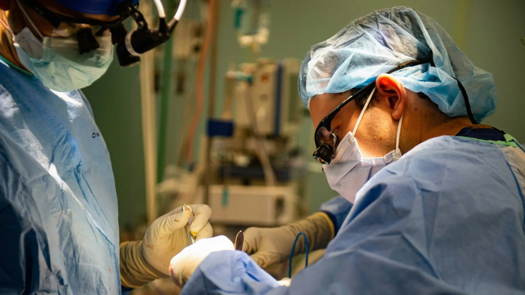
Lymphoma Surgery
The surgeon's job here is to name the disease. Drugs cure it. Which is why the biopsy, not the operation, is the thing worth getting right.
About This Department
The surgeon's job in lymphoma is to name the disease, not to remove it. That sentence is the whole of this page and everything below is elaboration.
Lymphoma is a cancer of the immune system, which means it lives in the lymph nodes, the blood, the marrow and the spleen at once, and cutting out one swollen node treats the disease no more than emptying one drawer tidies a house. The analogy is not a kind one. What cures lymphoma is drug treatment, sometimes with radiotherapy. What surgery does is obtain the piece of tissue those drugs are chosen from, and the quality of that single piece decides how accurately everything that follows is aimed. Nothing else about the surgery matters as much.
Free consultation
If a biopsy has already been done, send the report before anything else
The single most useful document is the histopathology report naming the exact subtype, since more than seventy subtypes sit under the word lymphoma and each one has its own treatment. Send it with a note of which kind of biopsy produced it, needle or whole node, because that tells us how much confidence the diagnosis deserves. Add the imaging on a disc, the blood counts, the lactate dehydrogenase and the beta-2 microglobulin if they were measured, and any PET report you hold. Tell us how long the node has been enlarged, whether it fluctuates, and whether you have had fevers, night sweats or unexplained weight loss. If a needle biopsy came back inconclusive, say so plainly, because that is a common and fixable situation and it changes what we advise first.
Why cutting it out does not work
Solid cancers begin in one place and spread outward from it, which is why a surgeon can cure them by removing the place they began along with a rim of normal tissue. Lymphoma does not work that way. The cells it arises from are lymphocytes, whose entire biological purpose is to circulate, and they travel through blood and lymph as a matter of routine long before anybody notices a lump. A node in the neck that has grown to the size of a grape is one visible expression of a process already present in nodes nobody can feel, in the marrow, and often in the spleen. Removing that one node removes the evidence and leaves the disease. This is not a limitation of surgical technique and it will not be solved by a better operation. It is the reason lymphoma became, decades ago, one of the first cancers to be treated systemically, and the reason it is among the most curable, since many aggressive lymphomas that would kill within months untreated are cured outright by six cycles of drug treatment, without a scalpel ever going near the tumour. A staging laparotomy, which used to be routine for Hodgkin lymphoma and involved opening the abdomen, removing the spleen and sampling nodes and liver, has been abandoned entirely because a PET scan now answers the same question in an afternoon. Nobody misses it.
Understanding this protects you from a specific and real risk. Some clinics will offer to remove a lymphomatous mass, and framed as removing the cancer it sounds reasonable to anybody who has not been told the above. It delays the treatment that works. It commits you to a recovery you did not need, and it does nothing measurable to the disease. If any surgeon anywhere proposes an operation to remove a lymphoma, the question to ask before consenting is which haematologist recommended it and what the drug plan is. There are three genuine exceptions to all this, and they are set out further down, but the exceptions are narrow and each of them is named. Read them before you decide.
The operation that actually matters
Everything hinges on getting enough of the right tissue. Diagnosing lymphoma is not a matter of finding malignant cells, which is where a needle sample stops. It requires seeing how the cells are arranged, because the architecture of the node is itself diagnostic. Follicular lymphoma is recognised by the pattern its follicles form. Hodgkin lymphoma is recognised by rare abnormal cells scattered through a very characteristic background of ordinary inflammatory cells, and a needle that misses those few cells returns a report saying reactive changes. Grading a follicular lymphoma means counting large cells across intact follicles.
None of it can be done on a fragment. Architecture is the diagnosis.
How often each method actually answers the question
A referral centre reviewed every lymph node biopsy processed over four years, 373 in total, of which 210 were image-guided core needle biopsies and 163 were surgical excisions. The diagnostic yield was 79 percent for the needle and 97 percent for the excision. Where a needle biopsy failed, the reason in most cases was insufficient or suboptimal tissue, and nothing subtler than that. The choice between the two methods had not depended on the patient's age, sex or how strongly malignancy was suspected, which is another way of saying it was often decided by convenience. Lymphoma itself was diagnosed equally well by both, and the needle held a specific advantage in large B-cell lymphomas, where the abnormal cells dominate the sample. The authors concluded that a needle is a good substitute when it is done properly, with flow cytometry available alongside it, and that the guidelines on when to use which need tightening.
Read plainly, roughly one needle biopsy in five comes back without an answer, against one excision in thirty-three. The consequence of a failed biopsy is rarely harm in itself. It is delay, because the patient goes back to the start and waits for a second procedure while the disease continues, and in an aggressive lymphoma a month is not a trivial amount of time. That is the calculation behind the general preference for taking the whole node when a node is accessible. Delay is the real cost.
A needle is nonetheless the correct choice in several situations and choosing it is not a compromise. When the abnormal tissue is deep in the chest or the abdomen and reaching it would need a major operation, a needle under scan guidance is far less to put a patient through. When a known lymphoma relapses and the question is simply whether it is the same disease, a needle usually answers it. When somebody is too unwell for anaesthetic, the needle is what is available.
What matters is that the choice is made deliberately, with the pathologist and the haematologist in the conversation, rather than by whoever has the earliest free slot. Somebody should be choosing.
This table scrolls sideways on a narrow screen. Swipe or drag to see every column.
| Method | What it gives, and where it falls short |
|---|---|
| Fine needle aspiration | A thin needle draws out loose cells. It can tell you that cells look abnormal and it is useful for confirming that a known cancer has spread to a node, but it destroys all architecture and cannot classify a lymphoma. Accepting a lymphoma diagnosis based on this alone is a mistake, and so is accepting a reassuring result from it. |
| Core needle biopsy | A wider needle takes several cylinders of tissue, keeping some architecture. Around four in five produce a diagnosis. It is the right choice for deep disease and for confirming relapse, and it works considerably better when several cores are taken and flow cytometry is running alongside. |
| Excision of the whole node | The entire node comes out intact with its capsule, which is what the pathologist wants. Around 97 percent produce a diagnosis. It needs a small operation and a scar, and for an accessible node in the neck, armpit or groin that is a modest price for the most reliable answer available. |
| Which node to take | Not the easiest one. Groin nodes are frequently enlarged from old foot infections and often disappoint, so a neck or armpit node is preferred where the choice exists, guided by which node the PET scan shows as most active, and never by which is nearest the surface. |
What the laboratory does with it
Patients are often surprised that a lymphoma diagnosis takes a week or two when a breast or bowel cancer diagnosis can be reported in days. The reason is that naming a lymphoma is a sequence of separate laboratory investigations run on the same piece of tissue, each answering a different question, and the sequence cannot be compressed much. Knowing what those steps are makes the wait easier to sit through and tells you what to check is present in the final report. Four steps, one specimen.
This table scrolls sideways on a narrow screen. Swipe or drag to see every column.
| Step | What it answers, and why it needs real tissue |
|---|---|
| Architecture under the microscope | Whether the node's normal structure has been effaced, and by what pattern. This is the first and most important question, it is answered in the first day or two, and it is precisely the question a fragmented sample cannot answer. |
| Immunohistochemistry | A panel of stains marking proteins on the cell surface, which is how a B-cell lymphoma is separated from a T-cell one and how the subtype is narrowed down. A dozen or more stains may be needed and each takes time, which is a large part of why the report is not immediate. |
| Flow cytometry | Fresh cells are passed through a laser and sorted by their surface markers. It is fast and highly informative, and it works only on unfixed tissue, so it has to be arranged before the operation and not requested afterwards. |
| Molecular and genetic testing | Specific rearrangements and translocations, such as those involving MYC, BCL2 and BCL6, separate lymphomas that look identical under the microscope but behave very differently and are treated with different regimens. This step adds days and it changes plans. |
Two practical points follow. Flow cytometry needs fresh tissue, so the theatre team must know before the operation that lymphoma is suspected, and a node that goes straight into formalin has quietly closed off one of the four steps. And the final report should name a specific entity from the WHO classification, with the immunohistochemistry panel listed. A report that says only malignant lymphoma is an incomplete report, and treatment should not be started on it. Ask for the entity by name.
The biopsy day, step by step
An excisional lymph node biopsy is a small operation and for a superficial node it is usually a day case lasting well under an hour.
Most people are surprised by how minor it is. Under an hour, and home the same day.
Choosing the node, and telling the laboratory
The PET scan decides which node is worth taking. At the same time the laboratory is warned that fresh tissue is coming, so that flow cytometry can be run instead of lost. That phone call is as much a part of the operation as anything done with a knife.
Anaesthetic
Local anaesthetic suits a node sitting just under the skin. A general anaesthetic is used for deeper nodes, for nodes close to important nerves in the neck, and for anyone who would rather be asleep, which is a perfectly reasonable preference and not a complication.
Taking the node whole
A short incision is made in a skin crease and the node is dissected out with its capsule intact. Keeping the capsule matters, since a node that arrives squashed or cut in half has lost part of the architecture the diagnosis depends on. Nearby nerves are identified and protected as the dissection goes.
Splitting the specimen and closing
Part of the node goes fresh to the laboratory for flow cytometry, part into formalin for the slides, and part is frozen where molecular work is likely. The wound is closed in layers with absorbable stitches under the skin. Most people go home the same day.
Risks are small and worth stating anyway. Bleeding and infection are the usual pair, uncommon and manageable. Numbness around the scar is normal for some months and usually recovers. Nerve injury is the one that matters and it depends entirely on where the node was, since a node in the posterior triangle of the neck sits near the nerve that lifts the shoulder, and one in the armpit sits near nerves supplying the chest wall. Lymph can also collect under the wound and occasionally needs draining. Being clear about the anatomy beforehand is the whole of the prevention.
Ask where the node sits.
The operation a drug replaced
Splenic marginal zone lymphoma is the closest thing lymphoma has to a surgical disease. It sits mainly in the spleen, it is slow, and for years removing the spleen was the standard treatment, which made intuitive sense and worked reasonably well. Then rituximab arrived, an antibody given by infusion, and the comparison was made properly.
The drug held its own.
Every Swedish patient over twenty years
All 358 people diagnosed with splenic marginal zone lymphoma in Sweden between 2000 and 2020 were studied. Median overall survival was 11 years and median age at diagnosis was 73. Eighty-six were started on watch and wait, 90 had rituximab alone, 47 rituximab with chemotherapy, 88 had their spleen removed, 37 had chemotherapy and 10 had both surgery and systemic treatment. Survival was worse only in the group given chemotherapy, and it was equal between rituximab, rituximab with chemotherapy and splenectomy. After adjusting for age, survival did not differ between the patients simply watched and those who had either surgery or rituximab. The authors concluded that watch and wait remains the most reasonable option for people without symptoms, that symptomatic patients should be offered rituximab alone first, and that surgery can be safely held back for those who do not respond to it.
There is a second finding that ought to be part of any conversation about this operation. A nationwide analysis of 30,619 patients with marginal zone lymphoma looked at how often the disease transforms into an aggressive large B-cell lymphoma, which happened in 2.08 percent overall and which roughly triples the risk of dying of the lymphoma. Having had a splenectomy for splenic marginal zone lymphoma was associated with double the risk of that transformation, at a hazard ratio of 2.04 with a confidence interval of 1.28 to 3.26. Whether the operation contributes to transformation or simply marks out patients whose disease was more aggressive to begin with cannot be settled by this kind of study, and the honest position is that nobody knows. It is a reason for caution rather than a prohibition.
Raise it before consenting.
Splenectomy has not disappeared and still has clear uses. It is done when the spleen is so large that it is physically uncomfortable or is destroying blood cells, when rituximab has failed, and occasionally to make a diagnosis when a spleen is the only abnormal thing and nothing else can be biopsied. It commits you to lifelong consequences worth knowing about, namely vaccination against encapsulated bacteria, a standing risk of overwhelming infection, and in many cases daily preventive antibiotics. Those are acceptable when the operation is needed. They are a poor trade when a drug would have done. Ask which was tried first.
The lymphoma antibiotics cure
Gastric MALT lymphoma is one of the strangest and most satisfying stories in oncology. It grows in the stomach lining, it is driven in most cases by chronic infection with Helicobacter pylori, and removing the bacterium with a two week course of antibiotics and an acid blocker makes the lymphoma regress in a large proportion of patients. No operation. No chemotherapy. Antibiotics.
Numbers are worth stating precisely here, because the effect depends entirely on whether the bacterium is actually there. In a review of 52 patients with stage IE gastric MALT lymphoma, Helicobacter was detected in 19 of them. All 19 of those, and eight of the 33 without detectable infection, were given eradication treatment. Complete remission was achieved in 63 percent of the infected group, being 12 patients out of 19, against 13 percent of the uninfected group, a single patient out of eight. Over a median follow-up of nearly seven and a half years, only two of the 12 who went into remission relapsed. The authors read this as strong support for antibiotics as first line treatment in infected patients, and as an argument against giving them to patients in whom no infection can be found, who should go to radiotherapy instead. Test for the bacterium first.
Endoscopic appearance also carries information. In a separate series of 114 patients, tumours with a superficial but sharply bordered appearance, resembling an early gastric cancer, went into remission after eradication in 34.3 percent of cases against 66.7 percent for the more diffuse gastritis-like pattern. Underlying that difference was a specific genetic rearrangement, the API2 to MALT1 fusion, which was present in about half the patients and which was the only independent predictor of whether antibiotics would work, at an odds ratio above twelve. Which is to say that if antibiotics fail, that failure was frequently predictable from the genetics, and the next step is radiotherapy or drug treatment rather than an operation.
Gastrectomy for this disease has been abandoned. Rightly so.
Where the scalpel is treatment
Primary testicular lymphoma is the exception that proves the rule, and it is worth understanding why the rule bends here. Chemotherapy drugs do not cross into the testis well, for the same reason they do not cross into the brain well, so a testis harbouring lymphoma is a sanctuary where disease can survive treatment that clears it everywhere else. Removing the testis is therefore both the way the diagnosis is made and a genuine part of the treatment. This is a disease of older men, usually a diffuse large B-cell lymphoma, and the operation is performed through the groin rather than through the scrotum. That detail matters.
The other testis is the second half of the story and it is the part patients are least often told about. Because the same sanctuary logic applies to the remaining side, prophylactic radiotherapy to the unaffected testis is standard. In a study of 235 patients with testicular diffuse large B-cell lymphoma drawn from Finnish and Danish registries, with full survival data on 189, treating the contralateral testis prophylactically was independently associated with better overall survival, at a hazard ratio of 0.514 with a confidence interval of 0.338 to 0.782. Intravenous chemotherapy directed at the central nervous system was similarly beneficial, at a hazard ratio of 0.419. Intrathecal chemotherapy, injected into the spinal fluid, had no effect on outcome at all despite being widely used, and the cumulative risk of the disease reaching the nervous system was 8.4 percent regardless of what was given. The authors interpreted the survival benefit as coming from better control of the disease throughout the body rather than from preventing spread into the nervous system. Systemic control did the work.
For a patient this translates into a short list of things to confirm. That the orchidectomy is being done through an inguinal incision. That a full course of immunochemotherapy is planned afterwards, since surgery alone is not treatment even here. That radiotherapy to the other testis is on the plan. And that central nervous system directed treatment has been discussed, with a clear preference for the intravenous route over the intrathecal one. A clinic offering the operation without the rest of that package is offering a fragment of the treatment. Ask for the whole plan.
Operations you might still need
Beyond the biopsy and the three exceptions, surgery enters lymphoma care in two other ways, and both are worth knowing about because they take patients by surprise. The first is that a lymphoma growing in the wall of the stomach or bowel has replaced normal tissue with tumour, and when chemotherapy makes that tumour melt away it can leave a hole. Perforation, bleeding and obstruction are uncommon but real, they tend to occur in the first cycles of treatment, and they need an operation quickly. Abdominal pain that is new and severe during the first weeks of chemotherapy for a gut lymphoma is not something to wait out overnight, and any unit treating you should have said so beforehand. The second is the operation almost every lymphoma patient actually has, which is the insertion of a central line or an implanted port for delivering chemotherapy. It is a small procedure under local anaesthetic and sedation, taking under an hour, and it spares the arm veins from months of repeated cannulation, which anybody who has had a difficult cannula put in during a long treatment will recognise as worth having. Patients rarely think of it as surgery and it is the only operation many of them will have. There is also a diagnostic procedure that is not quite surgery and is often confused with it, the bone marrow biopsy, taken from the back of the pelvis under local anaesthetic to establish whether the marrow is involved. Neither takes long.
A short test for any operation you are offered
Every legitimate operation in lymphoma falls into one of five boxes. It obtains tissue for a diagnosis. It removes a spleen that is causing symptoms or has resisted drug treatment. It removes a testis containing lymphoma, as part of a wider plan. It fixes a complication such as a perforation, an obstruction or bleeding. Or it inserts a line for chemotherapy. If an operation you are being offered does not sit in one of those five boxes, ask which haematologist recommended it and what the drug plan is, and ask for that in writing. This is a question, and no accusation is implied, and a good unit will answer it without hesitation.
Recovery, and what comes next
Recovery tracks the operation, and for the operations that matter here it is short. An excisional node biopsy is a day case with soreness for a few days, a return to desk work within two or three, and stitches that dissolve. A port insertion is a day case with a tender chest for a week. A laparoscopic splenectomy means two to four days in hospital and two to four weeks to full activity. An orchidectomy is one or two nights and about two weeks. Emergency surgery for a perforated gut lymphoma is a different matter, five to ten days in hospital, and it interrupts chemotherapy, which is its main cost. Lost weeks, not lost tissue.
More important than any of that, in lymphoma the operation is the beginning rather than the end, and the recovery that matters is the one after the drug treatment. Six cycles of immunochemotherapy is roughly four to five months of infusions every three weeks, with fatigue that accumulates, hair loss in most regimens, a period after each cycle when the immune system is low, and a considerable amount of ordinary life carrying on around it. Planning for that, more than for the biopsy, is where a patient's energy is best spent.
One thing above all should be arranged before treatment starts, which is the fertility conversation, since some regimens affect fertility and sperm or egg storage has to happen beforehand. Raise it early.
Coming to Istanbul
How long you need to stay in Istanbul depends on which part of the pathway brings you here. For a diagnostic biopsy with the pathology reported and a treatment plan agreed, plan on ten days to two weeks including hotel nights, since the laboratory work described above takes most of that time and leaving before the report exists defeats the purpose. For a splenectomy or an orchidectomy, two to three weeks. If chemotherapy is to be delivered here as well, that is a matter of months rather than weeks and it is a different kind of trip, usually best split between here and home. The first days go on review. Existing slides are re-read by our own haematopathologists where a biopsy has already been done elsewhere, imaging is reviewed or a PET scan arranged, blood counts and lactate dehydrogenase are checked, and the case is discussed at a haematology multidisciplinary meeting that includes the haematologist, the pathologist, the radiologist and, where an operation is proposed, the surgeon. In lymphoma that meeting is not a formality, since the treatment decision belongs to the haematologist and the surgeon's role is to serve it. That order is not negotiable.
Flying home after a node biopsy is reasonable within two or three days physically, and the argument for staying longer is the pathology rather than the wound. After a splenectomy or an orchidectomy, allow ten to fourteen days. Clot risk is raised after abdominal surgery and after a cancer diagnosis generally, so compression stockings, a defined period of anticoagulation where advised and moving about the cabin all matter. Anyone who has had a spleen removed leaves with a vaccination record, a written infection plan and, in most cases, a supply of antibiotics with instructions on when to start them. Interpreting is arranged in advance in English, Arabic, Russian, French and German, and you go home with the full pathology report, the imaging on a disc and a written treatment plan for your own haematologist. Check it before you fly.
What moves the cost
In lymphoma the surgery is usually the smaller part of the bill and the laboratory is the larger one, which is the opposite of most cancers and catches people out. A node biopsy is a short operation. The immunohistochemistry panel, the flow cytometry and the molecular testing that turn it into a diagnosis are where the work sits, and a quotation that covers the operation but not the pathology is quoting for the least significant half. The laboratory is the expensive part.
- Whether tissue comes from a needle under scan guidance or from an excision under anaesthetic, and whether the node is superficial or deep in the chest or abdomen.
- How large the immunohistochemistry panel has to be, which depends on what the first slides show and is not knowable in advance.
- Whether flow cytometry and molecular or genetic testing are needed, and how many separate tests that comes to.
- Whether outside slides are being re-read here, which avoids a second biopsy altogether and is frequently sufficient.
- Whether a PET scan is done here or has already been done to an acceptable standard elsewhere.
- Whether a bone marrow biopsy is required, which depends on the subtype.
- Whether an operation beyond the biopsy is planned, meaning a splenectomy, an orchidectomy or a port insertion.
- Whether any part of the drug treatment is to be delivered here rather than at home.
Ask any written quotation five questions. Does it include the complete pathology workup and not only the basic slides. Does it cover a repeat biopsy if the first one proves non-diagnostic, which happens in a minority and is nobody's fault. Does it include the multidisciplinary review and the written treatment plan. Does it cover a PET scan and a bone marrow biopsy if those turn out to be needed. And what happens to the figure if the diagnosis turns out to be something other than lymphoma, which is a real possibility and one of the reasons the biopsy is being done at all.
A quotation that answers those is a quotation. One that does not is a headline. Ask all five.
Follow-up once you are home
Lymphoma follow-up belongs to a haematologist and it runs for years. Aggressive lymphomas are watched closely for the first two to three years, when most relapses occur, and the risk falls steeply after that. Indolent lymphomas are watched indefinitely, since they behave as long term conditions that come back and are treated again rather than as diseases that are finished with. Surveillance is built around clinical examination and blood tests. Imaging is used when something on examination or in the bloods raises a question. Routine PET scanning of people in remission and feeling well is generally avoided, because it finds more false alarms than recurrences. Scanning healthy people has a cost.
Four documents make that follow-up practical, and you should leave with all of them.
- The full histopathology report naming the specific WHO classification entity, with the immunohistochemistry panel and any molecular results set out.
- The staging summary, including the PET report and the bone marrow result where one was taken.
- The written treatment plan with the regimen named, the number of cycles and what happens at the interim assessment.
- Where the spleen was removed, the vaccination record with dates, the antibiotic plan and written instructions on what to do with a fever.
Our team stays reachable for your haematologist's questions afterwards, and where a diagnosis is difficult we would rather review the slides again with you than have a plan built on an uncertain label.
Frequently asked questions about lymphoma surgery
Can lymphoma be cured by removing it surgically?
Is a needle biopsy enough, or does the whole node have to come out?
Why does the pathology report take one to two weeks?
Should I have my spleen removed for splenic marginal zone lymphoma?
Is it true a stomach lymphoma can be cured with antibiotics?
Why is the testis removed when other organs are not?
What are the risks of a lymph node biopsy?
How long should I plan to be in Istanbul?
References
- Assaf N, Nassif S, Tamim H, Bazarbachi A, Zaatari G, Chakhachiro Z. Diagnosing lymphoproliferative disorders using core needle biopsy versus surgical excisional biopsy, three-year experience of a reference center in Lebanon. Clinical Lymphoma, Myeloma and Leukemia. 2020;20(8):e455-e460.
- Junlén HR, Sonnevi K, Lindén O, et al. Splenic marginal zone lymphoma in Sweden 2000-2020, increasing rituximab use and better survival in the elderly. EJHaem. 2023;4(3):647-655.
- Sun X, Li H, Yang Y, et al. Transformation risk and associated survival outcome of marginal zone lymphoma, a nationwide study. Annals of Hematology. 2024;103(10):4211-4222.
- Laoruangroj C, Habermann TM, Wang Y, et al. Should all patients with stage IE gastric mucosa-associated lymphoid tissue lymphoma receive antibiotic eradication therapy? JCO Oncology Practice. 2024;20(8):1103-1108.
- Mannisto S, Vähämurto P, Pollari M, et al. Intravenous but not intrathecal central nervous system-directed chemotherapy improves survival in patients with testicular diffuse large B-cell lymphoma. European Journal of Cancer. 2019;115:27-36.
Editor's note
Written by the Biruni Hospital medical editorial team. Reviewed by Prof. Dr. Metin GÜDEN, Radiation Oncology.
Medically reviewed by
Prof. Dr. Metin GÜDEN
Radiation Oncology
Related Treatments
View All-
Bilateral Mastectomy
Whether you carry a BRCA mutation, have cancer in one breast or in both, bilateral mastectomy removes both breasts in one operation, and how much it helps depends on which of those three paths brought you to it.
-
Breast Conserving - Lumpectomy Surgery
Keeping the breast has never been the less thorough choice, and the largest recent studies now point the other way. This page explains who can have breast conserving surgery, what the margin report decides, and how long a woman traveling for it needs to stay.
-
Cytoreductive - Tumor Reduction Surgery
Patients cleared of every visible deposit lived 30 months in one gastric series. Those left with anything at all lived 7.3 months, however little remained.
-
Debulking - Ovarian Cancer Tumor Reduction
Two women with the same diagnosis on the same morning can get operations that share almost nothing. What decides it is who measured the disease first.
-
Debulking Surgery
Surgeons removed a median of 57 lymph nodes from women already cleared of visible disease. It bought no extra months, and tripled deaths within 60 days.