
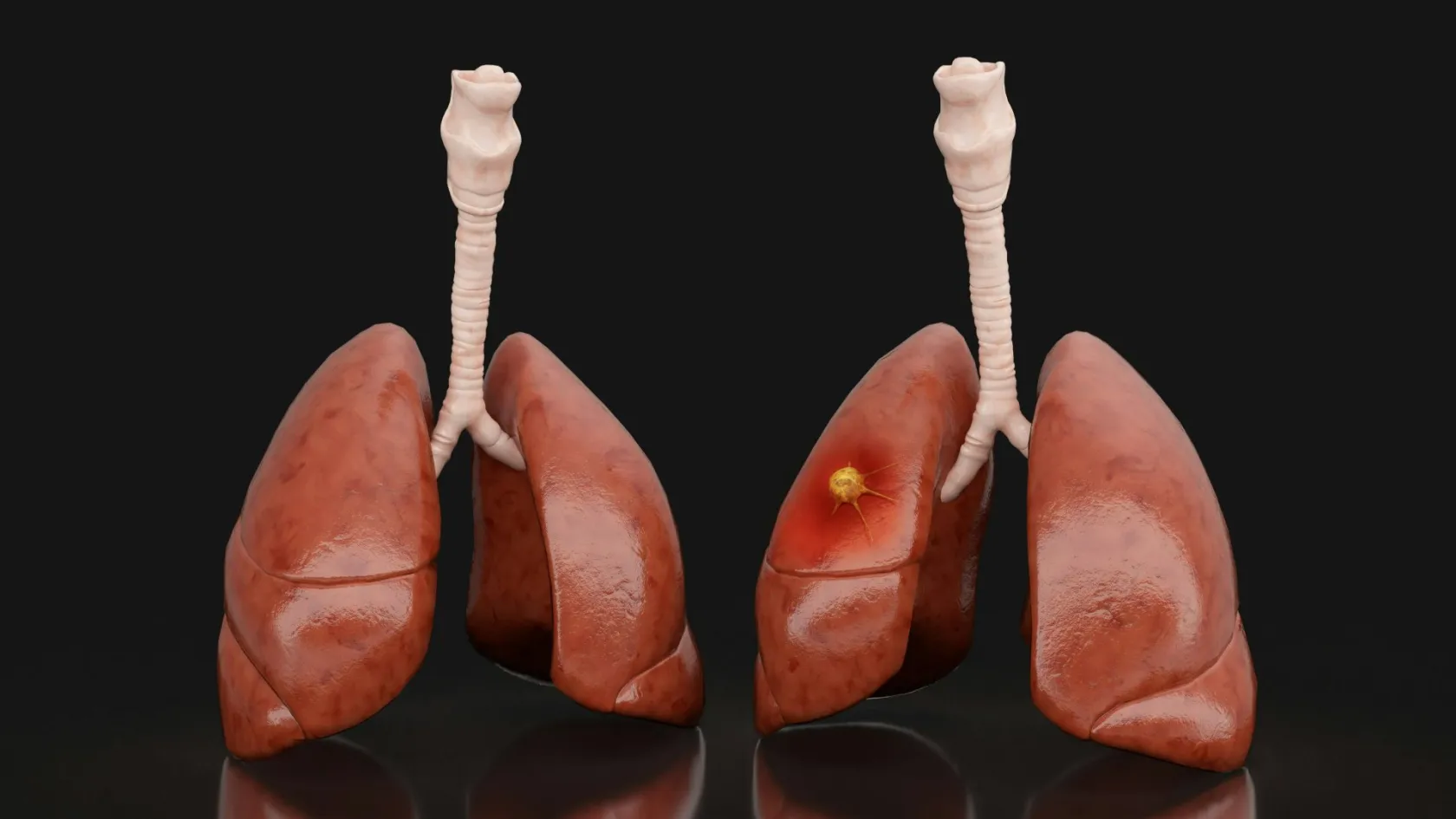
Lung Cancer Surgery
For thirty years the answer was always to remove a whole lobe. Two randomised trials reported in 2022 and 2023 and changed that for small tumours, and most published patient information has not caught up.
About This Department
Lung cancer surgery settles two separate questions, and patients are rarely told they are separate. The first is whether the tumour can be removed. The second is whether you can survive losing that much lung, and it is not a formality, because the smoking that produced most lung cancers also damaged the lungs that have to carry you through the operation. A tumour can be perfectly operable in a patient who is not. This page covers both questions, and it covers the change that has just altered the answer to the first one.
Free consultation
Send the CT and PET scan files together with your breathing tests
No thoracic surgeon can answer either question from a report. Send the CT and the PET-CT as image files, the pulmonary function test with the FEV1 and the DLCO figures on it, the biopsy pathology and any molecular results, and a note of your other medical conditions and how far you can walk before stopping. What comes back is an opinion on whether resection is realistic, which operation would be proposed, whether treatment before surgery would improve the odds, and whether radiotherapy would serve you better. The review costs nothing and carries no obligation.
Whether you can spare the lung
Two numbers dominate this assessment and you should learn them by name, because they will be quoted at you and nobody will stop to explain them. FEV1 is how much air you can force out in the first second, which measures how well the airways move air, while DLCO measures how efficiently oxygen crosses from the air sacs into the blood and is the number people with emphysema fail even when their FEV1 looks perfectly acceptable.
Neither figure is used raw. The surgeon calculates what each will be after the planned resection, by working out what proportion of your functioning lung is being removed, and it is that predicted postoperative value that decides the case.
This table scrolls sideways on a narrow screen. Swipe or drag to see every column.
| Test | What it measures, and why it matters here |
|---|---|
| Spirometry, giving FEV1 | How much air leaves the chest in one second. Low values mean narrowed airways and predict breathlessness after part of the lung is gone. |
| Gas transfer, giving DLCO | How well oxygen crosses into the blood. This is the better predictor of complications and death, and it is frequently the number that stops an operation. |
| Predicted postoperative values | Your current numbers minus the share the resected part contributes. This calculation, and not today's reading, is what the decision rests on. |
| Stair climb or shuttle walk | A cheap real-world test. Climbing several flights without stopping to catch your breath reassures a surgeon more than a borderline laboratory figure does. |
| Cardiopulmonary exercise testing | Measured oxygen uptake during exercise, reserved for borderline cases. The most accurate answer available, and the one that settles disagreements. |
Where the picture stays unclear, a perfusion scan is added, showing which parts of the lung are actually working, because a lobe destroyed by the tumour was contributing nothing and its removal costs you less than the arithmetic suggests.
Ask for your predicted postoperative FEV1 and DLCO as percentages. Those two figures summarise your risk more honestly than any adjective, and any thoracic surgeon can give them to you in a sentence.
The operations by name
Three lobes sit on the right and two on the left. Each lobe divides into segments with their own airway and blood supply, which is what makes anything smaller than a lobe possible in the first place.
Less than a lobe
Wedge resection cuts out the tumour with a rim of lung around it, ignoring anatomical boundaries, and it is the quickest option and the one that spares most tissue. A segmentectomy removes a whole anatomical segment along its own vessels and airway, taking the territory the tumour drains into, and it is a technically harder operation that demands the surgeon identify the right plane instead of simply stapling across the lung. Until recently both were regarded as compromises for patients too frail for a lobectomy. That has changed, and the next section explains why.
A lobe or more
Lobectomy removes an entire lobe with its lymph nodes and remains the standard operation for most lung cancers. A sleeve lobectomy goes further, taking the lobe plus a cuff of the main airway and then sewing the remaining airway back together, and it exists specifically to avoid removing the whole lung when a tumour sits at the origin of a lobar bronchus. Pneumonectomy takes the entire lung on one side. Surgeons work hardest to avoid this one, because losing a whole lung carries a considerably higher risk of complications and death and leaves a permanent limitation on what you can do, so a sleeve resection is preferred wherever the anatomy allows one, and being told that a pneumonectomy is unavoidable is a reasonable moment to ask whether a sleeve resection was considered and why it was ruled out.
The standard operation has changed
For thirty years lobectomy was the answer for every fit patient with a resectable lung cancer, on the strength of a single trial from 1995. Two large randomised trials reported in 2022 and 2023 and overturned that for small peripheral tumours. Most published patient information has not caught up.
Two trials, 1,803 patients between them
Japanese investigators across 70 institutions randomised 1,106 patients with peripheral tumours of 2 cm or less to segmentectomy or lobectomy. Five year overall survival was 94.3 percent after segmentectomy and 91.1 percent after lobectomy, and segmentectomy was not merely non-inferior but statistically superior. A North American trial randomised 697 patients with the same size limit to sublobar or lobar resection and found disease-free survival non-inferior, at 63.6 percent against 64.1 percent at five years, with overall survival of 80.3 against 78.9 percent. Neither trial found the smaller operation costing lives.
That trade is real, and it is local. Cancer came back at the original site in 10.5 percent after segmentectomy and 5.4 percent after lobectomy in the Japanese trial, so a smaller operation does leave more disease behind more often. Survival did not suffer, because a local recurrence caught on surveillance can be treated, and because patients who kept more lung died less often of everything else. In that trial 63 percent of the deaths after lobectomy were from causes other than lung cancer, against 47 percent after segmentectomy.
One qualification deserves stating out loud. A later analysis of the solid-appearing tumours in the Japanese trial, meaning the ones with no ground-glass component and therefore the more aggressive biology, confirmed the survival advantage for segmentectomy but found locoregional recurrence at 16 percent against 8 percent. The advantage held. The local control gap widened.
So the reasonable position is a narrow one. For a peripheral tumour of 2 cm or less with clear nodes, segmentectomy is now a first-choice operation and not a compromise, and it is the better choice in an older patient or one whose lungs are already marginal. Lobectomy remains standard for anything larger, for central tumours, for involved nodes, and where a segmentectomy could not achieve a clear margin.
Why the lymph nodes matter
Whether cancer has reached the lymph nodes in the middle of the chest changes the treatment more than the size of the tumour does, and finding out is a job that happens before the operation rather than during it.
PET-CT shows which nodes are metabolically active, and it is good at excluding disease and unreliable at confirming it, because inflammation lights up the same way. So a suspicious node gets sampled. Endobronchial ultrasound passes a bronchoscope with an ultrasound probe on its tip down the airway and takes needle samples of nodes sitting just outside the airway wall, under sedation and as a day case, while mediastinoscopy, a small incision at the base of the neck, is reserved for the stations that ultrasound cannot reach or for a result that comes back unconvincing.
This table scrolls sideways on a narrow screen. Swipe or drag to see every column.
| Investigation | What it can and cannot tell you |
|---|---|
| PET-CT | Shows which nodes are metabolically active. Reliable for ruling disease out, unreliable for confirming it, because infection and inflammation light up identically. |
| Endobronchial ultrasound | A bronchoscope with an ultrasound tip takes needle samples of nodes lying against the airway wall. Sedation, day case, and it gives tissue rather than a picture. |
| Mediastinoscopy | A small incision at the base of the neck, under general anaesthetic, reaching stations the ultrasound cannot. Reserved for a negative needle result that nobody quite believes. |
| Sampling during the resection | Systematic removal of the node stations, labelled by number. This is what fixes the final stage and decides what treatment follows the operation. |
Skipping that step is the shortcut that costs most.
A patient who goes straight to an operation with unsuspected disease in the mediastinal nodes has an operation that will not cure them, when treatment given first might have. During the resection itself the surgeon samples or removes the node stations systematically, which is what allows the pathologist to stage the disease properly and the oncologist to decide what follows. A resection reported without a node count is a resection nobody can act on.
Keyhole, robotic or open
Open lung surgery means a thoracotomy, an incision between the ribs with the ribs spread apart, and the rib spreading is what produces the pain that lung surgery is remembered for. Keyhole surgery uses one to four small incisions with a camera and no rib spreading at all. That single difference explains most of what follows.
What 503 randomised patients showed
British investigators randomised patients having lobectomy for known or suspected lung cancer to keyhole or open surgery, with patients kept unaware of which they had until discharge. Self-reported physical function at five weeks was better after keyhole surgery. Pain on the second day was lower, painkiller use around 10 percent lower, and the median hospital stay four days against five. Complications occurred in fewer patients, with a relative risk of 0.74 before discharge. Prolonged pain still requiring painkillers beyond five weeks was less common, at a relative risk of 0.82. Crucially for the cancer question, the proportion of patients found to have more advanced node disease than expected was the same in both groups, and survival was the same.
That last point is the one that matters. The worry about keyhole lung surgery was always that a surgeon working through small incisions would take fewer nodes and understage the disease, which would turn a gentler recovery into a worse cancer operation. The trial tested that directly and found no difference between the two groups. Robotic surgery does the same operation with articulated instruments and a three dimensional view, and it is increasingly used for segmentectomy in particular, where the dissection is finer. Open surgery keeps its place for large central tumours, for chest wall involvement, for sleeve resections in some hands, and for the moment a surgeon judges the view inadequate and converts.
Treatment before the operation
For a stage one tumour, surgery comes first and nothing precedes it. For larger tumours and for disease in the nodes, giving drugs before the operation has become standard in the last few years, and understanding why changes how you plan the trip.
What adding immunotherapy before surgery did
One phase three trial randomised patients with resectable stage IB to IIIA disease to chemotherapy alone or chemotherapy plus an immunotherapy drug before surgery. Median event-free survival was 31.6 months with the combination and 20.8 months with chemotherapy alone. More striking, 24 percent of those who had the combination had no viable cancer at all left in the removed lung and nodes, against 2.2 percent on chemotherapy alone. Adding immunotherapy did not increase serious side effects and it did not stop people getting to theatre. In fact slightly more of them had their operation, 83.2 percent against 75.4 percent.
What it does to the timetable
Three cycles, roughly nine weeks, then surgery a few weeks after that.
For an international patient that arithmetic decides everything about the trip. It means either a long stay or two journeys, and it means the drugs might reasonably be given at home while the surgery happens abroad, which requires the two teams to agree a plan in writing before anybody starts. Raise it at the first consultation, because discovering in week four that the operating date has moved by three months is the single most disruptive thing that happens to people in this position.
The operation and the drain
Two to four hours for a lobectomy. Longer for a sleeve resection, or where old infection has stuck the lung down.
One lung at a time
Your anaesthetist places a tube that lets the lung being operated on be deflated while the other keeps breathing for you. That collapsed lung gives the surgeon room to work. It also explains why the anaesthetic assessment cares so much about the state of the other one.
Dividing the artery, vein and airway
Each structure supplying the lobe is separated and divided with a stapler that seals as it cuts. Then the fissure between the lobes is opened, which is the fiddly part and the part that leaks afterwards. The specimen leaves in a bag through the largest of the small incisions.
The lymph node stations
Nodes are taken from defined positions around the airway and the great vessels, labelled by station number so the pathologist knows exactly where each came from. This is the part of the operation that determines your stage and therefore what treatment follows.
The water test, and the drain
Saline fills the chest and the lung is reinflated while everybody watches for bubbles, which is how air leaks are found before you leave theatre. Leaks that show are stitched or sealed. A drain then goes between the ribs into the chest cavity to remove air and fluid and let the remaining lung expand fully.
The drain is the thing that governs your discharge date, and nobody explains this in advance often enough. It stays until the lung has stopped leaking air and the fluid draining has fallen below a set volume. Most drains come out on day two or three. Where the lung continues to leak, and lung tissue does leak, the drain stays longer, and a prolonged air leak beyond five days is the commonest single reason a straightforward lung operation turns into a long hospital stay.
Emphysema makes it more likely, because the tissue is fragile and holds stitches badly. So does an incomplete fissure between the lobes. It is a nuisance more than a danger in most cases, and it is managed by waiting, sometimes with a portable drainage device that lets you go home with it in place where that is offered.
What goes wrong
Prolonged air leak is the commonest problem and it is described above. The rest fall into a short list. Individually uncommon, collectively not rare.
Pneumonia and sputum retention come next. A patient who cannot cough properly because coughing hurts fills the remaining lung with secretions, which is precisely why the physiotherapist appears on the first morning, and why the pain relief exists so that you can breathe deeply and cough hard, and never merely to make you comfortable lying still. Atrial fibrillation, an irregular heart rhythm, occurs in a meaningful minority after lung resection and is more likely after larger operations, and it is usually controlled with medication and settles. Then the uncommon and the serious. Bleeding needing a return to theatre. Injury to the recurrent laryngeal nerve, which runs beside the nodes on the left and produces a hoarse voice and a weak cough if it is bruised or divided. Bronchopleural fistula, where the stapled airway stump breaks down, which is rare, more likely after pneumonectomy, and serious. Empyema, an infection filling the space where the lung used to be, which needs drainage and a long course of antibiotics.
Chronic pain at the incision, sometimes with numbness, persists past three months in a proportion of patients, and keyhole surgery reduces it substantially without abolishing it, which the British trial measured directly. Death within 30 days is uncommon after a lobectomy in a properly assessed patient. Death runs several times higher after pneumonectomy, which is the clearest reason surgeons work so hard to avoid taking a whole lung.
Breathing afterwards
You will notice it. That is the first honest answer. Removing a lobe takes away part of your reserve permanently, and the loss shows up on hills and stairs, never sitting in a chair. Most people find the breathlessness at three months considerably better than at three weeks, and better again at a year as the remaining lung expands to fill the space and the chest wall settles. Segmentectomy costs less function than lobectomy, though the difference measured in the Japanese trial at one year was 3.5 percent of FEV1, which is smaller than patients expect and did not reach the threshold the investigators had set as clinically meaningful, so the case for the smaller operation rests on survival and on what happens over decades rather than on how much easier the stairs feel next spring, and a surgeon who sells segmentectomy purely on breathing is overselling the part of it that was measured smallest.
- Use the incentive spirometer every hour you are awake for the first weeks, because reinflating the lung base is what prevents pneumonia and nothing else does it as well.
- Take the pain relief before you need it, ahead of the pain, since the point of it is to let you cough and walk.
- Walk daily and add distance every few days, and expect hills to be the last thing that comes back.
- Do not smoke again, at all. A second primary lung cancer is a real risk in this population and stopping is the only thing that lowers it.
- Report a fever, increasing breathlessness, coloured sputum or new chest pain on the day, without waiting for the next appointment.
Driving comes back at around four weeks after keyhole surgery and six or more after a thoracotomy, judged by whether you could turn the wheel hard and brake sharply without hesitating. Office work follows at a similar point. Heavy physical work waits for three months.
How long you stay
Three to four weeks in the country for surgery alone, and considerably longer where drugs are given first.
Assessment takes four to six days and it is more involved than for most cancers, because the staging and the fitness testing run in parallel. CT and PET-CT, pulmonary function testing with gas transfer, an echocardiogram, endobronchial ultrasound where the nodes need sampling, bronchoscopy where the tumour is central, and a thoracic tumour board before a date is set. Node sampling adds days, and it is not a step to skip. Expect four to six days on the ward after keyhole surgery and six to ten after a thoracotomy, with the drain deciding the exact day. Add a week to ten days nearby afterwards for the wound check, the chest X-ray confirming the lung has stayed up, and the pathology discussion that sets the treatment for the next year, which is a conversation to have sitting in a room and not by reading a document forwarded to you after you have landed.
Flying deserves particular attention after lung surgery, for a physical reason. Cabin pressure at altitude is equivalent to standing at around 2,400 metres, so any air trapped in the chest expands. Nobody flies with a drain in or with a residual air pocket on the X-ray. Most surgeons clear travel at around three weeks after an uncomplicated keyhole resection, with a chest X-ray showing a fully expanded lung, no air leak, no fever, an oxygen saturation that holds up on walking, and an agreed plan for clot prevention. Get that chest X-ray done and read before you book the flight.
What drives the cost
Wedge resection and sleeve lobectomy are both lung cancer surgery and are priced nothing like each other. Eight things move the total, and one of them is peculiar to this operation.
- Which resection is planned, from wedge through segmentectomy and lobectomy to sleeve resection and pneumonectomy.
- The number of stapler cartridges the operation consumes, which is a genuine and substantial line item in lung surgery and varies with how complete your fissures are.
- The approach, since robotic and keyhole platforms carry instrument costs per case that open surgery does not.
- The staging work, including PET-CT, endobronchial ultrasound and mediastinoscopy, some of which sits outside a surgical quotation.
- How long the chest drain stays in, since a prolonged air leak converts a four day admission into a ten day one.
- Intensive care, planned or otherwise, which is more likely after pneumonectomy and in patients with marginal lung function.
- The pathology, including the node count and the molecular and PD-L1 testing that decides drug treatment afterwards.
- Chemotherapy or immunotherapy before or after the operation, which is the largest item of all and sits outside almost every surgical package.
Your own condition moves it as much as the operation. Emphysema, poor gas transfer, heart disease, diabetes, low body weight, continued smoking and previous chest surgery or radiotherapy each raise the chance of a longer stay and of care that nobody budgeted for.
A published package here typically buys the transfers, the tests before admission, the surgeon and anaesthetist, a stated number of theatre and ward days, translation, hotel nights and the appointments before departure. Excluded, almost always, are the air tickets, insurance, any night beyond the stated number, unplanned intensive care, the cost of a complication, and the whole of the drug treatment.
Six questions turn a headline into a quotation. Which resection does the figure assume, and what changes if the surgeon has to take a lobe instead of a segment. Are the staplers inside the figure or itemised. Is the endobronchial ultrasound and PET staging included. How many ward nights are covered and what an extra night costs, given that the drain decides. Is the molecular and PD-L1 testing inside the pathology fee. And what proportion of any chemotherapy or immunotherapy, if any, sits inside the quotation.
No figure means anything until a surgeon has read your scans and your breathing tests. That review costs nothing.
Once you are home
Surveillance after lung cancer surgery runs on CT and not on X-ray, at intervals of six months for the first two to three years and annually thereafter, continuing indefinitely. Two things are being looked for and they are different problems. A recurrence of the original cancer, and a second primary lung cancer in the remaining lung, which people in this group develop at a rate high enough that the annual scan never stops.
Take home the operative note naming the resection and the node stations sampled, the full pathology including the node count and the molecular and PD-L1 results, the imaging on disc, the pulmonary function figures before and after, and the surveillance schedule with dates. Get it in English. An oncologist at home cannot choose a drug without the molecular results, and requesting them six weeks later across a time zone wastes weeks that the treatment plan does not have. Expect a scarred lung to look odd on later imaging. Staple lines, scarring at the resection margin and shifted anatomy all read as abnormal to a radiologist who has not been told what was removed, so the operative note travelling with you prevents a round of scans and worry that nothing warranted, and the two minutes it takes to hand that note over at the first appointment save a fortnight of scanning and waiting later on.
Contact the team here for increasing breathlessness, a fever, coloured or bloody sputum, new or worsening chest pain, a wound that opens, or a swollen painful calf. A message with a photograph settles most questions within hours.
Frequently asked questions about lung cancer surgery
Do I have to lose a whole lobe?
How do they decide whether my lungs are good enough?
Why is the chest drain still in?
Will I be breathless for the rest of my life?
How long do I need to stay abroad, and when can I fly?
Would treatment before surgery help me?
References
- Saji H, Okada M, Tsuboi M, et al. Segmentectomy versus lobectomy in small-sized peripheral non-small-cell lung cancer (JCOG0802/WJOG4607L), a multicentre, open-label, phase 3, randomised, controlled, non-inferiority trial. The Lancet. 2022;399(10335):1607-1617.
- Altorki N, Wang X, Kozono D, et al. Lobar or sublobar resection for peripheral stage IA non-small-cell lung cancer. The New England Journal of Medicine. 2023;388(6):489-498.
- Hattori A, Suzuki K, Takamochi K, et al. Segmentectomy versus lobectomy in small-sized peripheral non-small-cell lung cancer with radiologically pure-solid appearance in Japan, a post-hoc supplemental analysis of a multicentre, open-label, phase 3 trial. The Lancet Respiratory Medicine. 2024;12(2):105-116.
- Lim E, Harris RA, McKeon HE, et al. Impact of video-assisted thoracoscopic lobectomy versus open lobectomy for lung cancer on recovery assessed using self-reported physical function, VIOLET randomised controlled trial. Health Technology Assessment. 2022;26(48):1-162.
- Forde PM, Spicer J, Lu S, et al. Neoadjuvant nivolumab plus chemotherapy in resectable lung cancer. The New England Journal of Medicine. 2022;386(21):1973-1985.
Editor's note
Written by the Biruni Hospital medical editorial team. Reviewed by Prof. Dr. Mustafa Teoman YANMAZ, Medical Oncology.
Medically reviewed by

Prof. Dr. Mustafa Teoman YANMAZ
Medical Oncology
Related Treatments
View All-
Bilateral Mastectomy
Whether you carry a BRCA mutation, have cancer in one breast or in both, bilateral mastectomy removes both breasts in one operation, and how much it helps depends on which of those three paths brought you to it.
-

Breast Conserving - Lumpectomy Surgery
Keeping the breast has never been the less thorough choice, and the largest recent studies now point the other way. This page explains who can have breast conserving surgery, what the margin report decides, and how long a woman traveling for it needs to stay.
-

Cytoreductive - Tumor Reduction Surgery
Patients cleared of every visible deposit lived 30 months in one gastric series. Those left with anything at all lived 7.3 months, however little remained.
-
Debulking - Ovarian Cancer Tumor Reduction
Two women with the same diagnosis on the same morning can get operations that share almost nothing. What decides it is who measured the disease first.
-
Debulking Surgery
Surgeons removed a median of 57 lymph nodes from women already cleared of visible disease. It bought no extra months, and tripled deaths within 60 days.