
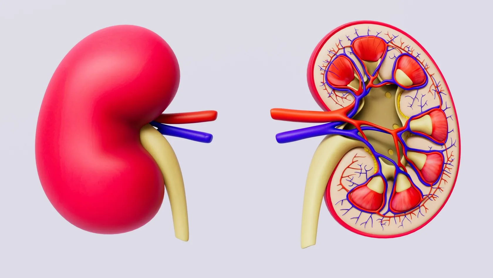
Kidney Stone Surgery
Three measurements on a CT decide which kidney stone procedure suits you, and none of them is a brand name. The stone free rate a clinic advertises is a different kind of number, because it depends on how hard that clinic looked afterward. This page covers shockwaves, ureteroscopy and percutaneous surgery, what the randomized trials found, and why half of stone formers come back.
About This Department
Your scan picks the operation. Their scanner picks the success rate.
Three measurements on a CT decide which procedure suits a kidney stone, and none of them is a brand name. The stone free rate a clinic advertises is a different kind of number, because it depends on how hard that clinic looked afterward. This page separates the two and tells you what the large trials found.
The scan makes the decision
Three numbers come off a low dose CT and between them they settle almost everything. How wide the stone is at its largest point, measured in millimeters. Where it sits, meaning the kidney or one of its lower pockets or somewhere along the tube down to the bladder. And how dense it is, reported in Hounsfield units, which tells you how stubborn it will be under shockwaves. Everything else follows from those three. A patient who understands them can hold a sensible conversation with any urologist in any country and can tell immediately when a recommendation has drifted away from the evidence. A patient who has only been told the name of a machine cannot. So this page is organized around the measurements first and the procedures second, which is the reverse of how most clinic pages do it and the same order a surgeon actually thinks in. Get those three numbers before you agree to anything. If nobody has given them to you, the assessment is not finished.
Which operation for which stone
Medicine offers four approaches here, and they do not compete across the whole range. Each one owns a part of the territory, and the overlap where two of them are both reasonable is narrower than the marketing suggests.
This table scrolls sideways on a narrow screen. Swipe or drag to see every column.
| Approach | Suits | How it is done | Main drawback |
|---|---|---|---|
| Watching and waiting | Stones under 5 mm in the tube, with pain controlled | Fluids, painkillers, a repeat scan, nothing else | Weeks of uncertainty and the chance it fails anyway |
| Shockwave lithotripsy | Softer stones under 15 mm, reachable by the shock head | Focused shockwaves from outside the body, awake or sedated | More repeat sessions and dense stones resist it |
| Ureteroscopy with laser | Almost anything in the tube, and kidney stones under 20 mm | A fine telescope up the natural passage, laser, no cuts | General anesthetic and frequently a stent afterward |
| Percutaneous surgery | Kidney stones over 20 mm and branched stones | A track through the back into the kidney, roughly 1 cm wide | Bleeding and fever risk, and a hospital stay |
The names that do not matter
Clinic pages fill up with trademarks. Laser types, scope brands, generations of machine, a suffix bolted onto a familiar word to make an ordinary procedure sound proprietary. Almost none of it changes what happens to you. A holmium laser and a thulium fiber laser both turn a stone into dust and the difference between them shows up in operating minutes rather than in whether you go home stone free. What changes your outcome is the size of the stone, where it sits, how dense it is, how experienced the surgeon is and whether anybody bothered to find out why you made a stone in the first place. Those five, in roughly that order. Everything on a brochure that does not touch one of the five is decoration. None of this means equipment is irrelevant, since a unit without a flexible scope cannot reach a lower pole stone and a unit without a modern shock head will treat you badly with an old one, and equipment of that kind is a floor every serious center cleared years ago, which makes it a poor thing to advertise.
How to read what follows
Sections three through seven run from doing nothing up to the biggest operation, in that order, so you can stop reading at the row of the table that matches your stone. Stop there. Sections nine through thirteen are the ones most pages leave out, and they are the reason a lot of people have this operation twice.
Most stones pass without help
Pain brings people to a hospital and the pain is genuinely severe, so the instinct to have something done immediately is understandable. Frequently that instinct is wrong. Anything of 4 mm or less sitting in the lower part of the tube passes on its own in the large majority of cases, given fluids, painkillers and a few weeks, and every procedure carries risks that doing nothing does not. Size and position again, before anything else. Stones in the lower third of the tube pass far more readily than one sitting where the tube leaves the kidney, and that single distinction changes the odds enough that a urologist who has not told you where yours sits has not given you the information you need to decide anything. Ask where it is.
The drug that does not do what it is given for
Somebody will probably prescribe you tamsulosin. The reasoning behind it sounds excellent, since relaxing the muscle in the wall of the tube ought to let a stone slide through more easily, and dozens of small studies reported exactly that. Then a properly powered trial across 24 hospitals randomly assigned 1,167 patients with a single stone of 10 mm or less to tamsulosin, to nifedipine or to a dummy tablet for four weeks, with nobody knowing who got which. The proportion who passed their stone without an operation came out at odds of 1.04 for the drugs against placebo, an absolute difference of 0.8 percent, and the confidence interval sat squarely across zero. Sensitivity analyses by sex, by stone size and by stone position changed nothing. Neither drug beat the other either. The trial authors added a line that deserves quoting in spirit. They noted the need to investigate why large high quality trials keep producing smaller effects than pooled analyses of many small low quality ones. That pattern is not unique to kidney stones and it is the single most useful thing a patient can carry into any consultation.
Taking the tablet harms very little and we will not argue if your own doctor has started it. Believing it is doing the work is the part to avoid, because a stone that has not moved in four weeks needs a decision and not another month of the same prescription.
When waiting stops being reasonable
Four situations end the argument and move a stone into the operating list regardless of its size. Infection behind the blockage comes first and it comes with a fever. Urine trapped above a stone that has become infected turns into a surgical emergency measured in hours, treated by draining the kidney immediately with a tube or a stent and dealing with the stone later, once the infection has gone. Pain that will not settle on proper painkillers is the second. A kidney that is losing function on the scans, or a patient with only one working kidney, is the third, since time spent blocked is function that does not come back. The fourth is simply the passage of weeks with no movement, because a stone parked in one spot for a month starts to scar the tube around it. Outside those four, the calendar is yours. A stone sitting quietly in a lower pocket of the kidney, causing nothing, found on a scan done for something else entirely, does not oblige anybody to book a flight. We will say so plainly when that is the situation, and we say it fairly often.
Shockwave lithotripsy
Shockwaves focused from outside the body onto the stone crack it into pieces small enough to wash out over the following days and weeks. Nothing enters you. Most people have it awake with sedation, walk out within a couple of hours and go back to work the next day, and for the right stone it remains the gentlest thing in urology. Working on the stone at a distance is its weakness, and it means some stones refuse on the stone at a distance and some stones refuse. Density above roughly a thousand Hounsfield units predicts failure. Distance from the skin to the stone matters, so a heavier build reduces the odds. Stones in the lowest pocket of the kidney fragment perfectly well and then sit there, because gravity gives the pieces nowhere to go. And the fragments still have to pass, which means the pain of passing a stone is deferred and not avoided, a point rarely made before somebody consents.
What the head to head trial found
613 patients with a single stone in the tube were randomly assigned to shockwaves or to a telescope and followed for six months, in a trial designed to test whether the non invasive option was good enough. Sixty seven of 306 in the shockwave arm needed further treatment, which is 22.1 percent, against 31 of 307 in the telescope arm, or 10.3 percent. The gap of 11.7 percentage points favored the telescope and still fell inside the margin the investigators had set, so shockwaves passed the test they were given. Read that carefully, because it means both answers are defensible. The telescope clears stones more reliably in one visit. Shockwaves avoid an anesthetic, avoid a stent and cost less, at the price of a one in five chance of coming back for more.
Ureteroscopy and the laser
A telescope thinner than a pencil lead travels up the natural passage from the bladder to the stone, a laser fiber passes down its working channel, and the stone is either dusted into sand or broken into fragments small enough to pull out in a basket. No incision anywhere. The flexible version reaches into the kidney itself, which is what made this the workhorse of modern stone surgery and pushed shockwaves toward a narrower role. Two developments did most of the work. Scopes became thin and steerable enough to turn back on themselves and look into the lower pockets of the kidney, and lasers became fine enough to pass down a channel that narrow without stiffening the instrument, so a procedure once reserved for the tube now reaches almost anywhere a stone can hide.
Two honest drawbacks sit alongside the high clearance rate. General anesthetic comes first, and shockwaves usually avoid it. The stent comes second, a soft plastic tube left from kidney to bladder to keep the passage open while swelling settles, and it is the single thing patients report as the worst part of this operation. It causes an ache in the flank when you pass urine, a need to go frequently, blood in the urine and a dull discomfort that ends the day it comes out. A stent is not always needed. Whether you will have one, and when it comes out, belongs in the conversation before surgery and not in a discharge letter, and anyone who leaves without knowing the removal date has been let down. Where the stone sat in the tube, where the passage was not traumatized and where nothing was left behind, we leave no stent in and we say so in the notes, since a good half of the misery people report after this operation belongs to the tube and not to the surgery it followed.
Percutaneous surgery for large stones
Above roughly 20 mm, and for the branched stones that fill the drainage system like coral, the telescope stops being efficient and a direct route wins. A needle passes through the back into the kidney under imaging, the track is widened to the width of a drinking straw, and the stone is broken and removed through it in one sitting. Count this as real surgery with a hospital stay, and it clears large stones better than anything else available. Positioning varies between units, with some operating face down and others on the back or the side, and that argument matters far less than whether the surgeon making the puncture does a reasonable number of these every year, because gaining safe access to a kidney through a needle is the part of this operation that experience most obviously improves.
Smaller tracks, and what they buy
Narrower versions of the same operation exist, using a track closer to the width of a pen refill, and they bleed less. They also take longer and clear large volumes of stone more slowly, so the sensible reading is that a smaller track suits a moderate stone while a full sized track still suits a large one. Anyone offering the smallest possible track for the largest possible stone is optimizing the wrong variable. The same caution applies to the drainage tube afterward. Leaving no tube shortens the stay and hurts less, and it suits a clean single puncture with no bleeding and nothing left behind, while a difficult case with several tracks or a fragment still sitting somewhere deserves a tube and a second look, with the early discharge that reads well on a brochure given up. Tubes come out the following morning in most of these cases anyway, so very little is actually lost.
What happens on the day
Taking ureteroscopy as the example, since it is the commonest of the three.
- A urine culture before anything else. Operating through infected urine is how a routine procedure turns into sepsis, so a positive culture postpones the date and gets treated first.
- General anesthetic, and around 45 to 90 minutes. Longer for a kidney stone than for one in the tube, and longer again if the passage is tight and needs a preliminary stent.
- Laser, then retrieval or dusting. Fragments are either lifted out in a basket or reduced to sand fine enough to wash away, and the choice affects how much you will pass afterward.
- A stent, or no stent. Decided at the end on what the passage looks like, with the removal date written down before you leave.
- Home the same day or the next. Blood in the urine for several days is expected, and burning when passing urine is expected, and a fever above 38 degrees is not.
Living with a stent
Drink more than feels necessary, expect the flank ache when your bladder empties, and take the painkillers on a schedule without waiting for the discomfort. Most come out between four days and two weeks, in a clinic, in under a minute, through the passage they went in by. The relief is immediate and out of proportion to how small the procedure is, and a surprising number of people describe that moment as the real end of the treatment. Some units now leave a thread attached to the stent so you can remove it yourself at home on a stated date, which suits people who have flown a long way and would rather not fly back, and it works provided somebody has explained clearly how and when. Ask for the thread if the geography suits it.
Stone free is a claim about a scanner
Why two clinics can quote wildly different numbers and both be telling the truthStone free means no fragments were seen. Seen how, and when, and how small is small enough to ignore. A plain X ray at six weeks misses a great deal that a CT would catch, and a clinic checking with X ray will report a higher stone free rate than the identical surgeon checking with CT. Move the threshold from any fragment at all to fragments under 4 mm and the number climbs again. Check at one day instead of three months and it changes a third time. None of this involves dishonesty. It means the figure on a brochure cannot be compared with the figure on another brochure unless both state the imaging and the cut off, and almost none of them do.
The fragments called insignificant
Leftover pieces of 4 mm or less, causing no symptoms and carrying no infection, have a name in the literature that tells you what the profession assumed. Clinically insignificant residual fragments. A group in Cleveland and Istanbul decided to follow them instead of assuming, taking 430 patients through percutaneous surgery with an overall stone free rate of 74.5 percent and residual fragments in 22 percent at three months, then tracking 38 of those patients for at least two years. Ten of the 38, which is 26.3 percent, had a symptomatic episode that needed treatment. The fragments grew in eight patients, stayed put or shrank in 27, and passed by themselves in three. Metabolic testing did not predict which ones would grow. Struvite showed up in three of the eight stones that enlarged. That fits, since fragments seeded by infection carry the bacteria that grow them and nothing short of complete clearance stops that. For every other composition nobody can say in advance which small fragment will sit still for a decade and which will double in a year.
Risks in the order you meet them
The first two weeks, from commonest to most seriousBlood in the urine for several days, which is normal after all three procedures. Burning and frequency, worse with a stent. Renal colic as fragments pass, especially after shockwaves. Urine infection, and the culture beforehand is what prevents it. Fever above 38 degrees with shivering, which is the one that means contacting somebody the same day rather than waiting. Injury to the wall of the tube, uncommon and usually managed with a stent. Bleeding needing transfusion, which belongs almost entirely to percutaneous surgery and rises with stone size. A stent that stayed in too long and encrusted, which is a complication of administration rather than of surgery and is entirely preventable by writing the removal date down.
The number that should worry you most
Sepsis is rare. It is the reason every item above concerning urine cultures and fevers is written the way it is. Infected urine under pressure seeds the bloodstream within hours, and the people who do badly are almost always the ones who waited a day to see whether the fever would settle. If you develop a temperature with shaking chills after any stone procedure, in any country, that is an emergency department visit and not a message. Send us the notes afterward and we will read them, and we prefer an unnecessary hospital visit to the alternative. Two things reduce the risk before it arrives. A urine culture taken early enough for the result to be back before the operating date, and antibiotics chosen against what actually grew instead of against what usually grows, both of which sound too obvious to state until you count how many stone procedures worldwide are performed without either.
What your stone is made of
- Calcium oxalate. The large majority. Managed with fluid volume, sodium restriction, normal dietary calcium and attention to oxalate in the diet.
- Uric acid. Forms in acidic urine, shows up faintly or not at all on plain X ray, and is the one stone type that can genuinely be dissolved with alkalinizing medication.
- Struvite. Grows out of a urine infection, forms the branched stones that fill a kidney, and returns quickly unless every fragment is cleared.
- Cystine. Inherited, starts young, recurs relentlessly, and needs lifelong specialist management rather than general advice.
- Calcium phosphate. Points toward a defect in how the kidney handles acid, or toward a parathyroid problem worth a blood test.
Four of those five change the treatment plan. A clinic that removes a stone without telling you which one you had has finished the operation and left the illness unexamined.
Stopping the next one
Two trials sit at the center of this and they point in opposite directions, and that is why the section exists.
Wide table. Scroll sideways on a narrow screen to reach every column.
| Feature | Drinking more water | A thiazide tablet |
|---|---|---|
| Patients and follow up | 199 first time stone formers, 5 years | 416 recurrent stone formers, median 2.9 years |
| What was compared | High water intake against no advice at all | Three doses of hydrochlorothiazide against placebo |
| Recurrence | 12 of 99 against 27 of 100 | 49 to 62 percent on drug against 59 percent on placebo |
| Time to the next stone | 38.7 months against 25.1 months | No dose response at all |
| Harms | None reported | More low potassium, gout, new diabetes and skin allergy |
Water halved the recurrence rate over five years and delayed the next stone by more than a year. The tablet, tested at three doses against a dummy in 416 people with recurrent stones, produced recurrence in 49 to 62 percent of patients depending on the dose against 59 percent on placebo, with no relationship between dose and effect, while causing measurably more harm. That result unsettled a lot of practice when it appeared and it has not been explained away. Fluid remains the intervention with the best evidence behind it, and the amount that counts is the volume of urine you produce and not the number of glasses you drink, and that is why the target is stated in liters of output, checked and not guessed. None of that makes medication useless. It makes medication something to aim at a finding, so a patient losing too much calcium in the urine or running a urine that is persistently too acidic has something specific to treat, and a patient whose tests came back unremarkable has been given side effects and very little else.
The number that outranks the success rate
Kidney stones are a chronic condition that presents as an acute event, and almost every page written on them describes the event and ignores the condition. Roughly half of people who form one stone form another within five to ten years. That figure is larger than the difference between any two operations on this page, larger than the difference between any two lasers, and it belongs at the front of the conversation and not in a leaflet handed over on the way out. Nobody advertises it. A page selling an operation has no reason to lead with the news that the operation treats an episode of a condition it does not cure, and the omission is so consistent across the industry that its absence has stopped being noticeable.
What a complete treatment contains
The stone analyzed. Blood tests covering calcium, uric acid, kidney function and parathyroid hormone where the picture suggests it. A 24 hour urine collection in anybody who has formed more than one stone, or who is young, or whose stone is not the common type. A written plan naming the fluid target, the dietary changes that apply to your stone type and any medication with its reason. And a date for the next scan. Our summaries contain all five, addressed to the doctor who will see you at home, and if a clinic quoting you a price has not mentioned any of them, you are being quoted for the operation and not for the treatment. That distinction is the most expensive one on this page, because the second operation costs what the first one cost and the flights cost what they cost, and a workup that might have prevented it would have added a modest sum to a single invoice.
Cost, timing and flying home
Length of stay follows the procedure. Shockwave treatment needs two to three nights in the country for the session and a check. Ureteroscopy needs four to six, covering the assessment, the operation, one night and a review, and longer again if a stent is coming out before you travel. Percutaneous surgery needs seven to ten, since it involves two or three nights in hospital and a postoperative scan. Build in slack at the end rather than at the beginning. Assessment and surgery run to schedule far more reliably than recovery does, and the days people wish they had bought are always the ones after the operation.
Fitness to fly, and the stent question
Flying itself puts no strain on a treated kidney. What decides the date is whether you are free of fever, passing urine comfortably and past the window in which bleeding would appear, which for most people means 24 to 48 hours after ureteroscopy and several days after percutaneous surgery. Stents catch people out. Flying home with one in place is entirely possible and frequently sensible, and it means arranging removal at home and confirming that arrangement before you leave, because a stent left in for months encrusts and turns a one minute clinic procedure into another operation. We write the removal date on the discharge summary, we send it to whoever will do it, and we ask you to confirm to us that it came out, which sounds excessive until you meet somebody who has carried one for a year.
Four things move the cost. Which procedure is done and whether one session will clear the stone. Whether a stent is placed and whether it is removed here or at home. Whether the stay is day case or inpatient. And whether the metabolic workup and stone analysis are included, which they should be and frequently are not. We quote in writing once the CT has been read, and we publish no figures on this page, since a price offered before anyone has measured your stone is a number chosen to win an inquiry. Follow up once you are home costs nothing. Send the scans, send the stone analysis if it was done elsewhere, send the 24 hour urine result, and we will read them and reply.
Coming to Istanbul
Send the scan first, before you discuss dates or prices or anything else at all. The table below sets out what makes a remote opinion useful.
On a narrow screen the table below scrolls sideways. Drag it to reach both columns.
| Send | What it settles |
|---|---|
| The CT images themselves, rather than the report | Size, position and density, which choose the procedure |
| Any previous stone analysis | Whether dissolving is possible and what prevention applies |
| Kidney function and calcium blood tests | Fitness for surgery and whether a gland needs investigating |
| A recent urine culture | Whether the date holds or the infection is treated first |
| Your operative notes, if you have had stones before | What was already tried and how the anatomy behaved |
Kidney stone surgery FAQ
Most first messages contain at least one of these.
Which procedure is best?
Does tamsulosin help a stone pass?
Shockwaves or a telescope for a stone in the tube?
How can one clinic claim 95 percent stone free and another 75?
Are small leftover fragments really harmless?
Will it come back, and can a tablet stop it?
How long do I need in Istanbul, and can I fly with a stent?
References
- Pickard R, Starr K, MacLennan G, Kilonzo M, Lam T, Thomas R, et al. Use of drug therapy in the management of symptomatic ureteric stones in hospitalised adults. The SUSPEND trial. Health Technology Assessment. 2015;19(63):1-171.
- Dasgupta R, Cameron S, Aucott L, MacLennan G, Thomas RE, Kilonzo MM, et al. Shockwave lithotripsy versus ureteroscopic treatment as therapeutic interventions for stones of the ureter (TISU). A multicentre randomised controlled non-inferiority trial. European Urology. 2021;80(1):46-54.
- Xue W, Pacik D, Boellaard W, Breda A, Botoca M, Rassweiler J, et al. Management of single large nonstaghorn renal stones in the CROES PCNL global study. Journal of Urology. 2012;187(4):1293-1297.
- Altunrende F, Tefekli A, Stein RJ, Autorino R, Yuruk E, Laydner H, et al. Clinically insignificant residual fragments after percutaneous nephrolithotomy. Medium-term follow-up. Journal of Endourology. 2011;25(6):941-945.
- Borghi L, Meschi T, Amato F, Briganti A, Novarini A, Giannini A. Urinary volume, water and recurrences in idiopathic calcium nephrolithiasis. A 5-year randomized prospective study. Journal of Urology. 1996;155(3):839-843.
- Dhayat NA, Bonny O, Roth B, Christe A, Ritter A, Mohebbi N, et al. Hydrochlorothiazide and prevention of kidney-stone recurrence. New England Journal of Medicine. 2023;388(9):781-791.
Editor's note
Written by the Biruni Hospital medical editorial team. Reviewed by Prof. Dr. Barış NUHOĞLU, Urology.
Medically reviewed by

Prof. Dr. Barış NUHOĞLU
Urology
Related Treatments
View All
Bladder Cancer Surgery
Bladder cancer is treated by two operations that share almost nothing. One scrapes tumor off the inside wall through the urethra in twenty minutes. The other removes the bladder and rebuilds the route urine takes out of the body. The short one is treated everywhere as a minor procedure, and how well it is done decides the staging, the recurrence rate and whether the long one ever becomes necessary.
Fusion Biopsy Prostate - MRI-Guided Prostate Biopsy
Software builds a model of your prostate from the MRI and another from the live ultrasound, then deforms one onto the other so the lesion lands somewhere the needle can reach. Every step of that carries a few millimeters of error. This page covers where the error comes from, what the trials comparing the three targeting methods found, who draws the outline the needle aims at, and why good units still take cores from the rest of the gland.

Inflatable Penile Prosthesis
An inflatable penile prosthesis has three parts, and only one of them gets discussed. The reservoir sits inside the abdomen, decides where the operation gets difficult, and causes most of the reoperations men end up needing. This guide covers the hydraulics, the move from the space of Retzius to high submuscular placement, the evidence on palpability and auto inflation, the link between blood sugar control and infection, recovery week by week, and what the operation means in Istanbul.
Kidney Cancer Surgery - Renal Cancer Surgery
Kidney cancer surgery means two different operations, and the choice between them is made from a scan before anyone knows what the tumor is. The only randomized comparison found that sparing the kidney protected kidney function and did not extend life. This page works through that result, the clamp and the clock, biopsy before surgery, surveillance, and what changed when the disease has already spread.
Laparoscopic Prostatectomy
Keyhole removal of the prostate inflates your abdomen with carbon dioxide, tips you head down for hours and works through straight rods that cannot bend at the tip. This page covers what each of those three does to your body, which route through the abdomen suits you, how to read the continence figure a clinic quotes, and where the randomized trials favor a robot instead.