
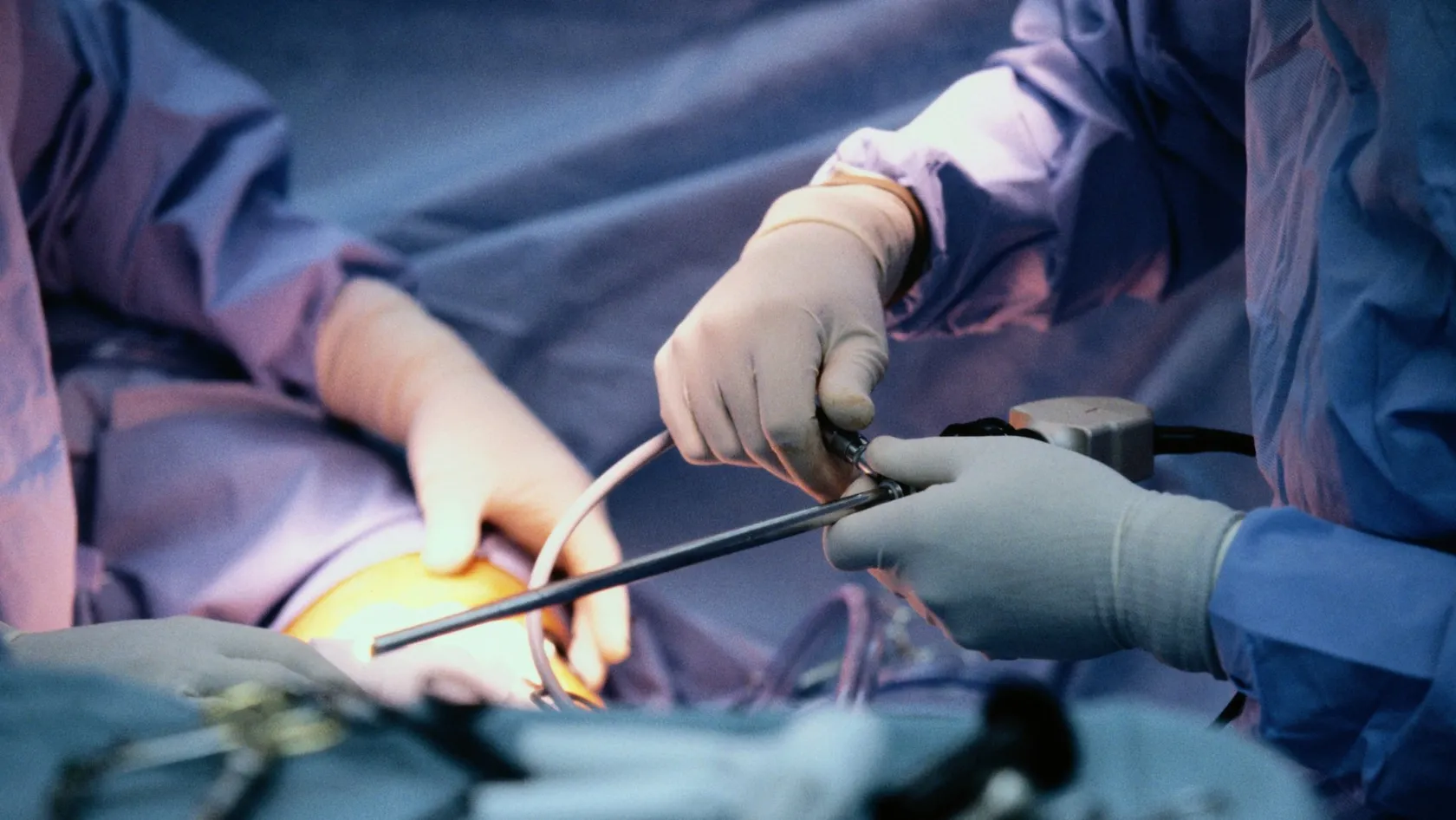
Fusion Biopsy Prostate - MRI-Guided Prostate Biopsy
Software builds a model of your prostate from the MRI and another from the live ultrasound, then deforms one onto the other so the lesion lands somewhere the needle can reach. Every step of that carries a few millimeters of error. This page covers where the error comes from, what the trials comparing the three targeting methods found, who draws the outline the needle aims at, and why good units still take cores from the rest of the gland.
About This Department
You are aimed at a lesion on a picture taken weeks ago. Fusion is the arithmetic that puts it on a live one.
Software builds a model of your prostate from the MRI and another from the live ultrasound, then deforms one onto the other so the lesion coordinates land somewhere the needle can reach. Every part of that sentence can go wrong by a few millimeters. This page covers where the error comes from, what the trials comparing the targeting methods actually found, and why experienced units still take cores from the rest of the gland.
What the word fusion describes
Your MRI was taken on a different day, in a different room, with you lying flat and nothing pressing on you, while the biopsy happens with a probe in your rectum, your position changed, your bladder and rectum holding different amounts, and the gland itself squashed out of the shape the scanner recorded. Those two pictures do not match, and the whole business of a fusion biopsy consists of making them match well enough to aim through. Software takes the outline somebody drew around your prostate on the MRI, builds a three dimensional model from it, builds a second model from a live ultrasound sweep taken at the start of the procedure, and then stretches one onto the other until the surfaces agree. The lesion coordinates ride along with that stretch and appear on the live screen as a colored blob. The needle goes where the blob says. Two things in that sentence deserve stating out loud. The blob is a calculation and not an observation, and nothing on the screen tells the operator how good the calculation was.
Nothing in that sequence is magic and every step of it carries an error.
Three ways to hit a lesion
Cognitive, software, or inside the scanner
Clinics advertise fusion as though it were the only option, and three approaches are in routine use, the first of which is cognitive registration, where the operator studies the MRI, forms a mental picture of where the lesion sits, and aims the needle there by eye. Software fusion is the process described above, with a tracked probe and a screen showing the overlay. In-bore biopsy puts the man inside the MRI scanner, places a needle guide, takes a picture with the needle in position, and confirms directly that the tissue being sampled is the tissue that was suspicious. Each one trades cost and time against certainty. Clinics use the word fusion loosely enough that it covers the first two of those approaches in ordinary conversation, so a man told he is having a fusion biopsy has learned less than he thinks, and the useful question to put out loud is which of the three he has actually been booked for.
This table scrolls sideways on a narrow screen. Drag or swipe to see every column.
| Feature | Aiming by eye | Software fusion | Inside the scanner |
|---|---|---|---|
| Equipment needed | An ultrasound machine and a good memory | A tracked probe and a fusion platform | An MRI scanner and compatible instruments |
| Confirmation the needle reached the lesion | None | On screen, and only as good as the registration | Direct, by imaging the needle in place |
| Typical time in the room | Shortest | Longer, and it shortens with operator experience | Longest, and it occupies a scanner |
| How widely available | Everywhere | Common in larger centers | A small number of centers worldwide |
| What it depends on most | The operator's spatial skill | The contour drawn on the MRI and the registration | Scanner access and radiologist time |
Most of what follows concerns the middle column of that table, since software fusion is what almost every clinic offering this procedure actually means by the word.
What happens when they are compared
Dutch investigators randomized the question properly. 665 men with a previous negative biopsy and a persistent suspicion of cancer had a 3 Tesla scan, and the 234 of them with a lesion scoring 3 or above were assigned at random to one of the three methods. Overall cancer detection came out at 49 percent with software fusion, 44 percent with aiming by eye and 55 percent inside the scanner, with a p value of 0.4, and serious cancer came out at 34, 33 and 33 percent, with a p value above 0.9. The authors noted that fewer men than expected had a suspicious lesion, which left the trial underpowered for its main question.
The pooled picture across 43 studies
A systematic review gathered 43 studies covering all three methods, with 34 of them running a standard ultrasound guided biopsy alongside for comparison, and set against that standard biopsy, targeting under MRI guidance found the same amount of cancer overall, more of the cancer that matters, and less than half as much of the harmless kind. Between the three targeting methods the picture was thinner. Biopsy inside the scanner beat aiming by eye for overall detection. Neither of the other two head to head comparisons reached significance, and for serious cancer specifically no method came out ahead of any other.
Wide table. Scroll sideways on a narrow screen to reach the last column.
| Comparison | Result | What it means for you |
|---|---|---|
| Targeting against standard biopsy, all cancer | Relative risk 0.97, confidence interval 0.90 to 1.07 | The same total number of cancers, differently chosen |
| Targeting against standard biopsy, serious cancer | Relative risk 1.16, confidence interval 1.02 to 1.32 | More of the disease worth treating |
| Targeting against standard biopsy, harmless cancer | Relative risk 0.47, confidence interval 0.35 to 0.63 | Less than half as much overdiagnosis |
| Inside the scanner against aiming by eye | Significant advantage, p equal to 0.02 | Direct confirmation beats memory |
| Software fusion against the other two | Neither comparison reached significance | Fusion sits between them, closer to the scanner |
How to read a tie like that oneA comparison that fails to separate two methods can mean the methods are equivalent, and it can also mean the study was too small, the operators too varied or the outcome too crude. All three explanations are live here. Aiming by eye in the hands of somebody who has done it two thousand times is a different procedure from aiming by eye in the hands of somebody who has done it twice, and a trial that randomizes men between hospitals blends both into one number. The practical conclusion is modest and defensible. Under MRI guidance you will find more serious cancer and less trivial cancer than with the old untargeted approach, and which targeting technology your unit owns matters far less than who is using it and what the scan was like.
The study that tested all three on the same men
Randomizing between patients hides differences that a paired design exposes, and somebody in Los Angeles ran the paired version. 300 men coming for a first biopsy took part. 248 of them had a visible lesion on MRI and each of those men had three biopsies in a single sitting, first a systematic set, then cores aimed by eye, then cores aimed by software fusion. A further 52 men with no visible lesion had systematic cores alone, which gave the study a measure of how often the scan misses. Designs like that one stay rare because they ask a great deal of the men who agree to them, and they are the only designs capable of separating methods whose differences are smaller than the differences between patients.
Four numbers from one sittingAiming by eye found serious cancer in 47 percent of the 248 men, 116 of them. Systematic cores found it in 60 percent, 149 men. Software fusion found it in 60 percent as well, 154 men. Combining systematic cores with targeted cores found it in 70 percent, 174 men, which is more than any single method managed on its own. Among the 52 men whose scan showed nothing, systematic cores still turned up serious cancer in 15 percent, and that figure is the honest measure of what a clear MRI is worth. Higher lesion scores and higher PSA density both raised detection, and a larger prostate lowered it.
The thirteen point gap between software fusion and aiming by eye is the number to keep, since it appeared in the same men, on the same day, with the same scan and the same operator, which removes almost every explanation except the method itself. A trial randomizing men between hospitals could never have seen it.
Locations of the cancers also differed between methods, so the three techniques were finding partly different tumors inside the same gland.
Why the gland will not hold still
Registration is the word for making the two pictures agree, and it comes in two flavors. Rigid registration lines up the outer surfaces of the two models and then treats the inside as fixed, which works when the gland has not changed shape, while elastic registration warps the interior as well, stretching some regions and compressing others to account for the deformation the probe causes. Elastic sounds better and frequently is, with one awkward property. A warp that has gone wrong looks exactly like a warp that has gone right, since the software will always produce a smooth answer and nothing on the screen announces when that answer is wrong. That property explains a good deal of what follows on this page. No alarm sounds. Registration failure arrives with no error message and no visible sign, so the only protection against it is an operator who checks the overlay against anatomy he can recognize on both pictures, the bladder neck, the urethra, a calcification sitting somewhere obvious, and who registers again from the beginning when those landmarks refuse to line up.
Four things move the gland between the scan and the needle. The probe pressing on the back of it. A rectum holding a different amount than it did in the scanner. A bladder that has filled or emptied. And the man himself, who breathes and occasionally shifts. Experienced operators reduce all four deliberately, by using light probe pressure, by checking the overlay against landmarks they can see on both images, and by re-registering partway through when something stops looking right. Press lightly. Probe pressure is the largest of the four and the easiest to get wrong, since pressing harder produces a clearer picture and a more deformed gland at the same moment.
Nobody can tell you the error on your own registration, because measuring it would require a second scan with the needle in place.
Lesion size against targeting error
Here is the arithmetic that decides whether targeting works for you, and it takes a single line, because a needle aimed with a few millimeters of error will hit a large lesion whatever happens and can miss a small one entirely. Everything else on this page is a footnote to that sentence. That is the whole story. The size of your lesion appears on the MRI report in millimeters, and it belongs in the conversation before anybody discusses which platform the hospital bought.
This table scrolls sideways on a narrow screen. Drag or swipe to see every column.
| Lesion diameter | How much aiming error it tolerates | What a careful unit does |
|---|---|---|
| Under 6 millimeters | Very little, and a clean result proves less than you would like | More cores into the lesion, and a plan for what happens if they are clear |
| 6 to 10 millimeters | Some, and the registration quality starts to show | Three to five targeted cores and a careful overlay check |
| 10 to 15 millimeters | Reasonable tolerance in competent hands | Standard targeted set plus systematic cores |
| Above 15 millimeters | Generous, and almost any method reaches it | Sample the edges as well as the middle, where grade often differs |
What a clean result means on a small lesion
Sampling a small lesion three times and finding nothing leaves two possibilities standing, namely that the lesion held nothing and that the needle went past it. Those two look identical on paper and they have opposite consequences. Any unit worth using will say which of the two it believes and why, will record how confident the overlay looked at the moment the cores were taken, and will agree a follow up plan in the same conversation instead of leaving you with a reassuring letter.
Who draws the outline
Before any software can fuse anything, somebody has to trace the edge of your prostate and the edge of the lesion on the MRI images, a step called segmentation that takes a few minutes and leaves the needle aiming at whatever shape it produced. When the radiologist who reported the scan draws it, the contour carries his judgment about where the lesion actually stopped. When somebody in the biopsy room redraws it from the written report an hour beforehand, the contour is a reconstruction of a description, and the difference between those two situations never appears in any brochure. Some platforms now outline the gland automatically and invite the operator to correct the result, which saves several minutes and moves the error without removing it, so an automatic outline accepted without review becomes a machine's opinion of where your prostate ends, and the needle will follow that opinion precisely.
- Who segmented the gland and the lesion, and were they the same person who read the scan.
- Was the segmentation done on the original images, or on a copy that had been compressed for transfer over the internet.
- Does the unit review the contour with the radiologist when a lesion sits at a difficult border.
- Will the contour and the overlay be saved, so that a repeat biopsy later can be compared against this one.
The ceiling nobody can raise
Fusion equipment cannot improve a scan.
A study done on an older machine, or reported by somebody who reads a handful of prostate examinations a month, sets a limit that no platform lifts, since the software is faithfully aiming at a contour drawn around whatever that reader believed he saw. Sending the images themselves rather than the report is the single most useful thing a man traveling for this procedure can do, because a second reading costs a morning and occasionally changes the entire plan.
Why systematic cores are still taken
Men frequently arrive asking for targeted cores only, on the reasonable logic that the point of a scan is to avoid sampling tissue at random, and a very large American study answered that logic with the one endpoint that settles arguments, which is what the whole gland showed after it had been removed. 2,103 men with a visible lesion had both targeted and systematic biopsies. Cancer turned up in 1,312 of them, and 404 went on to have the prostate removed, which allowed the biopsy grade to be checked against the truth, and very few studies in this field ever get that check, so this one deserves close reading.
- Targeted cores found fewer of the harmless grade group 1 cancers and more of the dangerous grade group 3 to 5 cancers than systematic cores did, and both differences were significant.
- Take both sets. Doing both diagnosed cancer in 208 more men, close to a tenth of the whole study, than either approach managed alone.
- Doing both raised the grade in 458 men, better than a fifth of the study, compared with what one approach alone would have reported.
- Targeted cores on their own would have got the grade wrong in 8.8 percent of the men whose cancer was grade group 3 or above.
The cleanest figure came from the 404 removed prostates. Judged against the whole gland, the grade was revised upward to grade group 3 or higher in 3.5 percent of men after combined biopsy, in 8.7 percent after targeted cores alone and in 16.8 percent after systematic cores alone. A man deciding between surgery, radiation and surveillance is deciding on the strength of that grade, so getting it right on the first attempt justifies the extra ten minutes and the extra cores.
The learning curve nobody advertises
A New York group looked back at 1,813 biopsies across four years and found their own detection of serious cancer in men with a strong lesion rising by 26 percent over that period, with no change in the equipment. Then they looked at men who came back for a second fusion biopsy because the suspicion had not gone away. Among those with a strong lesion, 53 percent of the repeat biopsies disagreed with the first one in a way that changed the clinical picture, against 23 percent in men whose lesion had been weak. The equipment never changed. The most recent biopsies missed and undergraded far less cancer than the earliest ones did. Read that 53 percent figure twice. It describes men whose first fusion biopsy had already been performed on a visible lesion, with the same equipment, inside the same unit, and the second attempt still disagreed with the first often enough to change what happened next in more than half of them.
German investigators watched a single beginner instead, comparing his first 42 fusion biopsies with his next 42 and with 42 done by the unit's expert, and crude cancer detection across those three blocks came out at 64, 62 and 62 percent, which says nothing at all. The measure of how reliably the targeted cores actually hit cancer rose from 0.33 in the first block to 0.75 in the second, and median procedure time fell from 45 minutes to 25, against the expert's 24.
What a fusion report should contain
Reports from a serious fusion unit read differently from reports produced by a unit that bought the machine last year. A good one names each lesion and its score, states how many cores went into each one, lists the systematic cores separately with the region each came from, records which registration method was used, and gives the name of the person who performed the procedure. Pathology then attaches to that map and never floats free of it, and a repeat biopsy two years from now becomes far more useful when somebody can open the file and see exactly where the first needles went.
Request the file. Keep all of it, including any screenshots of the overlay.
Questions to put to a unit
Four questions sort the units that understand this procedure from the units that own the equipment. How many fusion biopsies has the named operator performed, and how many in the past year. Who draws the lesion contour. How many targeted cores go into a lesion of my size, will systematic cores be taken alongside them, and what happens if those targeted cores come back clear while the lesion is still sitting there on the scan.
Reading the replies
Units that answer all four inside a day, giving numbers and not adjectives, are describing how they actually work, while a unit answering with the brand name of its fusion platform has answered a question nobody asked. The platform matters less than the contour, the contour matters less than the scan, and all three matter less than the number of times the person holding the probe has done this before.
The appointment, and why it runs longer
Add fifteen to twenty five minutes to whatever you were expecting. The extra time goes on loading your images, checking the contour, sweeping the probe to build the live model, waiting for the overlay to settle and verifying it against landmarks before a single core is taken. Setup is not padding. None of that is wasted, and a rushed setup produces a confident looking overlay that is quietly wrong, after which cores come in a defined order, targeted first in most units so that the freshest registration serves the lesion, systematic afterward. Order matters more than it sounds. Registration drifts as a procedure goes on, because the probe has been pressing on the gland for longer and the tissue has changed slightly under the anesthetic, so cores taken late sit on a slightly older map than cores taken early. Units that go to the lesion first have thought this through. Working through the systematic set and finishing with the lesion says the opposite.
What you feel
The same as any prostate biopsy, since the needle and the anesthetic are identical. Pressure, a series of loud clicks, and a procedure that sounds worse than it feels, with the setup stage involving nothing beyond the probe already being in place, so the added minutes pass quietly. Speak up. Say something the moment the anesthetic feels inadequate, since more of it can go in at any point.
The days afterward
Recovery follows the pattern of any prostate biopsy and the number of cores makes only a small difference to it, so, with our prostate biopsy page already setting out the timetable for bleeding and the warning signs in detail, this section covers only what changes when the biopsy was a targeted one.
When the result comes
Seven to ten working days for the pathology. The useful conversation happens once the report and the MRI are open side by side. Cancer found in the targeted cores and nowhere else tells a different story from cancer found across the gland, and both differ again from a clear targeted set with disease in a systematic core somewhere else entirely. Those three results lead to three different plans, and a phone call reading out a grade without the map attached has skipped the part that decides what happens next.
Cost, travel and flying home
Five to seven nights is the sensible window, a night or two longer than a plain biopsy needs, and the extra time exists because a fusion biopsy is only as good as the scan feeding it, and a scan arriving from abroad frequently needs re-reading, occasionally needs repeating and sometimes changes the plan completely.
Fitness to fly
Nothing about a targeted biopsy changes the flying rules. Forty eight hours within reach of the hospital, because infection and bleeding declare themselves in that window and treatment is minutes away rather than continents away, then fly whenever you like. Book a ticket you can move. Take our direct number, the antibiotic we send you with, and a copy of the procedure record in case any doctor at home needs it before the pathology arrives.
What moves the cost
Three things. Whether the MRI is repeated here or your existing scan is used, how many cores are planned in total, and whether pathology sits inside the quoted figure or arrives separately afterward. Get all three in writing. Every one of them is knowable before you fly. One coordinator runs the whole visit and stays on WhatsApp once you are home, which counts for more here than in most procedures, since the result lands after you have left. English, Arabic, French, Russian, Serbian, Romanian and Spanish are spoken in the building, anything else is interpreted on request, a companion can attend every appointment, hotel and transfers are arranged around the date, halal, vegetarian and diabetic meals are ordinary here, a prayer room sits on the ground floor, and visa invitation letters go out roughly ten days ahead. When the pathology arrives, a urologist here goes through it with you on a call at no charge, with the MRI and the core map open in front of him, and not the summary line alone.
No prices appear on this page. A figure quoted before a radiologist has opened your images is a number chosen to win an inquiry.
Fusion biopsy FAQ
Seven questions arrive in almost every message about this procedure.
Is fusion better than a normal biopsy?
Can I have targeted cores only?
Does the brand of fusion platform matter?
My targeted cores were clear. Am I in the clear?
Can you use the MRI I had at home?
Is aiming by eye acceptable?
How long do we need to be in Istanbul?
References
- Elkhoury FF, Felker ER, Kwan L, Sisk AE, Delfin M, Natarajan S, Marks LS. Comparison of targeted versus systematic prostate biopsy in men who are biopsy naive, the PAIREDCAP study. JAMA Surgery. 2019;154(9):811-818.
- Ahdoot M, Wilbur AR, Reese SE, Lebastchi AH, Mehralivand S, Gomella PT, et al. MRI-targeted, systematic, and combined biopsy for prostate cancer diagnosis. New England Journal of Medicine. 2020;382(10):917-928.
- Wegelin O, Exterkate L, van der Leest M, Kummer JA, Vreuls W, de Bruin PC, et al. The FUTURE trial, a multicenter randomised controlled trial on target biopsy techniques based on magnetic resonance imaging in the diagnosis of prostate cancer in patients with prior negative biopsies. European Urology. 2019;75(4):582-590.
- Wegelin O, van Melick HHE, Hooft L, Bosch JLHR, Reitsma HB, Barentsz JO, Somford DM. Comparing three different techniques for magnetic resonance imaging-targeted prostate biopsies, a systematic review. European Urology. 2017;71(4):517-531.
- Meng X, Rosenkrantz AB, Huang R, Deng FM, Wysock JS, Bjurlin MA, et al. The institutional learning curve of magnetic resonance imaging-ultrasound fusion targeted prostate biopsy, temporal improvements in cancer detection in 4 years. Journal of Urology. 2018;200(5):1022-1029.
- Mager R, Brandt MP, Borgmann H, Gust KM, Haferkamp A, Kurosch M. From novice to expert, analyzing the learning curve for MRI-transrectal ultrasonography fusion-guided transrectal prostate biopsy. International Urology and Nephrology. 2017;49(9):1537-1544.
Editor's note
Written by the Biruni Hospital medical editorial team. Reviewed by Prof. Dr. Barış NUHOĞLU, Urology.
Medically reviewed by

Prof. Dr. Barış NUHOĞLU
Urology
Related Treatments
View All
Bladder Cancer Surgery
Bladder cancer is treated by two operations that share almost nothing. One scrapes tumor off the inside wall through the urethra in twenty minutes. The other removes the bladder and rebuilds the route urine takes out of the body. The short one is treated everywhere as a minor procedure, and how well it is done decides the staging, the recurrence rate and whether the long one ever becomes necessary.

Inflatable Penile Prosthesis
An inflatable penile prosthesis has three parts, and only one of them gets discussed. The reservoir sits inside the abdomen, decides where the operation gets difficult, and causes most of the reoperations men end up needing. This guide covers the hydraulics, the move from the space of Retzius to high submuscular placement, the evidence on palpability and auto inflation, the link between blood sugar control and infection, recovery week by week, and what the operation means in Istanbul.
Kidney Cancer Surgery - Renal Cancer Surgery
Kidney cancer surgery means two different operations, and the choice between them is made from a scan before anyone knows what the tumor is. The only randomized comparison found that sparing the kidney protected kidney function and did not extend life. This page works through that result, the clamp and the clock, biopsy before surgery, surveillance, and what changed when the disease has already spread.
Kidney Stone Surgery
Three measurements on a CT decide which kidney stone procedure suits you, and none of them is a brand name. The stone free rate a clinic advertises is a different kind of number, because it depends on how hard that clinic looked afterward. This page covers shockwaves, ureteroscopy and percutaneous surgery, what the randomized trials found, and why half of stone formers come back.
Laparoscopic Prostatectomy
Keyhole removal of the prostate inflates your abdomen with carbon dioxide, tips you head down for hours and works through straight rods that cannot bend at the tip. This page covers what each of those three does to your body, which route through the abdomen suits you, how to read the continence figure a clinic quotes, and where the randomized trials favor a robot instead.