
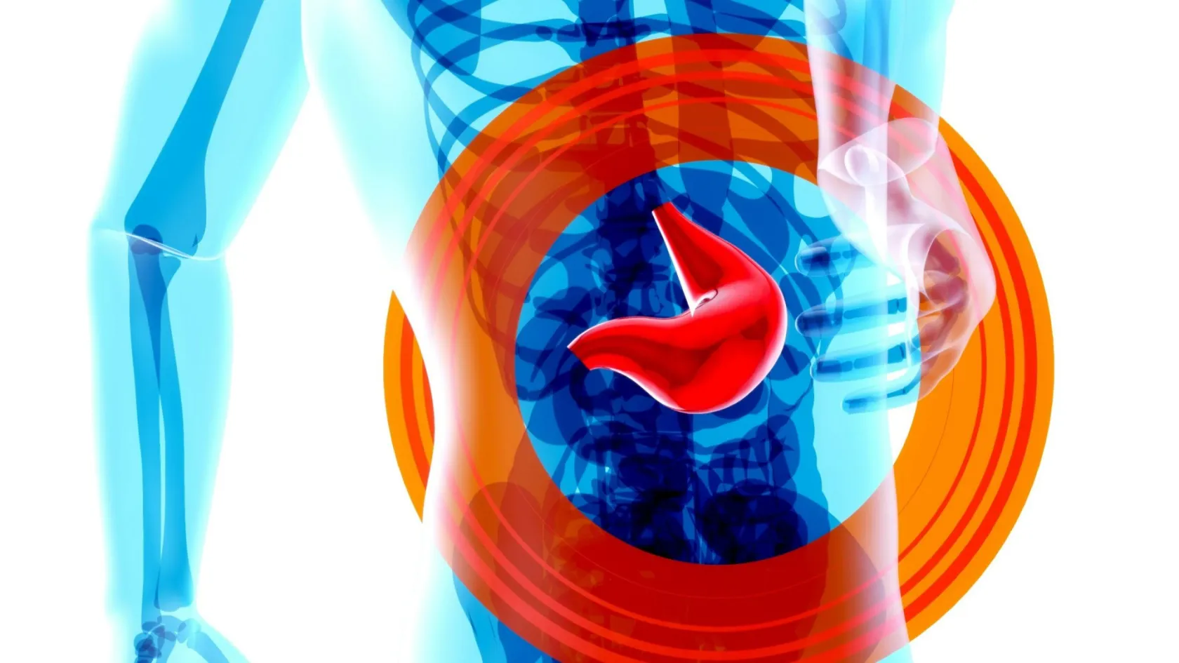
Hepatectomy - Liver Resection
Two drains after a liver resection used to be automatic. Seven randomised trials later, the evidence says most people do better without one. This is what actually happens during a hepatectomy, and what has quietly changed.
About This Department
Two drains after a liver resection used to be automatic. Seven randomised trials later, the pooled evidence says most people do better without one. That is one of several things about this operation that changed quietly while the descriptions on the internet stayed where they were, and it is a reasonable place to start, because a patient who has been told they need a hepatectomy wants to know what will actually be done to them and what the days afterwards look like. This page is about the operation itself rather than about choosing it.
Free consultation
Send the scan files and the clotting and liver blood tests
What a surgeon needs to plan a resection is the imaging itself, because the operation is designed around which vessels the lesion touches. Send the CT or MRI as files, the bilirubin, albumin, INR and platelet count, hepatitis results if you have them, any biopsy report, and a list of your medicines including anything that thins the blood. What comes back is a description of the operation that would be proposed, how long it would take, whether intensive care is planned, and how many nights on the ward to expect. The review costs nothing and carries no obligation.
The reasons it is done
Cancer accounts for most hepatectomies, and a good deal of the surrounding literature assumes that is the only reason. Not so. A significant minority of these operations are done on people who do not have cancer at all, and the conversation with those patients is a different one, because the operation has to justify itself against simply leaving the problem alone.
Hepatocellular adenoma is the clearest example. These behave benignly, occur mostly in women, carry an association with the contraceptive pill, and come out when they grow past about 5 cm or when they turn up in a man, because of the risks of bleeding and of malignant change. A large haemangioma is the opposite case and gets left alone, since it is harmless, and surgery is reserved for one causing genuine symptoms or growing rapidly. Focal nodular hyperplasia is almost never removed once the diagnosis is secure. Then the reasons that have nothing to do with tumours at all. A hydatid cyst that has failed medical treatment. Stones and strictures in the ducts inside one part of the liver, causing repeated infections that will not settle. Trauma, where a torn liver has to be dealt with. Caroli disease affecting one lobe. And living donation, where a healthy person gives part of their liver to somebody else and undergoes precisely this operation with no benefit to themselves.
Technical steps below stay the same in all of these. What changes is the amount of risk a surgeon and a patient will accept when the alternative is simply leaving the problem alone.
The fortnight before
Most of what determines how this operation goes has already happened by the time you reach the anaesthetic room.
Blood-thinning medication is stopped on a schedule that depends on which drug it is, and this is the single item patients most reliably get wrong when they are organising themselves from another country. Clopidogrel and the newer oral anticoagulants have different windows, aspirin is frequently continued, and the instruction has to come from the team doing the operation, never from a general rule. Diabetes medication gets adjusted. Iron goes in where the haemoglobin is low, since arriving anaemic for an operation with a real transfusion risk is a fixable problem that too often is not fixed. Stopping smoking matters more here than most patients expect, because the commonest complication in the first week is not in the abdomen at all. The base of the right lung collapses under a painful upper abdominal incision, and smokers get it far more. Aim for four weeks, and take any period over none. Alcohol stops completely, and where the liver is already damaged that is not a suggestion.
Nutrition gets attention that surprises people. Weight loss and a low albumin before an operation predict a poor recovery after it, and units running modern pathways will spend two weeks building somebody up instead of operating on schedule. Physiotherapy assessment and breathing exercises are taught before the operation, while you can still concentrate, because a patient who has already practised is a patient who can do it while sore. Nothing dramatic happens the night before. No bowel preparation, which is not needed for liver surgery. Clear fluids until a few hours before, and in most units a carbohydrate drink instead of a long fast. Blood gets cross-matched and held. You will meet the anaesthetist, and it is worth asking them directly about the epidural or nerve block, because how the pain is managed decides how early you walk and how well your lungs behave.
The operation, in order
Three to six hours, depending on how much liver is coming out and whether the approach is open or keyhole. What follows sets out the sequence for an open resection, since that is the version most patients are picturing when they ask what happens to them.
Anaesthetic and lines
General anaesthetic, a breathing tube, a large drip in the neck for measuring pressures and giving fluid quickly, an arterial line in the wrist reading blood pressure beat by beat, and a urinary catheter. An epidural or a block in the abdominal wall goes in for pain relief. All of this takes 45 minutes to an hour before anything surgical begins.
Opening, and looking properly
An incision runs across the upper abdomen, sometimes with a short extension upwards, and keyhole surgery replaces it with four or five small ports and one slightly larger opening to get the specimen out. The surgeon then frees the liver from its attachments and puts an ultrasound probe directly onto it. That scan finds lesions no preoperative imaging saw and shows exactly where the veins run, and in a meaningful minority of operations it changes the plan on the spot, which is one reason a surgeon will describe what they intend to do as an intention.
Controlling the inflow
Surgeons find and tie off the artery and the portal vein branch feeding the part being removed. That side changes colour at once. The line where the colour changes marks the plane to cut along. A soft tape goes around the whole inflow too, ready to squeeze shut.
Dividing the liver
Dividing the liver goes slowly and takes most of the operating time. The surgeon breaks down liver tissue while leaving the vessels and ducts running through it standing, then clips, ties or seals each one before dividing it, working with an ultrasonic aspirator, a stapler, energy devices or a crushing clamp, and the choice between those instruments comes down largely to what the surgeon was trained on and not to any proven superiority of one over another. The specimen comes out in one piece.
Checking the cut surface, and closing
Somebody inspects the raw surface under pressure, looking for bleeding and for bile. Some units run a leak test, pushing fluid or dye up the bile duct and watching where it appears, which is the only way to find a small duct that has been divided without anybody noticing. Anything found gets stitched. A haemostatic material may be laid on the surface. Then the abdomen is closed in layers, the epidural is checked before you are woken, and the question of whether a drain is left behind is settled in the theatre by the surgeon looking at the surface, which is a decision that has changed a good deal and is covered below.
The scan that gets done in the room
Intraoperative ultrasound remains the step patients never hear about and surgeons would least like to give up. A probe placed directly on the liver, with no skin, fat or gas between it and the tissue, resolves detail that no scan taken through the abdominal wall can match. It finds small deposits that were invisible beforehand, it shows precisely where each vein runs relative to the planned line of division, and it confirms whether a lesion sitting near a major vessel can be taken with a clear margin. In a meaningful minority of operations it changes what is done, which is why the plan you consented to is described as a plan.
Where the operation is done through keyhole or robotic ports, every one of those steps still happens. The instruments differ and the incision differs.
How the bleeding is controlled
Roughly a quarter of everything the heart pumps goes to the liver, through two separate systems, and it drains into the largest vein in the body. Blood loss during transection is the strongest single predictor of how the rest of the recovery goes, so most of the craft of this operation is about preventing it rather than treating it.
Two techniques do most of the work, and both have been tested more rigorously than patients would guess.
Clamping the inflow, and how long for
Squeezing the whole inflow shut for short spells while cutting is called the Pringle manoeuvre, and the worry has always been that starving the liver of blood and then restoring it does damage of its own, which would be an odd price to pay for a technique whose only purpose is to keep the surgical field dry. Combining two randomised trials and following 176 patients with liver cancer, the group in whom intermittent clamping was used had five year overall survival of 72.1 percent against 58.1 percent in the group where it was not, and clamping remained favourable on multivariable analysis. The benefit was clearest in cirrhotic patients, and the useful clamp interval was 16 to 30 minutes. A separate randomised trial of 270 patients found that a single dose of dexamethasone before the clamp went on reduced liver enzyme release, bilirubin, inflammatory markers, major complications and length of stay.
The second technique keeps the pressure in the veins low while the liver is being cut, so that the cut surface oozes less. Anaesthetists have achieved that for decades by restricting fluid, and it works. It also makes anaesthetists uneasy, because a dry patient can become unstable.
That assumption has now been tested for keyhole and robotic surgery, where the pressure of the gas in the abdomen already suppresses bleeding. Randomising 112 patients to low venous pressure or no manipulation of it at all, investigators found the same blood loss in both groups, the same complications, and less haemodynamic instability in the group left alone. For minimally invasive resections, the old rule appears not to apply, and units are dropping it.
The drain question
Almost every patient description of this operation mentions two tubes coming out of the abdomen, one under the diaphragm and one below the liver, put there to carry away blood and bile. The reasoning sounds obvious. A raw liver surface weeps, so give the fluid somewhere to go and you will detect a leak early and prevent a collection.
Tested properly, that reasoning does not survive.
Seven randomised trials, and 1,064 patients
Pooling every randomised comparison of drainage against no drainage after liver resection, patients who had a drain had significantly more overall complications, with a relative risk of 1.37, and more than twice the rate of wound-related complications at a relative risk of 2.29. Against that, there was no significant reduction in bile leak, no reduction in intra-abdominal collections, no reduction in collections that needed a radiologist to drain them, and no difference in how long anybody stayed in hospital. A separate analysis covering 5,050 patients undergoing major resections found the bile leak rate higher in the drained group, more patients needing an interventional drain afterwards, and a hospital stay a day longer. The authors of the randomised pooling put it plainly, recommending against routine drainage after an uncomplicated hepatectomy.
A drain still earns its place in specific situations, and all of them are judgements made in the theatre and not decisions taken in advance.
This table scrolls sideways on a narrow screen. Swipe or drag to see every column.
| Situation | Why a drain is reasonable there |
|---|---|
| Bile duct joined to bowel | A new join between duct and intestine can leak, and the authors of the pooled analysis name this as the exception to their own recommendation. |
| Bile seen at the end of the operation | A surface that is still weeping bile after everything visible has been stitched is a different proposition from a dry one. |
| Very large cut surface | Extended resections and complex two-sided clearances leave far more raw area than a wedge does. |
| Cirrhosis with ascites | A liver that already leaks fluid into the abdomen behaves differently after surgery, and the judgement here is individual. |
What none of those situations describes is putting a drain in every patient because that is the routine.
So it is fair to ask before your operation. Not to argue with the answer, but because a surgeon who can say why they do or do not drain, and can name the evidence, is telling you something about how the rest of your care will be decided.
Waking up, and what is attached
You wake in recovery and go from there to intensive care or a high dependency unit for the first night, sometimes two after a major resection. That reflects monitoring rather than any sign that something is wrong, and families should be told in advance, because seeing somebody in intensive care after a planned operation frightens people who were not told to expect it.
This table scrolls sideways on a narrow screen. Swipe or drag to see every column.
| What it is | What it is for | When it comes out |
|---|---|---|
| Neck line | Measuring pressures, giving fluid and drugs quickly, taking blood without further needles. | Day two or three, once you are drinking and stable. |
| Wrist line | Continuous blood pressure and frequent blood gases during and after the operation. | Usually when you leave intensive care. |
| Urinary catheter | Measuring urine output hour by hour, which is one of the early signs of trouble. | Day one or two, or when the epidural comes out. |
| Epidural or block | Pain relief that lets you breathe deeply and walk, which is the whole point of it. | Day two to four, replaced by tablets. |
| Abdominal drain, if used | Carrying away fluid where the surgeon judged it necessary, rather than as a routine. | When the volume falls and the fluid is clear of bile. |
A nasogastric tube is no longer routine and comes out at the end of the operation where one was used at all. The oxygen mask stays for the first day or so.
Day by day on the ward
Modern liver units run enhanced recovery pathways, and the practical meaning of that phrase is that almost nothing is withheld from you, since you drink on the day of surgery, eat the following day, and are expected out of bed and walking early. Resting in bed is not part of the treatment. Day one you sit out and walk a short distance, take sips and then more, and start the breathing exercises you practised beforehand. Day two you move to the ward, walk further, and the catheter and the neck line come out. Day three the epidural is weaned onto tablets and you are eating normally, and the blood tests taken every morning start to tell a clear story. Days four and five the bilirubin and clotting are settling, you are walking the corridor several times, and the conversation turns to discharge. Days five to eight, depending on the extent of the resection and on whether it was open or keyhole, you go home.
Patients understand those daily blood tests least and should ask about them most. Bilirubin and the clotting time are how the remaining liver reports on itself, the pattern matters far more than any single value, and both of them rise for the first day or two after a major resection before falling away. A bilirubin that keeps climbing on day three and four, with clotting that stays deranged, is the pattern the team is looking for, and finding it early is why blood is taken every morning rather than every other day.
Pain bites for the first few days and then fades faster than most people expect. You feel it at the incision and often at the tip of the right shoulder, which is referred pain from the diaphragm and alarms patients who were not warned about it.
What the team is watching for
Four things, in the order they tend to appear.
In the first days
Bleeding declares itself in the first hours, through a falling blood pressure, a rising pulse, a haemoglobin that drops on consecutive tests, or blood in a drain where one was placed. It happens rarely, and it is treated by going back to theatre or, occasionally, by a radiologist blocking the vessel.
Chest problems arrive next, around day two or three, as the base of the right lung collapses under a painful incision and then becomes infected. Chest trouble outranks everything else as the commonest complication of this operation, and it is also the one most influenced by things within your control, meaning stopping smoking, doing the breathing exercises, taking the pain relief so that you can breathe deeply, and getting out of bed.
From the end of the first week
Bile leak appears from day three to the end of the second week. Bile escaping from small ducts on the cut surface collects, causing pain, fever and a raised inflammatory count, and it is confirmed on a scan and treated by a radiologist placing a drain into the collection. Most settle over days to weeks. A leak from a major duct needs an endoscopic stent or an operation, and it is rare.
Liver failure sits behind everything else in the assessment, and it shows itself as jaundice that deepens rather than clears, clotting that will not correct, fluid gathering in the abdomen and confusion. After a properly assessed resection it is uncommon. The point of measuring the remaining volume beforehand is precisely to keep it that way.
Beyond those four, the ordinary hazards of major abdominal surgery apply, meaning wound infection, clots in the legs or lungs, and a hernia at the incision months later.
The first three months
Tiredness dominates and it surprises people who braced themselves for pain. A liver rebuilding itself is doing metabolic work, and four to six weeks of needing an afternoon sleep is normal and not a sign of a problem.
- Walk every day and build the distance instead of resting until you feel better, because the deconditioning after major surgery is real and is reversed by movement.
- No alcohol at all until you are told otherwise, and no paracetamol beyond the written dose you were given, since the remaining liver is metabolising everything through less tissue than before.
- Eat little and often. Appetite returns unevenly and a full meal is difficult for several weeks.
- Nothing heavy for six weeks and no strenuous abdominal work for three months, which is what protects the incision from becoming a hernia.
- Have the blood tests your team asks for, at the intervals they ask for them, and send the results back rather than filing them.
Weeks six to twelve
The rule for getting back behind the wheel is not a date. The test is whether you could stamp on a brake pedal, hard, without flinching or hesitating, and after an open incision that takes longer to arrive than after keyhole ports. Office work tends to become possible around the same time. Anything physical waits until the three month mark.
Your liver runs well ahead of you. Most of the volume returns within three months, and a scan a year later looks nothing like the one taken before your operation, which is a fact your own radiologist needs telling before they report it.
How long you stay
Three to four weeks in the country for a straightforward resection, and the arithmetic is easier to trust once you have seen it.
Here is where the days go.
- Four to six days of assessment, covering the specialist liver imaging and volumetry, the full liver function panel, hepatitis screening, anaesthetic review and the multidisciplinary discussion.
- One day for the operation, with the first night in intensive care or high dependency.
- Four to seven further nights on the ward after a keyhole resection, or seven to twelve after an open or extended one.
- A week to ten days nearby after discharge, covering the wound check, the pathology discussion and the blood tests that confirm the remaining liver is behaving.
Flying gets cleared at around three weeks, and later after a major resection or where a drain is still in place. What has to be true first is a healed wound, a bilirubin that is normal or clearly falling on consecutive tests, no fever, no undrained collection, and a written plan for clot prevention on the flight.
Resist the temptation to leave at day ten because you feel well. Bile leak, the complication most likely to send an international patient back to a hospital that has never seen their operation, appears most often in the second week, and feeling well on day ten predicts very little about day fourteen.
What drives the cost
One word, hepatectomy, covers a wedge taken through keyhole ports in ninety minutes and an extended resection with vascular reconstruction that occupies a theatre for most of a day. Seven things move the total.
- How much liver is coming out, since theatre time, staffing and the blood held on standby all scale with it.
- The approach, because robotic and laparoscopic platforms carry instrument costs per case and open surgery does not.
- Which transection device is used, and how many staplers or sealing cartridges the operation consumes.
- Whether intensive care is planned for one night or for several, which after theatre time is normally the largest single variable in a quotation.
- Blood products, held ready for every case and transfused in a minority.
- Whether a vessel or bile duct has to be reconstructed, which lengthens everything downstream.
- The pathology on the specimen, including any molecular testing that guides treatment afterwards.
Your own condition moves it as much as the operation does, through cirrhosis, diabetes, heart and lung disease, obesity, anaemia, blood thinning medication, previous abdominal surgery and poor nutrition, each of which raises the chance of a longer stay and of care that nobody budgeted for.
A published package for this operation typically buys you the airport pickup, the tests done before admission, the operating fee for surgeon and anaesthetist, a stated number of theatre and ward days, translation, hotel nights for you and one companion, and the appointments before departure. Sitting outside it, almost always, are the air tickets, travel insurance, any night beyond the stated number, unplanned critical care, blood products past an allowance, whatever a complication costs to treat, and every form of drug therapy. Read the exclusions before the inclusions, because that is where a quotation either holds up or falls apart. Five questions turn a headline into a quotation. Which resection does the figure assume, named by segments. How many intensive care nights are inside it, and what an additional night costs. Are blood products included or charged separately. What happens financially if a bile leak keeps you here an extra fortnight and needs a radiologist to place a drain. And whether the specimen pathology, molecular testing included, sits inside the figure.
No quotation means anything until a surgeon has read your imaging and named the operation. That review costs nothing.
Follow up once you are home
Whoever operated on you should hand over a written plan and not merely a discharge letter, and the two are not the same document. What you need is the operative note naming the segments removed and describing what was reconstructed, the pathology report in full, the volumetry, the imaging on disc, the blood test schedule with the actual intervals, the wound care instructions, and the name and contact details of somebody here who will answer a message. Ask for all of it in English, collect it before you leave the building, and check that the pathology is attached and not merely referred to, because a summary that says the results were discussed with the patient is worth nothing to a doctor in another country trying to plan what happens next.
Blood tests continue after you fly. Liver function and clotting are checked at intervals your team sets, more closely in the first month, and the results have to travel back to the surgeon who operated, instead of sitting in a folder at home. A bilirubin drifting upward six weeks later means something, and it means it only to somebody who knows what was taken out. Watch the incision for months rather than weeks. An incisional hernia after upper abdominal surgery typically appears between six months and two years as a soft bulge that becomes obvious when you cough or stand, and it is repairable, but the repair is easier when it is small. Report a new bulge instead of waiting to mention it at an annual appointment.
Contact the team here directly for yellowing of the eyes or skin, a temperature, pain in the abdomen that worsens instead of settling, a swollen abdomen, unusual drowsiness or confusion, or a wound that opens or discharges. A message with a photograph answers most questions within hours, and the ones it cannot answer get you sent somewhere local with a clear instruction rather than a guess.
Frequently asked questions about hepatectomy
How long does a hepatectomy take?
Will I have drains after the operation?
Why will I be in intensive care after a planned operation?
Why does my right shoulder hurt after liver surgery?
How long do I need to stay abroad, and when can I fly?
Is a hepatectomy only done for cancer?
References
- Hajibandeh S, Hajibandeh S, Raza SS, Bartlett D, Dasari BVM, Sutcliffe RP. Abdominal drainage is contraindicated after uncomplicated hepatectomy, results of a meta-analysis of randomized controlled trials. Surgery. 2023;173(2):401-411.
- Dezfouli SA, Unal UK, Ghamarnejad O, et al. Systematic review and meta-analysis of the efficacy of prophylactic abdominal drainage in major liver resections. Scientific Reports. 2021;11(1):3095.
- Lee KF, Chong CCN, Cheung SYS, et al. Impact of intermittent Pringle maneuver on long-term survival after hepatectomy for hepatocellular carcinoma, result from two combined randomized controlled trials. World Journal of Surgery. 2019;43(12):3101-3109.
- Huang Y, Xu L, Wang N, et al. Preoperative dexamethasone administration in hepatectomy of 25-min intermittent Pringle's maneuver for hepatocellular carcinoma, a randomized controlled trial. International Journal of Surgery. 2023;109(11):3354-3364.
- Teoule P, Dunker N, Debatin J, et al. Reduction of central venous pressure in elective robotic and laparoscopic liver resection, the PRESSURE trial. Annals of Surgery. 2025;282(2):210-218.
Editor's note
Written by the Biruni Hospital medical editorial team. Reviewed by Prof. Dr. Özcan YILDIZ, Medical Oncology.
Medically reviewed by

Prof. Dr. Özcan YILDIZ
Medical Oncology
Related Treatments
View All-
Bilateral Mastectomy
Whether you carry a BRCA mutation, have cancer in one breast or in both, bilateral mastectomy removes both breasts in one operation, and how much it helps depends on which of those three paths brought you to it.
-

Breast Conserving - Lumpectomy Surgery
Keeping the breast has never been the less thorough choice, and the largest recent studies now point the other way. This page explains who can have breast conserving surgery, what the margin report decides, and how long a woman traveling for it needs to stay.
-

Cytoreductive - Tumor Reduction Surgery
Patients cleared of every visible deposit lived 30 months in one gastric series. Those left with anything at all lived 7.3 months, however little remained.
-
Debulking - Ovarian Cancer Tumor Reduction
Two women with the same diagnosis on the same morning can get operations that share almost nothing. What decides it is who measured the disease first.
-
Debulking Surgery
Surgeons removed a median of 57 lymph nodes from women already cleared of visible disease. It bought no extra months, and tripled deaths within 60 days.