
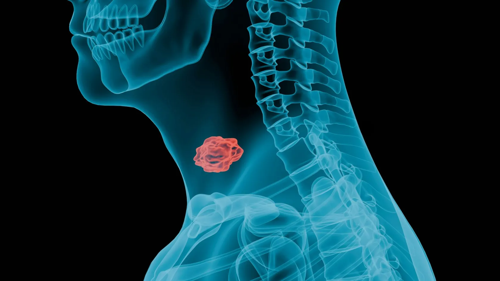
Head and Neck Cancer Surgery
The operation that removes the cancer is the shorter half of the day. This is what the rebuild involves, and why it decides how you speak and eat for the rest of your life.
About This Department
Removing the cancer is the shorter half of the day. Head and neck surgery takes tissue out of the places you use to talk, to eat and to show a face to other people, so the operation that puts something back is usually the longer one and it is the one that decides how the rest of your life goes. Any page that describes the resection and stops has told you about half of what happens to you. This one gives the rebuild the space it actually occupies in the operating theatre.
Free consultation
Send the biopsy, the scans and a photograph of your teeth
The last of those surprises people and it should not. If radiotherapy follows the surgery, dental work has to happen before it and not after, so the state of your mouth changes the order of your entire treatment. Send the biopsy report with the p16 or HPV result if one was done, the CT or MRI of the head and neck and the chest, any PET report, a list of what you take, and a note of whether you smoke, drink, or have had radiotherapy to this area before. What comes back is an opinion on which operation fits, whether a flap is likely, and what the first three months would realistically look like. The review costs nothing and carries no obligation.
Not one disease
Head and neck cancer is an administrative category more than a diagnosis. It gathers together tumours of the mouth, the throat behind the mouth, the voice box, the space behind the nose, the sinuses and the salivary glands, and those tumours differ from one another in cause, in behaviour, in what the operation involves and in how likely you are to be cured, which means that a survival figure you find online for head and neck cancer as a whole describes a population you are almost certainly not part of, and the number that applies to you comes from the site, the stage, the p16 result and your own general health rather than from a headline. A cancer of the tonsil driven by a virus and a cancer of the tongue driven by forty years of smoking sit in the same chapter of the same textbook and share almost nothing else.
So the first useful thing anybody can tell you is where yours is.
This table scrolls sideways on a narrow screen. Swipe or drag to see every column.
| Site | What the operation tends to involve |
|---|---|
| Oral cavity | Tongue, floor of mouth, gum, cheek lining. Surgery leads here in nearly every case. Small tumours come out through the mouth and close directly. Larger ones need a flap, and where the jawbone is involved a segment of it comes out and is rebuilt with bone from the leg. |
| Oropharynx | Tonsil and base of tongue, and the site where virus-driven cancer now dominates. Surgery and radiotherapy are both reasonable first treatments, which is unusual and is covered in the next section. Robotic access through the mouth has replaced splitting the jaw. |
| Larynx and hypopharynx | Early tumours are removed with a laser through the mouth and the voice is largely kept. Advanced ones force the hardest conversation in this field, between chemoradiotherapy to preserve the voice box and removing it outright with a permanent breathing hole in the neck. |
| Nasopharynx | The exception that proves the rule, treated with chemoradiotherapy first because it responds extremely well and sits somewhere surgery reaches badly. An operation here is generally a salvage procedure for disease that came back. |
| Salivary glands | Surgery leads, and the parotid gland has the facial nerve running straight through it, which makes preserving the movement of your face the central technical problem of the operation. |
| Sinuses and skull base | Rare, and increasingly reached endoscopically through the nostril by a joint team of head and neck surgeons and neurosurgeons rather than through the face. |
Two words on your pathology report change the picture more than almost anything else. If a tumour of the tonsil or the base of the tongue is p16 positive, meaning it is driven by human papillomavirus, it behaves as a distinct disease with markedly better outcomes than the same-looking tumour in a lifelong smoker, and it is staged on a separate system for exactly that reason. Look for that line. If it is absent from a report on an oropharyngeal tumour, ask why.
Surgery or radiotherapy
For most cancers described on this site the question is which operation. Here, for a substantial group of patients, the question is whether an operation is the right instrument at all, because radiotherapy can cure the same tumour without cutting anything. That comparison has been run properly, and the result is not the one a surgical department would choose to advertise.
The trial that compared them head to head
ORATOR randomised 68 patients with early oropharyngeal cancer to radiotherapy or to robotic surgery through the mouth with neck dissection, and measured swallowing a year later on a validated questionnaire. Radiotherapy scored 86.9 and surgery 80.1, a difference that reached statistical significance while falling short of the ten-point gap the investigators had defined as clinically meaningful. The pattern of side effects diverged sharply. Radiotherapy produced more hearing loss, tinnitus and low white cell counts, surgery produced far more jaw stiffness, and there was one death from bleeding after surgery. The authors concluded that patients should be told about both options.
Read that carefully, because it is easy to misread in either direction. Robotic surgery did not turn out to be the swallowing-sparing alternative it was widely assumed to be. It also did not turn out to be worse in any way that a patient would notice, and it avoids six weeks of daily radiotherapy along with the hearing loss and the dry mouth that come with it.
What tips the decision is something other than the average.
- Surgery appeals when the tumour is small and reachable through the mouth, when avoiding radiotherapy altogether looks plausible, and when you are young enough that decades of dry mouth are a real cost.
- Radiotherapy appeals when closing the defect would need a flap, when the neck disease is extensive enough that radiation is coming regardless, and when a long anaesthetic is a bad idea.
- Getting both is what everybody is trying to avoid, and it happens when the pathology after surgery turns up features that demand radiation.
So the discussion before the operation has to include a straight estimate of how likely that third outcome is for you, and a surgeon who cannot put a rough number on it has not thought about your case in enough detail.
Getting it all out
A margin is the rim of normal tissue around the tumour. In the mouth and throat there is very little room to take a generous one, since a centimetre in any direction is the difference between a tongue that works and a tongue that does not, and the surgeon is negotiating with anatomy the whole way round.
Frozen section is how that negotiation is settled during the operation, with a pathologist examining tissue while you are still asleep so that more can be taken if the edge is not clear. There are two ways of doing it and the difference between them is not a technicality.
Sampling the specimen against sampling the hole
A meta-analysis of nine studies covering 1,240 patients with oral cavity cancer compared taking the frozen section from the removed specimen itself against taking it from the bed the tumour came out of. The specimen-driven approach led to more re-resection during the operation, a significantly lower rate of positive margins on the final pathology, and less local recurrence. Overall recurrence and survival did not differ significantly between the two methods.
In plain terms, checking the piece you removed finds problems that checking the empty space misses, and finding them while the patient is still on the table is the only moment when fixing them is easy. Which method a unit uses is a fair thing to ask about. A positive margin on the final report is not a catastrophe and it is not nothing. It generally means radiotherapy is added, sometimes with chemotherapy, and occasionally it means going back to take more. Perineural invasion, meaning tumour tracking along a nerve, and extranodal extension, meaning disease that has burst out of a lymph node, are the two other findings on that report that most often change the plan after the operation, and both of them are reasons to read the pathology yourself rather than accept a summary of it over the phone, since the words on the page determine what happens for the next six months and a paraphrase loses exactly the detail that decides it.
What happens to the neck
Almost every operation for a mouth or throat cancer includes an operation on the neck, and patients frequently arrive not knowing this. Scans miss small deposits in lymph nodes reliably enough that a normal-looking neck cannot be trusted.
Whether to operate on a neck that looks clear was argued about for decades until somebody settled it.
Operating early against waiting to see
A randomised trial of 500 patients with early lateralised oral cancers and no visible nodal disease compared clearing the neck at the time of the primary operation against watching and clearing it only if disease appeared later. At three years, overall survival was 80.0 percent in the group operated on early and 67.5 percent in the group that waited, with a hazard ratio for death of 0.64. Disease-free survival was 69.5 percent against 45.9 percent. Serious complications occurred in 6.6 percent and 3.6 percent of the two groups respectively.
Twelve percentage points of survival is about as decisive as surgical trials get, and it explains why the neck is now cleared routinely rather than watched. Modern neck dissection is selective, meaning the surgeon removes the specific groups of nodes that this particular tumour drains into and leaves alone the muscle, the large vein and the nerve that older radical operations sacrificed. The nerve that lifts your shoulder runs through the field and is preserved, though it is frequently bruised, which produces a stiff and aching shoulder for some months and responds well to physiotherapy started early rather than started once the stiffness has set in, which is the mistake most people make with it. Shoulder exercises begin in the first fortnight and continue for months, and they are boring in a way that makes them easy to abandon. Numbness of the ear lobe and the skin of the neck is expected and permanent in patches.
For very small oral cancers, sentinel node biopsy offers a smaller version of the same thing, tracing the first node the tumour drains into and clearing the rest of the neck only if that node is involved. Whether that is offered to you depends on the size and site of the tumour and on whether the unit performs enough of them to be good at it, and that is a question to put directly.
The second operation
Here is the part most pages skip. When a defect is too large to close directly, tissue is taken from somewhere else on your body, moved to the head with its artery and vein, and plumbed into vessels in the neck under a microscope. That is a free flap, and it is a second full operation performed by a second team, frequently at the same time as the first so that the total anaesthetic stays reasonable.
This table scrolls sideways on a narrow screen. Swipe or drag to see every column.
| Donor site | Used for, and what it costs you where it came from |
|---|---|
| Forearm | Thin, pliable skin for the tongue, floor of mouth or cheek lining, with long reliable vessels. Leaves a skin graft on the inner forearm that is visible for life and a patch of numbness over the thumb. |
| Outer thigh | A thicker sheet for larger soft tissue defects, and the workhorse where bulk is needed. The donor wound closes directly, leaving a long scar down the thigh and no functional loss worth naming. |
| Fibula | Bone for rebuilding the jaw, long enough to be cut into segments and shaped into a mandible, and strong enough to take dental implants later. Leaves a scar down the outer calf and a leg that takes a fortnight to walk confidently. |
| Shoulder blade region | Bone and soft tissue together on one blood supply, useful for complex three-dimensional defects of the midface and jaw. Positioning on the operating table is harder, so the two teams cannot always work at the same time. |
Choosing where it comes from
Choice comes down to what the defect needs, whether bone is part of it, and what your own vessels look like on a scan of the limb, because roughly one patient in ten has anatomy that rules out the obvious option and has to be found out beforehand rather than in theatre.
Raising and shaping it
While the first team removes the tumour, the second raises the flap with its blood supply intact. Where a jaw is being rebuilt, the fibula is cut into segments and assembled into the curve of a mandible, and this is increasingly planned on a computer beforehand with cutting guides printed to match, which shortens the time the tissue spends without a blood supply.
The join
Under a microscope, an artery a couple of millimetres across is sewn to an artery in the neck and a vein to a vein, with stitches finer than a hair. This is the step everything depends on. It takes as long as it takes, and a surgeon who is rushing it is doing the wrong thing.
Watching it
For the first two or three days a nurse checks the flap every hour, looking at its colour and pricking it to see how it bleeds. Almost every flap that is going to fail declares itself in that window, and almost every one caught early can be saved by going straight back to theatre. It is what the staffing on those nights exists for.
How often the flap fails
A systematic review pooling 45 studies and 9,663 head and neck free flap reconstructions reported flap failure in 2.5 percent, venous clotting in 3.8 percent and a return to theatre in 5.6 percent. The review compared two ways of joining the vein to the neck circulation and found no significant difference between them in any of those outcomes, which is a useful reminder that technique preferences at this level are frequently preferences rather than evidence.
Roughly nineteen flaps in twenty are entirely uneventful. When one does fail the operation is not over and the reconstruction is redone with different tissue, which lengthens everything.
Donor sites deserve more attention than they get, because nobody warns patients about them properly. A forearm flap leaves a skin graft and a scar you will see every day. A fibula flap leaves a leg that needs a fortnight before it walks confidently. Ask what the donor site will look like and how it will feel, and ask before the operation.
Speech, swallowing and the team behind them
Whether you speak clearly and eat normally in a year depends on the surgeon, and it depends nearly as much on people whose names most patients never learn.
Speech and language therapists assess you before the operation, so that afterwards there is something to compare against, and they run the swallowing rehabilitation that decides how quickly the feeding tube comes out. Dietitians keep your weight up during a period when eating is difficult and calories matter more than they ever have. Dentists check your mouth before radiotherapy, because a tooth extracted after radiation heals badly and can lead to the jawbone dying, which is a far worse problem than losing the tooth early would have been. A unit without those three services is not a head and neck unit. Meet the speech therapist before you agree to anything, because the quality of that conversation tells you more about where you have landed than any brochure will, and a therapist who cannot describe what your swallowing rehabilitation will consist of week by week is a warning about the whole department.
There is good news, and it belongs to the smaller operations.
Function five years later, after a small oral cancer
Eighty-one patients whose early oral cancers were removed through the mouth without a flap, with sentinel node biopsy for the neck, were assessed a median of five years afterwards using standardised speech testing and validated swallowing questionnaires. Scores across every assessment landed in the top ten percent of the scales. Tumour size, tumour site, age and nodal stage made no significant difference. The two things that did lower swallowing scores were having gone on to a completion neck dissection, or having had radiotherapy.
Notice what that last sentence identifies. The operation on the tumour was not what cost these patients function. The additional treatment was, which is one more argument for catching these cancers small and one more reason the added radiotherapy question matters so much at the planning stage.
Larger resections are a different conversation and an honest one has to include that some function is permanently changed. What surgery and rehabilitation together are aiming at is a mouth that manages ordinary food and speech that strangers understand on the telephone, and for most patients with reconstructed tongues and jaws that target is reached.
The first week
Patients find the first week harder to picture than the operation itself, so here it is plainly.
- You wake with a tracheostomy if the mouth or throat has been operated on extensively, because swelling makes the airway unreliable for a few days. It comes out again in the great majority of cases. You cannot speak through it initially, and writing or a phone is how you communicate.
- A feeding tube through the nose delivers nutrition while the reconstruction heals and swallowing is unsafe. Uncomfortable, yes. Permanent, no.
- Drains sit in the neck for several days, and the flap is checked hourly around the clock by somebody who will wake you to do it.
- Sitting up and walking start on day one or two even with all of that attached, because chest infections and clots are the complications that actually threaten people at this stage.
- A speaking valve goes on within the first week, and the tracheostomy comes out shortly after that, once the swelling settles enough for the airway to look after itself.
Swelling of the face and neck peaks around days two to four. It alarms families who were not warned. It settles, and the face you see at three weeks is not the face you saw at three days.
Pain falls short of what people expect after an operation of this size, and the dominant complaints are the tube in the nose, the inability to speak and thirst, in that order, which is not what anybody prepares for. Anyone travelling with you should know all of this before they walk into the room, because their reaction in the first forty-eight hours affects you more than anybody plans for.
What follows the surgery
The pathology report arrives about a week to ten days after the operation, and it decides whether anything else is needed.
When radiotherapy is added
Positive or very close margins, several involved nodes, disease that has broken out of a node, perineural invasion or a deeply invasive primary all point towards radiotherapy afterwards, and extranodal extension or a positive margin brings chemotherapy alongside it. Treatment starts around six weeks after surgery. It then runs daily for six weeks. Waiting longer than about six weeks to start is associated with worse control, which matters enormously if you are travelling and have to arrange it at home.
What radiotherapy costs you
Dry mouth is the lasting one and it is more disruptive than it sounds, affecting taste, sleep, dental health and the pleasure of eating for years. Sore throat, thickened saliva, fatigue and a skin reaction dominate the six weeks themselves, and the fatigue in particular arrives late, deepens through the final fortnight and takes a couple of months after the last session to lift properly. Swallowing needs active work during treatment and afterwards, because muscles that stop being used stiffen, and the therapists will push you to keep swallowing through the worst of it precisely for that reason, which feels unreasonable at the time and is the single intervention that most reliably separates people who eat normally at a year from people who do not. All of which argues that the surgical plan and the radiotherapy plan should be made together, at the same meeting, by people who are both in the room, since a surgeon planning a resection without knowing whether radiation is coming afterwards is planning half an operation.
How long you stay
Three to five weeks in the country for anything involving a flap, and two to three weeks for a smaller resection with neck dissection. These are the longest stays of any operation described on this site, because the reconstruction has to prove itself and the swallowing has to be re-established before you get on an aeroplane.
Assessment runs four to seven days. Examination under anaesthetic with biopsies where the extent is unclear, cross-sectional imaging of the head, neck and chest, a PET scan in selected cases, vessel imaging of the donor site for a flap, dental assessment, speech and swallowing baseline, nutritional assessment and a multidisciplinary meeting. That meeting is the point of the whole week and it should include a radiation oncologist in the room rather than consulted afterwards by letter, since the decision about what happens after the operation shapes what the operation itself should be. The hospital stay runs seven to fourteen days for a flap reconstruction, with the first two or three in a high dependency setting, and three to five days for a smaller operation. Then one to two weeks nearby. That fortnight covers the wound checks, the removal of drains and tubes, the pathology discussion that determines whether radiotherapy is coming, and the first sessions of swallowing therapy with somebody who will still be reachable by video call after you fly. None of that week is idle and very little of it compresses, so book your flights around this timetable and not around an optimistic estimate somebody gave you on the telephone.
When you are cleared to fly
Flying is cleared once the wounds are dry, the tracheostomy is out and closed, the flap is stable and you are taking enough nutrition by mouth or through a tube you can manage yourself. For a straightforward flap that is around two and a half to three weeks. Anybody flying with a tracheostomy still in place needs the airline told in advance, along with equipment and an escort. All of that is arrangeable. None of it is arrangeable the day before.
What drives the cost
Of every operation on this site, this one produces the widest spread, and two patients with the same diagnosis can end up in genuinely different places. Eight things account for most of the spread.
This table scrolls sideways on a narrow screen. Swipe or drag to see every column.
| Driver | Why it moves the figure |
|---|---|
| A free flap | Brings a second surgical team, a much longer theatre session and high dependency nights. This single yes or no moves the total more than everything else on this list combined. |
| Which flap | A fibula reconstruction planned on a computer with printed cutting guides sits well above a forearm flap, and the planning is billed separately by some units. |
| The access method | Robotic, laser through the mouth, endoscopic through the nose, or open. Console time is charged where a robot is used. |
| The neck | One side or both, and how many levels are cleared, which changes theatre time and drain days. |
| Airway and feeding | Whether a tracheostomy and a feeding tube are needed, and how many days each of them stays in. |
| Pathology | Frozen section during the operation, and the extent of the final report including p16 testing and any molecular work. |
| Length of stay | Driven here by the flap and by swallowing rather than by pain, which is why it is longer than patients expect. |
| Teeth and prosthetics | Dental work before radiotherapy, and any obturator or prosthesis made for the mouth afterwards. |
The parts that sit outside the surgical figure
Adjuvant radiotherapy or chemoradiotherapy is a separate course of treatment lasting six weeks, and where you have it is a decision in itself. Speech and swallowing therapy continues for months and is the part left out of a quotation most consistently, even though it is running long after the surgical bill has been settled and it is doing more for your daily life at that point than anything else on the list. Nutritional support, dental restoration and, for some, a voice prosthesis after laryngectomy carry on well past the point where the surgical episode is formally closed. Packages here ordinarily cover the transfers, the assessment week, the operating fees for both teams, the stated hospital and high dependency nights, an interpreter, accommodation and the appointments before you leave. Outside them sit the flights, insurance, additional nights, the treatment of a complication, radiotherapy and everything named in the sentences above it, which taken together can amount to a larger sum than the operation, spread across a longer period and paid in instalments nobody quotes for. Find out early where the radiotherapy is expected to happen, because the answer changes the arithmetic completely and it is the question that goes unasked most consistently until the pathology report has already arrived.
Six questions to ask before you accept a figure
Does the figure assume a flap, and what happens to it if the frozen section forces a larger resection than planned. How many hospital nights and how many high dependency nights are included, and what an additional night costs. Is a return to theatre for flap trouble covered. Are speech therapy and dietetic input included for the whole of the stay rather than as a single visit. Is the dental assessment included, and any extraction that follows from it. And is radiotherapy quoted at all, or assumed to be happening somewhere else.
Nothing here means anything until a surgeon has seen your scans and your pathology. That review is free, and it is the only thing that turns a website number into a number about you.
Once you are home
Follow-up here is closer than for most cancers, because recurrences in this region are found by a doctor putting a scope through the nose and looking rather than by a scan. Expect review every one to two months through the first two years, stretching to twice yearly and then annually out to five years, which is a demanding schedule to run from another country and is the single practical detail most worth settling before you travel rather than after. A baseline scan three months after treatment finishes is standard, giving everybody a picture to measure future ones against, and imaging after that is driven by symptoms or by something felt on examination.
Five documents should travel home with you, in English, since your care will be run from them by people who were not in the room.
- The operative note, naming what was removed, which flap was used and which neck levels were cleared.
- The full pathology, with margins in millimetres, perineural invasion, node counts and extranodal extension stated explicitly.
- The multidisciplinary recommendation on radiotherapy, stating plainly the date by which treatment should have started.
- The speech and swallowing assessment, with the exercises you were given.
- The dental report, with anything that needs doing before radiation.
Two things need reporting quickly wherever you are. Any new lump, ulcer that fails to heal within three weeks, persistent pain in one ear or change in voice, all of which are how recurrence announces itself. And jaw pain or exposed bone after radiotherapy, which needs assessing promptly rather than watched. Get in touch here for either, and for anything about the flap, the tube feeding or the swallowing exercises.
Stopping smoking and stopping alcohol change your odds of a second cancer more than any drug anybody can offer you. That is a blunt thing to put on a hospital page. It is also the truest sentence in this article, and the one most likely to still matter in ten years.
Frequently asked questions about head and neck cancer surgery
Will I be able to speak and eat normally afterwards?
Is robotic surgery better than radiotherapy for tonsil and tongue base cancer?
Why operate on my neck when the scans are clear?
What happens if the flap fails?
Will I need a tracheostomy and a feeding tube?
How long do I need to stay abroad, and when can I fly?
References
- D'Cruz AK, Vaish R, Kapre N, et al. Elective versus therapeutic neck dissection in node-negative oral cancer. The New England Journal of Medicine. 2015;373(6):521-529.
- Nichols AC, Theurer J, Prisman E, et al. Radiotherapy versus transoral robotic surgery and neck dissection for oropharyngeal squamous cell carcinoma, ORATOR, an open-label, phase 2, randomised trial. The Lancet Oncology. 2019;20(10):1349-1359.
- Caraway J, Millay D, Zarrella D, et al. Intraoperative margin assessment methods in oral cavity squamous cell carcinoma, a systematic review and meta-analysis. Otolaryngology, Head and Neck Surgery. 2026;174(4):881-891.
- Costantino A, Uralov D, Festa BM, et al. End-to-side venous anastomosis in head and neck free flap reconstruction, a systematic review and meta-analysis. Microsurgery. 2025;45(8):e70148.
- Romer CAE, Broglie Daeppen MA, Mueller M, Huber GF, Guesewell S, Stoeckli SJ. Long-term speech and swallowing function after primary resection and sentinel node biopsy for early oral squamous cell carcinoma. Oral Oncology. 2019;89:127-132.
Editor's note
Written by the Biruni Hospital medical editorial team. Reviewed by Prof. Dr. Metin GÜDEN, Radiation Oncology.
Medically reviewed by
Prof. Dr. Metin GÜDEN
Radiation Oncology
Related Treatments
View All-
Bilateral Mastectomy
Whether you carry a BRCA mutation, have cancer in one breast or in both, bilateral mastectomy removes both breasts in one operation, and how much it helps depends on which of those three paths brought you to it.
-
Breast Conserving - Lumpectomy Surgery
Keeping the breast has never been the less thorough choice, and the largest recent studies now point the other way. This page explains who can have breast conserving surgery, what the margin report decides, and how long a woman traveling for it needs to stay.
-
Cytoreductive - Tumor Reduction Surgery
Patients cleared of every visible deposit lived 30 months in one gastric series. Those left with anything at all lived 7.3 months, however little remained.
-
Debulking - Ovarian Cancer Tumor Reduction
Two women with the same diagnosis on the same morning can get operations that share almost nothing. What decides it is who measured the disease first.
-
Debulking Surgery
Surgeons removed a median of 57 lymph nodes from women already cleared of visible disease. It bought no extra months, and tripled deaths within 60 days.