
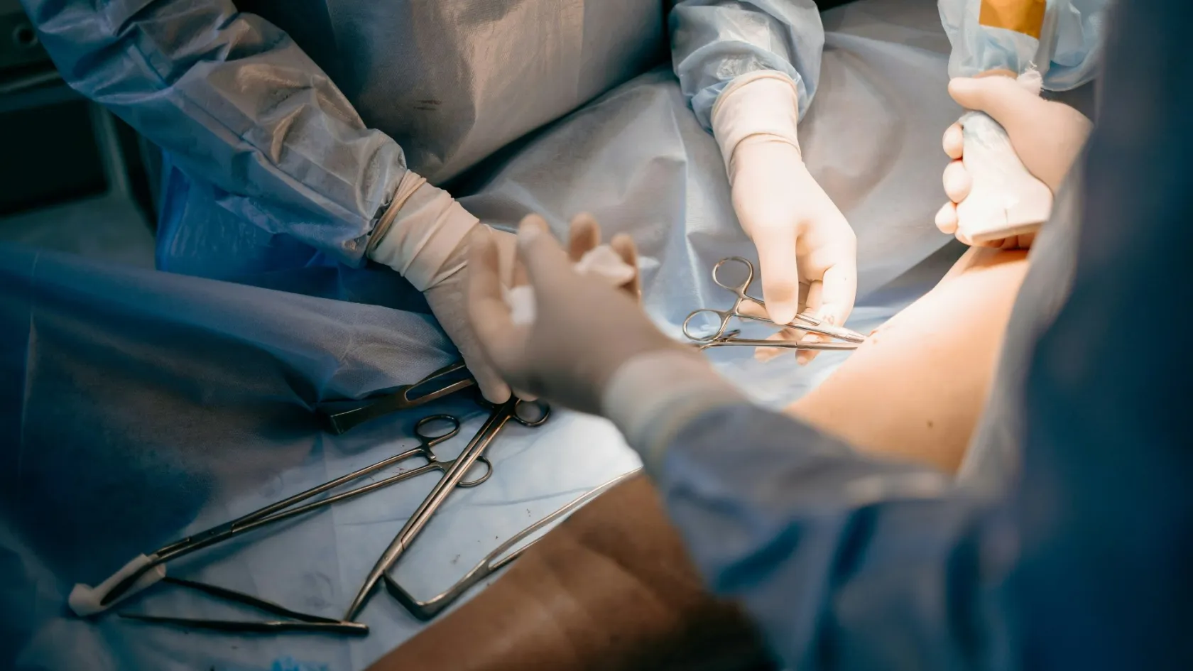
Free Flap Reconstruction
There are two verdicts on a free flap. One arrives in theatre when the clamps come off and the tissue changes colour. The other arrives on the third morning, and only the second one counts.
About This Department
Living tissue is cut away from its blood supply, carried across the body and plumbed back in somewhere else. That single sentence explains everything else about this operation, including why it takes so long, why the nights afterwards are staffed the way they are, and why nobody will tell you it worked until three days have passed. There are two verdicts on a free flap. One arrives in theatre when the clamps come off. The other arrives on the third morning.
Free consultation
Tell us about the vessels, not only the defect
Whether this operation is possible depends on the plumbing at both ends, which means the vessels at the defect and the vessels wherever the tissue will come from. Send the imaging of the affected area, any previous operation notes covering that region, a note of any radiotherapy given to it, and details of smoking, diabetes, blood pressure and any circulation problem in the arms or legs. Add any angiogram or duplex you already have. What comes back is an opinion on whether a free flap is the right answer, which donor site your own anatomy supports, and what the first seventy-two hours will involve. The review costs nothing and carries no obligation.
What makes a flap free
Free here has nothing to do with cost and everything to do with detachment, and a flap is any piece of living tissue moved while its blood supply stays intact. Most flaps stay attached at one edge and swing into position like a door on a hinge, which limits how far they travel and keeps them tethered to where they started. A free flap is cut loose entirely, artery and vein divided, and carried to a site that may be on the other side of the body.
Detachment buys the freedom. Detachment also creates the entire risk.
This table scrolls sideways on a narrow screen. Swipe or drag to see every column.
| Method | How it survives, and what that means for you |
|---|---|
| Skin graft | Has no blood supply of its own and must grow one from the bed underneath. Quick and simple where the bed is healthy, and useless over bare bone, exposed metal or tissue that has been irradiated. |
| Pedicled flap | Stays connected to its original vessels and rotates into place. No microsurgery, shorter operation, no monitoring of an anastomosis afterwards. Restricted to defects within reach, and unavailable when the nearby tissue has itself been damaged. |
| Free flap | Detached completely and reconnected under a microscope, bringing healthy circulation into a place that had none. Reaches anywhere, matches the tissue to the job, and costs a longer operation plus three days of intensive watching. |
Perforator flaps changed the field. Instead of taking a whole muscle to carry the blood vessels, the surgeon traces individual vessels the width of a pencil lead as they pierce through the muscle, dissects them out and takes only skin and fat, leaving the muscle where it was and still working. Modern abdominal breast reconstruction therefore leaves the abdominal muscles in place, and why donor sites recover far better than they did a generation ago.
The clock that starts
From the moment the artery is divided, the tissue in the surgeon's hands has no blood going into it and none coming out. The tissue is alive on borrowed time. Skin and fat tolerate this reasonably well for a couple of hours. Muscle tolerates it considerably worse, which is one of the quieter arguments for perforator flaps over muscle-carrying ones. Everything the team does between dividing the vessels and releasing the clamps is shaped by keeping that interval short, and it is why the tissue is shaped and positioned before the join rather than after it, and why the two surgical teams work simultaneously wherever the anatomy allows. None of this is visible from outside the theatre, and all of it is the difference between a flap that survives comfortably and one that spends its first night struggling. Ask how long your operation is expected to take. A team that has thought about the answer will give you a range rather than a shrug.
When the clamps come off, the flap changes colour within seconds. Watching it happen counts as the first verdict, and it is a good one to be present for.
None of which ends anything. Blood flowing at the end of the operation says the join is open now, and says nothing about whether it will still be open at four in the morning, which is the question the rest of this page is about.
The join
Two vessels get sewn together, each about the width of the lead in a pencil, using thread finer than a human hair and viewed through an operating microscope. Everything else in this operation is preparation for that.
Finding vessels to join to
Recipient vessels get prepared first, and finding good ones is frequently harder than raising the flap, because radiation, previous surgery, infection and arterial disease all leave vessels that look perfectly adequate and bleed poorly. A surgeon who abandons a vessel and goes looking for a better one is doing exactly the right thing.
The artery
Sewn by hand, stitch by stitch, usually eight to twelve of them around the circumference. Arteries are thick-walled and hold their shape, which makes them the more forgiving of the two despite being the one patients worry about.
The vein
Thin-walled, collapsible and slow-flowing, so the vein and not the artery is where most trouble begins. Many units now join it with a coupler, a pair of small rings that lock the vessel ends together, instead of stitching it.
Release, and inset
The clamps come off, the flap fills, and only then is it stitched into its final position, and that insetting is done slowly because a kinked or compressed vessel undoes everything that came before it, and the commonest late disaster in this operation is a perfectly good join strangled by the closure.
Rings against stitches, in eighteen studies
A meta-analysis screened 3,184 articles and pooled eighteen comparative studies of venous joins made with a coupler device against those sewn by hand. Flap loss ran at 1.53 percent with couplers and 2.32 percent with stitches, and clotting at 2.91 against 3.17 percent. The clearest difference was time, at 7.5 minutes for the coupler against 32.2 minutes for hand sewing. In the head and neck subgroup the gap widened, with flap loss of 0.66 percent against 3.08 percent. All eighteen studies were retrospective.
Twenty-five minutes saved on a single join sounds like a convenience. It is also twenty-five minutes less that the tissue spends without blood, which is the reason the difference in outcomes exists at all.
Seventy-two hours of watching
Almost everything that will go wrong with a free flap goes wrong in the first three days, and almost all of it is fixable if somebody notices in time. Those two facts together explain why the monitoring runs as relentlessly as it does. Somebody comes every hour, and for the first day or two that includes every hour of the night. What they are doing looks unglamorous and remains the best test available. They look at the colour. They press the skin and count how long it takes to pink up again. They feel the temperature. Where a paddle of skin is visible they prick it with a needle and watch both what colour of blood appears and how quickly it comes, because bright red after a couple of seconds is a healthy flap while dark blood appearing instantly is a congested one, and no machine yet built reads that distinction as reliably as a nurse who has seen it before.
Machines assist and have not replaced this, and the simplest of them is a handheld probe that finds the pulse in the flap. An implantable probe can be wrapped around the vessel itself and left in place to give a continuous signal, and there has been an argument for years about whether it belongs on the artery or the vein.
Artery or vein, for the implanted probe
A systematic review and meta-analysis pooled seven studies covering 763 flaps, with probes on 527 arteries and 388 veins. Arterial monitoring reduced the risk of a false alarm by 74 percent and the risk of losing the signal altogether by 63 percent. What it did not change was anything that matters to a patient. Take-backs, salvage rates, flap failures, sensitivity and specificity were all statistically indistinguishable between arterial and venous probes, and the authors say so plainly.
Fewer false alarms matters, because a false alarm at three in the morning means an unnecessary trip to theatre. Nobody should be told that a particular probe placement will save their flap.
Your part in all of this is small and it is not nothing, so stay warm, stay hydrated, keep the flap free of pressure, and mention immediately if the area feels different, because patients occasionally notice a change before the chart does.
When it goes wrong
Two things can block, and they look completely different.
Arterial blockage starves the flap, which goes pale, cool and slack, refill after pressing becomes sluggish, and a needle prick produces very little. Venous blockage happens more often and looks far more dramatic. Blood arrives and cannot leave, so the flap turns purple, swells, feels tense, refills instantly and bleeds dark blood the moment it is pricked. Either finding sends you back to theatre, and speed is the whole game. The clot is removed, the join is redone or a different vessel is found altogether, and flaps re-explored quickly do considerably better than flaps re-explored late, which is the entire reason for the hourly checks and the reason nobody waits until morning to see whether it settles. Return to theatre after a free flap is not a sign that something was done badly. The system is working as designed, and knowing that beforehand takes most of the fear out of somebody wheeling you off the ward at midnight. Raise it before the operation, while it is still an abstraction, rather than in the moment.
Where congestion persists and no surgical fix is possible, one very old treatment remains genuinely useful and horrifies almost everybody who is offered it.
What the leech evidence actually shows
A systematic review of medicinal leeching in breast surgery gathered eighteen studies with a combined 28 patients, most of them reconstructive cases. Typical treatment was two leeches at a time, three sessions a day, for three days. Leeching prevented the loss of 75 percent of the tissue transfers it was used on. The complication rate was 81.14 percent, dominated by infection and anaemia. The reviewers conclude that it works and must be used judiciously within the clinical context of the patient.
Both halves of that finding deserve stating, because a leech secretes a powerful anticoagulant while it feeds and keeps the wound bleeding for hours afterwards, which drains a congested flap long enough for new small vessels to grow in. Three flaps in four were saved. Four patients in five had a complication, most often needing antibiotics or a transfusion, and anybody presenting leech therapy as a quaint curiosity without mentioning that second figure is not describing the treatment honestly.
Which flap, from where
Around a dozen donor sites cover the great majority of what is done, and the choice follows from what the defect needs rather than from surgeon preference.
This table scrolls sideways on a narrow screen. Swipe or drag to see every column.
| What is needed | Where it comes from, and why |
|---|---|
| Thin, pliable lining | The forearm. Long, generous vessels and skin thin enough to line a mouth or resurface a tongue, at the price of a graft on the arm that stays visible. |
| Bulk and coverage | The outer thigh. The workhorse of modern reconstruction, adjustable in thickness, usually closed directly, and hidden under clothing. |
| Soft tissue for a breast | The lower abdomen. Skin and fat taken on perforating vessels while the abdominal muscles stay in place, giving a reconstruction that behaves like the tissue it replaced. |
| Bone for a jaw | The fibula. Long enough to be cut into segments and assembled into a mandible, strong enough to hold dental implants later, and losable without losing the ability to walk. |
| Bone with soft tissue | The shoulder blade region or the hip. Several tissue types on one blood supply, which suits complicated three-dimensional defects at the cost of harder positioning in theatre. |
| Working muscle | The inner thigh or the back, transferred with its nerve so that it can be reanimated. Used for facial paralysis and for restoring movement rather than merely filling space. |
Your own anatomy overrules that table, since imaging of the intended donor area before surgery shows the vessels feeding it, and in a meaningful minority of people those vessels are absent, too small or arranged unhelpfully, which is far better discovered on a scan than on the operating table.
Who it works in
Age on its own rules nobody out. Free flaps are performed successfully in people in their eighties, and fitness for a long anaesthetic matters far more than the number in the notes. What genuinely lowers the odds is anything that damages small blood vessels or the healing that follows. Smoking sits at the top of that list and is the one thing a patient can still change, since nicotine constricts precisely the vessels this operation depends on, and stopping four to six weeks beforehand is long enough to matter. Poorly controlled diabetes, established peripheral arterial disease, previous radiotherapy to the recipient area and a clotting disorder all raise the risk in ways that get weighed rather than automatically excluding anybody, and a surgeon who declines to operate on those grounds should be able to tell you which specific factor decided it. Take a vague refusal to somebody else. A specific one rarely needs a second opinion, because a surgeon who can name the vessel or the disease that stopped them has already done the work.
Questions that are fair to ask
How many free flaps does this unit perform in a year. Who monitors the flap overnight, and how often. Is there a surgeon able to take me back to theatre at three in the morning, present in the hospital rather than on call from home. What is the plan if this flap fails.
Any unit that answers those four without hesitating is telling you most of what you need to know.
The ward routine
Ward routine has changed in two ways over the last decade, and both will be done to you, so they are worth a paragraph each.
Getting up sooner
Patients used to be kept flat, still and nil by mouth for days on the theory that movement threatened the join, until structured recovery protocols replaced it with early feeding, early sitting, early walking and less sedation, and the results have been measured.
Before and after a structured protocol, in 283 patients
One hospital compared 169 patients having free flap reconstruction for head and neck cancer before it introduced an enhanced recovery protocol with 114 patients afterwards. Average hospital stay fell from 20 days to 13, and intensive care stay from 6 days to 5. Medical complications such as chest infections fell significantly. Surgical complications did not change, and neither did mortality at thirty days or six months. The gain came from fewer of the problems that follow lying still, rather than from anything about the flap itself.
Hanging the leg down
Where the flap is on a lower limb, it spends the first days elevated, and it cannot stay that way forever because you eventually have to stand up. The transition is managed with a dangle protocol, lowering the leg for a few minutes at a time and building up gradually so that the new circulation learns to cope with gravity without the flap becoming congested.
Starting earlier, in 103 patients
Researchers at a trauma centre compared 46 patients who began dangling on day four to six after lower limb free flap reconstruction with 57 who began on day seven or eight. Flap failure was 4.3 percent in the early group and 1.8 percent in the late group, a difference that was not statistically significant. Take-backs, partial necrosis, congestion, haematoma, infection and clot rates did not differ. Average hospital stay was 21.5 days in the early group against 25 in the late group. The authors call for prospective work before treating this as settled.
Which fairly summarises where the evidence sits, in that starting earlier appears to get people home sooner without measurably harming the flap, and the numbers involved are small enough that reasonable units still do it differently.
Recovery and the donor site
You have two wounds healing on two different timetables, and the one people ask least about is frequently the one that irritates them longest.
- The flap itself is numb, will stay numb for months, and may never regain full sensation. It also has no sweat glands working normally at first, which patients notice in warm weather.
- Swelling in the flap peaks in the first fortnight and continues settling for three months, so its final shape cannot be judged before then.
- The donor site sets the pace for walking, lifting and driving. A leg donor site needs a fortnight before it walks confidently, an abdominal one restricts lifting for six weeks, a forearm one needs a splint and hand therapy.
- A refinement operation to thin, reshape or revise the flap is planned rather than exceptional, and usually happens three to six months later.
- Scars at both sites redden and thicken between six weeks and three months before fading across the following year.
Nobody should be judging the appearance of any of this at six weeks.
One warning saves a good deal of distress, which is that a flap taken from the abdomen or thigh keeps the behaviour of the tissue it came from, which means it gains and loses weight with the rest of you. Substantial weight change after reconstruction alters the result, and that is worth knowing before rather than after.
How long you stay
Allow two and a half to four weeks in the country, and the range depends far more on the donor site than on the flap. An upper body reconstruction sits at the shorter end. A lower limb flap sits at the longer one, because the leg has to be taught to hang down again before you can walk to a departure gate. Assessment takes three to five days. Imaging of the donor vessels, which is the investigation most likely to change the plan, assessment of the recipient area including any previous radiotherapy, anaesthetic review, bloods and photographs, and a joint discussion between the reconstructive team and whoever is performing the resection where that is happening at the same operation. Very little of that week compresses, and the imaging in particular cannot be skipped in the interest of an earlier operating date, since the whole point of it is to find the problem that would otherwise be found halfway through the operation with the tissue already raised.
Hospital stay runs seven to fourteen days, with the first two or three in a high dependency setting where the hourly checks happen, followed by one to two weeks nearby for wound review, the removal of drains and sutures, mobilisation and the first work on the donor site. Those two weeks are not padding, and skipping it is the single commonest way an otherwise successful reconstruction turns into a problem managed by strangers.
When you are cleared to fly
Around three weeks. That clearance depends on wounds being dry, drains being out, the flap having been stable for a full week and the donor site allowing you to move about the cabin, and compression stockings and regular walking matter here more than after most operations, since a long procedure and a long flight are each a clotting risk and the two of them stack.
What drives the cost
Most of what makes this expensive happens outside the operating theatre, which is the opposite of what patients assume, and seven things move the figure.
- Theatre time, which for a free flap runs to most of a working day.
- High dependency nights, driven by the hourly monitoring rather than by how ill you are.
- The nursing establishment those hourly checks require, which is the single least visible item on any quotation.
- Consumables including coupler devices, microsurgical instruments and any implanted monitoring probe.
- Preoperative vessel imaging, and computer planning with printed guides where bone is being rebuilt.
- The provision for an unplanned return to theatre, which is a real event rather than a clause.
- The refinement operation that follows months later, which quotations routinely leave out.
Packages here ordinarily cover the transfers, the assessment, the operating fees, the stated hospital and high dependency nights, an interpreter, accommodation and the appointments before you fly. Outside them sit the flights, insurance, extra nights, later stages, garments and therapy, and the treatment of a complication.
Six questions to ask before you accept a figure
How many high dependency nights are included and what an extra one costs. Who pays if the flap has to be re-explored. What happens financially if the flap fails and a second one is needed. Is the donor vessel imaging inside the figure. Is a coupler or implanted probe itemised separately. And is the refinement stage quoted, or assumed to be somebody else's problem.
The second and third of those are the ones that separate serious quotations from optimistic ones.
Nothing here means anything until a microsurgeon has seen your imaging. That review is free, and it will tell you inside a week whether this operation is the right one for your defect and your vessels, which is more than any published figure can do.
Once you are home
By the time you fly, the dangerous window has closed. Follow-up from then on is about healing and shape rather than survival of the flap, with review at roughly six weeks, three months and a year, and the conversation about the refinement stage happening at the second of those appointments rather than the first.
Four documents travel with you, in English. Whoever sees you next was not in the theatre and will be working entirely from what you hand them.
- The operative note, naming the flap, the donor and recipient vessels, and how the vein was joined.
- A record of whether the flap needed re-exploration, since that changes how any future problem is interpreted.
- The donor site instructions, with the rehabilitation timetable and any weight or lifting restriction.
- Photographs taken before discharge, which is the only reliable baseline anybody will have later.
Report a flap that darkens, swells or feels tense, a wound that opens, spreading redness or fever, and any sudden increase in pain at either site. Send a photograph in daylight rather than describing the colour, because colour described over a message is close to useless and a picture answers the question in seconds, at any hour, from anywhere. Take it in daylight. Take it from the same angle each time, so that two photographs a day apart can actually be compared rather than argued about. Long term, this tissue is yours and behaves like it. It ages with you, tans with you, and if it came from your abdomen it will still know it came from your abdomen twenty years from now. Therein lies the strength of the operation, and the reason a procedure carrying three days of hourly observation has outlasted every simpler alternative proposed to replace it.
Frequently asked questions about free flap reconstruction
Why is the flap checked every hour, even at night?
Is the artery or the vein more likely to cause trouble?
Are they really going to use leeches?
Does a coupler device make the operation safer?
Am I too old, or too unwell, for a free flap?
How long do I stay, and when can I fly?
References
- Ohayon C, Gross N, Capucha T, Carasso S, Shkedy Y, Rachmiel A, Emodi O. Efficiency and outcomes in microvascular anastomosis, a meta-analysis of mechanical versus manual techniques. Journal of Cranio-Maxillo-Facial Surgery. 2025;53(10):1720-1730.
- Klifto KM, Milek D, Gurno CF, Seal SM, Hultman CS, Rosson GD, Cooney DS. Comparison of arterial and venous implantable Doppler postoperative monitoring of free flaps, systematic review and meta-analysis of diagnostic test accuracy. Microsurgery. 2020;40(4):501-511.
- Rajaram R, Cevik J, Bhindi N, Seth I, Rozen WM. The use of medicinal leeching in breast surgery, a systematic review. Journal of Clinical Medicine. 2024;13(5):1243.
- Nieminen T, Tapiovaara L, Bäck L, et al. Enhanced recovery after surgery protocol improves patient outcomes in free flap surgery for head and neck cancer. European Archives of Oto-Rhino-Laryngology. 2024;281(2):907-914.
- Nachabe AM, Messa GE, Schwartzberg HG, et al. Timing of dangle protocol initiation following lower extremity free flap reconstruction. Annals of Plastic Surgery. 2025;96(6S):S449-S454.
Editor's note
Written by the Biruni Hospital medical editorial team. Reviewed by Prof. Dr. Mustafa Teoman YANMAZ, Medical Oncology.
Medically reviewed by
Prof. Dr. Mustafa Teoman YANMAZ
Medical Oncology
Related Treatments
View All-
Bilateral Mastectomy
Whether you carry a BRCA mutation, have cancer in one breast or in both, bilateral mastectomy removes both breasts in one operation, and how much it helps depends on which of those three paths brought you to it.
-
Breast Conserving - Lumpectomy Surgery
Keeping the breast has never been the less thorough choice, and the largest recent studies now point the other way. This page explains who can have breast conserving surgery, what the margin report decides, and how long a woman traveling for it needs to stay.
-
Cytoreductive - Tumor Reduction Surgery
Patients cleared of every visible deposit lived 30 months in one gastric series. Those left with anything at all lived 7.3 months, however little remained.
-
Debulking - Ovarian Cancer Tumor Reduction
Two women with the same diagnosis on the same morning can get operations that share almost nothing. What decides it is who measured the disease first.
-
Debulking Surgery
Surgeons removed a median of 57 lymph nodes from women already cleared of visible disease. It bought no extra months, and tripled deaths within 60 days.