
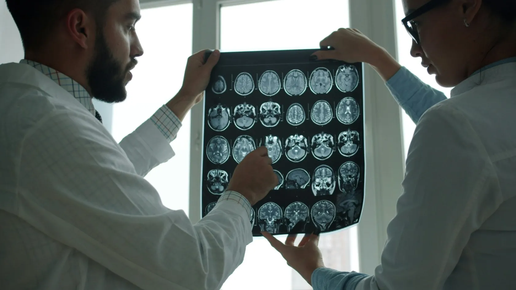
Screening & Diagnostic Imaging Center.
Scan a well person from head to toe and something turns up. Almost every time. The question is never what a scan can see, but whether finding it helps.
Let us call you
Send us your contact information and we will call you as soon as possible.
About This Center
Scan a well person from head to toe and something turns up.
Almost every time. A cyst on a kidney, a nodule in a lung, a lump on an adrenal gland, a spot on a liver, a disc that bulges the way most discs over forty bulge. The machine is not malfunctioning when it reports these things. It is doing precisely what it was built to do, which is to describe tissue in enormous detail without holding any opinion at all about whether the tissue matters, and supplying that opinion is the whole job, and it is the part that gets left out when imaging is sold as a product.
So the question this department exists to answer is never whether a scan can see something. It is whether finding it helps you.
Free consultation
Send the images, and tell us what question you want answered
Send previous scans on a disc or as the raw imaging files, since a report without the pictures cannot be re-read and a second opinion on a paragraph is worth very little. Include the oldest ones you have, because a nodule that has not changed in four years is a different object from the same nodule seen once. Tell us plainly what you are worried about, whether a symptom, a family history or a finding somebody has already mentioned to you, since that determines which scan is worth doing and frequently determines that none is. List your kidney function if you know it, any previous reaction to contrast, any implanted metal or device, and whether you have had a great deal of imaging already. If you are pregnant or might be, say so first.
Scan a well person and something turns up
Two things have to be true before a test is worth doing on somebody with no symptoms, and the second is the one that gets skipped. The first is that the test can find the disease. The second is that finding it earlier changes what happens to the person, which is a far higher bar than the first one and is cleared far less often, because a great many things a scanner can see either never would have caused trouble or cannot be helped by knowing about them sooner.
Imaging fails that second test unusually often, for a reason worth understanding. Modern scanners resolve small structures beautifully, and the human body turns out to be full of small structures that are simply there, have always been there, and were never going to do anybody any harm. A nodule in a lung, a cyst on a kidney, a small mass on an adrenal gland, a haemangioma in a liver, a thyroid nodule, a meniscus that looks frayed. Each one is real. Each one gets a sentence in a report, and each sentence, once written, has to be dealt with by somebody, which usually means another scan in six months, sometimes a biopsy, occasionally an operation, and always a period of the person believing they might have cancer.
That chain has a name in the literature. The cascade. It is the main harm imaging causes.
None of which is an argument against scanning. It is an argument for asking, before the scan rather than after it, what will be done with each of the plausible answers. If a finding would not change your treatment, would not change your surveillance and would not change a decision you are facing, the scan that finds it has cost you something and given you nothing. That question takes a minute to ask, and it is the single most valuable minute in this department.
The whole-body check-up
Head-to-toe MRI has become one of the most heavily promoted products in private medicine, sold directly to consumers by companies and not prescribed by clinicians, and marketed on an argument that sounds unanswerable. No radiation, one appointment, everything looked at, catch it early.
Here is what the radiology literature actually says about it.
What the reviewers found, and what they did not
A 2026 review in the clinical imaging literature examined whole-body MRI as a general screening tool for people without symptoms. It credits the technology fairly. Diffusion-weighted imaging and faster acquisition protocols have genuinely improved what these scans can see, there is no ionising radiation involved, and the technique has established value in specific high-risk groups and in following people who already have cancer. Then it sets out the problem. Adoption has been driven by public interest and by direct-to-consumer imaging companies rather than by evidence, and the review names them. Patients and clinicians alike are left uncertain how to manage the incidental findings these scans produce, which generates downstream testing and what the authors describe as the psychological burden of uncertain diagnoses. On the question that matters most, whether any of this results in people living longer or better, the review states that the evidence remains unclear, and concludes that the gap has to be closed before widespread adoption can be justified.
So we do not sell it. The reason has nothing to do with the machine being unavailable in this building. It is that a scan which reliably produces findings and unreliably produces benefit is a poor thing to sell to somebody who feels well and has flown a long way to be reassured.
There is a narrower version that is defensible. People carrying a genetic condition that predisposes them to multiple cancers, people with a strong and specific family pattern, and people already under cancer surveillance are a different population with a different prior probability, and for them whole-body imaging is a considered tool and not a retail product. That distinction is the entire argument, and it turns on who is being scanned, never on what the scanner can do.
Which scans earn their place
Some imaging screens are among the best-evidenced interventions in medicine, and refusing the whole-body scan is only credible from a service that says clearly which ones it does recommend and to whom.
This table scrolls sideways on a narrow screen. Swipe or drag to see every column.
| Scan | Who it is for | What we tell you first |
|---|---|---|
| Low-dose lung CT | Current and former heavy smokers inside the eligible age band, and nobody else. | This one works, and it works only in that group. It also produces false alarms in roughly one screen in twelve, and you should know that number before the first scan and not afterwards. |
| Mammography with tomosynthesis | Women in the screening age band, earlier and more often where family history or a known gene changes the calculation. | Dense tissue reduces what mammography can see, so density is reported and discussed, and supplemental ultrasound or MRI is decided on that basis and not on request. |
| Coronary calcium score | People at genuinely intermediate cardiovascular risk, where the result would tip a decision about preventive medication. | Useful precisely when a doctor is undecided. Of no value to somebody already on treatment or already known to be low risk, since the answer changes nothing either way. |
| Aortic ultrasound | Older men with a smoking history, and anybody with a first-degree relative who had an aortic aneurysm. | Quick, harmless, no radiation. For the right person it is among the highest-yield tests in this table, and a single normal scan in later life usually settles the question for good. |
| Whole-body MRI | People with a cancer predisposition syndrome or under formal cancer surveillance. | Declined for everybody else, for the reasons set out above, and the refusal is about the population being scanned and never about the machine standing in the room. |
| Full-body CT check-up | Nobody. There is no group for whom this is the right test. | Declined outright. It combines the incidental finding problem of whole-body MRI with a radiation dose, which is the worst available combination. |
That lung row deserves its numbers, because it is the one screen on this page that has moved from evidence into national policy in the last two years and because its harms are real and quantified instead of hand-waved.
A national pilot programme in Israel ran from 2021 to 2024 across all four of the country's health funds, screening 5,395 current and former heavy smokers with low-dose CT. Just over half of eligible people took part. Lung cancer was found in 63 of them, which is 1.2 percent, and 57.9 percent of those cancers were caught at stage one or two, which is the point of the entire exercise since that is where the disease is still curable. Among scans flagged as suspicious, the proportion that turned out to be cancer was 11.6 percent for the lower suspicion categories and 54.2 percent for the highly suspicious ones, meaning false positives ran at 8.4 percent and 0.7 percent respectively. Nobody suffered an immediate serious complication. The median wait from an abnormal scan to a diagnostic procedure was 6.4 weeks. On the strength of it, the country added lung screening to its national list of health services.
Here is what a screen looks like when it works. Note that the harms are stated, counted and small, and note that the eligibility is narrow.
One efficiency is worth mentioning, since it costs nothing extra. The same low-dose chest CT that looks for lung cancer also shows calcium in the coronary arteries, and a 2026 review in the radiology literature makes the case for reporting it. The people eligible for lung screening are, almost by definition, people carrying substantial cardiovascular risk, and the information is already in the images with no additional scan, radiation or inconvenience. The review is careful about how far that goes. No prospective trial has yet shown that acting on calcium found this way reduces cardiac events, although long-term follow-up of a randomised screening trial found that reporting it was associated with lower cardiovascular mortality, most likely because it prompted people into preventive treatment they were not already receiving.
What follows an abnormal result
Everything in this section belongs before the booking of any scan, because the scan is never the end of the process and the next step carries most of the risk.
Take the commonest example in imaging, a suspicious lung nodule under two centimetres, where the obvious move is to biopsy it with a needle through the chest wall under CT guidance, which is standard practice everywhere and sounds like the end of the story. A 2026 study from Turkish centres looked at how well it performs.
The number that changes how a negative biopsy should be read
Ninety-five patients with suspicious nodules of two centimetres or less underwent CT-guided needle biopsy between 2021 and 2024. Every biopsy produced enough material to examine. Overall accuracy was 89 percent, sensitivity 83 percent and specificity 94 percent. When the biopsy said cancer it was right 98 percent of the time, which is why a positive result can be acted on with confidence. When the biopsy said benign it was right only 65 percent of the time, meaning roughly a third of the nodules called benign were malignant after all. Pneumothorax, air leaking into the chest cavity, occurred in 30 percent of procedures, and about a quarter of those needed a chest drain. The authors' conclusion is unambiguous, which is that a negative needle biopsy cannot be treated as safe on its own and that these nodules need continued imaging follow-up or a surgical biopsy regardless.
Three lessons come out of that, and they generalise well beyond the lung.
A test that is excellent at confirming disease is frequently poor at excluding it, and the two properties are separate, which is why reassurance is consistently harder to obtain from any investigation than alarm is. Alarm comes easily. And a procedure with a 30 percent complication rate is not something to walk into because a scan you did not need found something you were not looking for, which is precisely the sequence a whole-body screen sets in motion.
What we do instead of the reflex biopsy is boring and effective. Old images are hunted down and compared, since stability over two or more years settles most nodules without anybody being touched, and established size and growth criteria then decide the interval for any repeat imaging, so that where a biopsy genuinely is needed the route is chosen for the lowest complication risk and never for the fastest booking, and you are told the pneumothorax number in advance of consenting to anything.
Radiation, counted honestly
Radiation from a CT scan is a known carcinogen. The risk from any single scan is small. Both sentences are true at once. The reason the second does not cancel the first is volume.
One year of scanning, counted forward
Researchers took organ-specific radiation doses from a multicentre CT dose registry, scaled them to the 93 million CT examinations performed on 61.5 million patients in the United States in 2023, and projected the lifetime cancers those examinations would eventually cause. The figure was approximately 103,000, with an uncertainty range of 96,400 to 109,500. Risk per scan was highest in children and adolescents, but because adults have very many more scans than children do, adults accounted for 91 percent of the total. The commonest resulting cancers were lung, colon, leukaemia and bladder, with breast second commonest among women. Abdominal and pelvic CT in adults produced 37 percent of the projected cancers from 32 percent of the examinations, and chest CT another 21 percent. Sensitivity analyses shifted the total between 80,000 and 127,000 without changing the picture. The authors noted that if current practice continues, CT-associated cancer could eventually account for around 5 percent of all new cancer diagnoses each year.
Read that carefully and the message is not that CT is dangerous. It is that CT is enormously useful and enormously overused, and that the fix is not fear but selection.
Age changes the arithmetic more than most people expect. A separate 2026 analysis of preoperative planning CT before shoulder replacement estimated lifetime attributable cancer risk by age and sex and found it falling steeply as patients got older, from 73.1 per 100,000 in women in their fifties to 9.6 in women in their eighties, with thyroid and lung risk significantly higher in women than in men in the intermediate age groups. The practical translation is that a scan which is essentially free of consequence in a person of eighty is a real decision in a person of thirty-five, and that the same request from two patients deserves two different answers. So four habits follow. Ultrasound and MRI are used wherever they answer the same question, and for a great many abdominal, pelvic, musculoskeletal and paediatric problems they do. Dose is set by the question and not by a default protocol, since a follow-up looking at a known lung nodule needs a fraction of the dose of a first diagnostic study. Repeat scans are checked against previous ones before being ordered, because a surprising proportion of imaging repeats something done elsewhere within the year, and in anybody young the conversation about whether the scan is necessary is a longer one, which is exactly as it should be.
Contrast deserves a line. Iodinated contrast used in CT is very safe in people with reasonable kidney function, and the older belief that it commonly damages kidneys has been substantially revised as better-controlled studies separated the contrast from the illnesses that led to the scan. Gadolinium used in MRI carries a different set of considerations, and the modern agents are chosen accordingly. Neither is given without checking kidney function and previous reactions, and neither is withheld out of vague caution when the diagnosis genuinely requires it.
Reading the report you were handed
Radiology reports are written by one doctor for another, in a register built for precision rather than for reassurance, and patients now read them first. That mismatch causes an enormous amount of avoidable fear.
This table scrolls sideways on a narrow screen. Swipe or drag to see every column.
| The phrase | What the radiologist meant by it |
|---|---|
| No acute abnormality | Nothing here needs attention today. It is not a statement that everything is normal, and it does not cover things the scan was not designed to show, which is why a normal head CT does not exclude a great many causes of headache. |
| Cannot be excluded | A possibility exists that this scan cannot rule out, which is usually a statement about the limits of the test and not a suspicion about you. Almost every scan cannot exclude something. |
| Clinical correlation advised | The pictures alone do not settle it, and the answer depends on your symptoms and examination. It is a request to the referring doctor and not a warning to you. |
| Probably benign, follow-up suggested | Very likely harmless, with the repeat scan existing to confirm stability and not because anybody is worried. This category is where most incidental findings live and where most anxiety is generated. |
| Technically limited study | Movement, body habitus, metal or timing degraded the images, so a negative result carries less weight than usual. This sentence matters more than almost any other and is the one most often skipped over. |
| Correlate with prior imaging | The radiologist did not have your old scans and is telling you the interpretation would change if they did. Sending the old images is frequently worth more than repeating the new one. |
Every report issued here comes with a plain-language summary written for you alongside the technical report written for your doctor, and the two say the same thing. Where a finding needs explaining, that happens in a conversation and never in a paragraph.
Where the software actually helps
Artificial intelligence in radiology has been oversold for a decade and is now, in a small number of narrow tasks, genuinely good. Breast screening is the clearest of them, and the evidence is unusually strong because somebody ran a proper randomised trial.
The Swedish MASAI trial randomised 105,934 women to screening with software support or to standard double reading by two radiologists without it, then followed them for interval cancers, meaning the cancers that appear between screening rounds and represent what screening missed. Interval cancer rates were 1.55 per thousand with software and 1.76 without it, a non-inferior result on the trial's own pre-specified margin. Sensitivity was significantly higher with software, 80.5 percent against 73.8, and the advantage held across ages, across breast densities and for invasive cancer. Specificity came out identical at 98.5 percent in both arms. The extra detection cost nothing in recalls of healthy women. The intervention arm also had fewer interval cancers of the more aggressive kinds, and the reading workload fell.
Genuinely good, and bounded. A systematic review published in 2025 draws the boundary.
Reviewing 31 studies covering more than two million screening examinations, the authors found that software used as a second reader or as a triage tool maintains or modestly improves sensitivity while preserving specificity, and can cut reading volumes by between 40 and 90 percent. Standalone software achieved accuracy comparable to radiologists in real-world cohorts. But they were explicit that certainty is limited by inconsistent study designs, by reliance on enriched datasets in some accuracy estimates, and by incomplete interval cancer follow-up in several major studies, and they concluded that transparent reporting, standardised evaluation and long-term population studies are still needed before software should be considered as a standalone reader.
Which is exactly how it is used here. Software assists a radiologist and prioritises the worklist. It does not sign a report.
What we do not offer
Executive check-up packages are among the largest medical travel products sold out of Istanbul, and a page about screening that avoids the subject is being evasive.
We do not sell a bundle of scans and blood tests to somebody with no symptoms and no risk factors. The commercial logic of these packages is that more tests justify a higher figure, and the clinical consequence is that a well person is exposed to a set of investigations chosen by a price list instead of by their history. What we do instead is an appointment in which somebody takes that history, works out which of the screens in the table above you are actually eligible for, and orders those, which for many people is fewer tests than they expected and occasionally none at all.
Nor do we perform whole-body CT on well people, at any age, for any package. The reasoning is set out above and it does not bend for a request.
We do not run tumour marker panels as screening. These blood tests were designed to monitor people who already carry a diagnosis, they rise in a long list of entirely harmless conditions, and turned loose on a well population they generate very many more false alarms than they do cancers. Each alarm buys a scan, and the scan finds something incidental, and the sequence is by now familiar.
And we do not issue a scan without an interpretation attached. Images with a one-line report and no conversation is a product rather than a service, and it is how somebody ends up on a flight home carrying a sentence about a lesion nobody has explained. Every study here is reported by a subspecialist in that body area, discussed with the referring clinician where a decision hangs on it, and explained to the patient in language they can repeat to their own doctor.
One boundary is worth naming for completeness. Screening that is not imaging belongs elsewhere in the hospital, so cervical screening, hereditary cancer gene testing and the assessment of a family history all sit with the services built for them, and this department images what those services ask it to image.
What a visit involves
Less scanning than most people expect, and more talking beforehand.
The question, written down before anything is booked
What you are worried about, what symptoms exist, what has already been imaged and when. Then the decision about which scan answers that question, and whether one is needed at all. A meaningful share of enquiries end here with advice and no appointment.
Old images retrieved and loaded
Every previous study you can obtain is uploaded before the appointment, because comparison is the single most powerful tool in this department and a stable finding needs no further action at all. Chasing images from another hospital is tedious and is done by us, never left to you.
Safety checks that take four minutes
Kidney function, previous contrast reactions, pregnancy where relevant, and for MRI a careful account of implanted metal, pacemakers, cochlear implants, aneurysm clips and retained fragments. Claustrophobia is asked about in advance instead of discovered in the scanner, and it is manageable when it is known about.
The scan itself
Ultrasound takes fifteen to thirty minutes. CT takes a few. MRI runs anywhere between twenty and fifty depending on what is being examined and how many sequences the question needs. Protocols are set for the question instead of run from a default, and a radiologist is available during the study so that an unexpected finding can be pursued immediately instead of at a second appointment.
Results, in two versions
The formal report for clinicians, and a plain summary for you that states what was found, what it means and what if anything happens next with a date attached. You leave with the images themselves in a standard format any hospital in the world can open, which matters more than most people realise until they need a second opinion.
Coming to Istanbul
Imaging is the most portable service in the hospital, which cuts both ways.
A second opinion on scans you already hold needs no travel at all. Upload the images, and a subspecialist radiologist re-reads them and issues a fresh report, which is worth doing when a finding is ambiguous, when a report and a clinical picture disagree, or before consenting to an operation on the strength of a single reading. Where new imaging is genuinely needed, two to three days covers a consultation, the scans and a results appointment in person, and where a biopsy under imaging guidance follows, allow five to seven days so that the procedure and its check are both done here rather than one of them being done at an airport. Most nationalities enter Turkey visa-free or on an electronic visa completed online in minutes, with a letter of invitation issued where an application needs one. Interpreters cover Turkish, English, Arabic, Russian and German as standard and other languages by arrangement, and reports are issued in English and Turkish with a translated plain summary for the doctor who will act on it. Airport transfer and a nearby hotel are arranged alongside the appointments so that scans, results and any follow-up procedure fall inside one working week. A companion is welcome, and for anyone who will be sedated for a procedure a companion is required.
One honest caution about flying for imaging. If the scan you want is available at home, having it at home and sending us the images is usually the better path, since it avoids a journey and keeps the pictures in the system that will manage you afterwards. Travel earns its place when the expertise is the thing being sought rather than the machine, which in practice means complex reporting, subspecialist second opinions, image-guided procedures and situations where a scan and the treatment decision need to happen in the same week.
What moves the cost
Quotations come per scan, which sounds simple and conceals almost everything that matters.
The largest driver is how many body regions are examined and how many sequences each one takes. An MRI of one knee and an MRI of the whole spine share a word and nothing else, and the same is true of a chest CT against a chest, abdomen and pelvis study. Contrast is the second driver, since a study with contrast requires the agent, a cannula, a period of observation afterwards and kidney function checked beforehand. Sedation or general anaesthesia, needed for some children and for severe claustrophobia, changes the cost structure entirely, since it brings an anaesthetist, a recovery period and a second set of consent conversations into what was going to be a twenty-minute outpatient appointment. An image-guided procedure is priced as a procedure. Then there are the parts that are easy to leave out of a quotation and that surface later. Whether the report is included, which sounds absurd until you meet a package where reporting is billed separately. Whether a subspecialist reads it or a general radiologist does. Whether you receive the images themselves or only a report, which decides whether a second opinion is possible anywhere else. Whether a follow-up scan, if the first one recommends one, is inside the figure. Whether the consultation that decides what to scan is charged. And whether translation of the report is included.
Your own circumstances move it as well. A single-region MRI in somebody who lies still is a straightforward study. The same request in a person who cannot lie flat, has a pacemaker, weighs more than the table's stated limit or needs sedation is a different piece of work, and quoting the two identically would be misleading.
Four questions turn a figure into something comparable. Name the exact study, with the regions and whether contrast is used. Ask who reports it and what their subspecialty is. Ask whether you receive the raw images. And ask what a recommended follow-up scan would cost, since roughly one incidental finding in a handful generates one. We answer all four in writing before anything is booked.
Once you are home
An imaging episode generates an obligation, and it is nearly always an obligation to do something later and not today.
If a finding needs a repeat scan, you leave with the date, the modality, the exact region and the reason written on a single sheet a radiology department anywhere can act on. That sheet exists because the commonest failure in this whole field is not misdiagnosis but the follow-up that never happened, where a probably benign nodule was found, a six-month scan was recommended, the recommendation lived in the fourth paragraph of a report, and nobody ever booked it. We also write to your own doctor with the same information, and we send a reminder at the interval. Keep the images, and keep them in the standard format rather than as photographs of a screen. They are the comparison that makes every future scan easier to interpret, and a nodule with four years of stable prior imaging behind it is a non-event while the identical nodule seen for the first time is a work-up. Storage costs you nothing, and it saves the next radiologist a great deal of the guesswork that turns a five-minute read into a six-month follow-up.
If a scan here settles a question, the correspondence says so explicitly, because a report that merely lists findings leaves the referring doctor to infer a conclusion and inference is where things go wrong. And where a finding needs treatment, the handover to the relevant specialty happens from here and is never left to you to arrange from another country.
Frequently asked questions
Should I have a whole-body MRI as a check-up?
How much radiation is a CT scan, really?
Does lung cancer screening work?
My biopsy came back benign. Is that the end of it?
Is artificial intelligence reading my scan?
My report says a finding cannot be excluded. Should I worry?
Can you review scans I already have without me travelling?
How long should I plan to stay in Istanbul?
References
- Smith-Bindman R, Chu PW, Azman Firdaus H, et al. Projected lifetime cancer risks from current computed tomography imaging. JAMA Internal Medicine. 2025;185(6):710-719.
- Lee J, Cyphers E, Beutler BD. Brief communication, the clinical opportunities and ongoing challenges with whole-body MRI for screening. Clinical Imaging. 2026;138:110907.
- Maya R, Liora V, Dorith S, et al. Feasibility assessment of implementing lung cancer screening among current and former heavy smokers, a national pilot program in Israel 2021-2024. Lung Cancer. 2026;219:109536.
- Cengiz SK, Demirer E, Baysal T, et al. Diagnostic utility and limitations of CT-guided transthoracic fine-needle aspiration biopsy in Lung-RADS 4 pulmonary nodules of two centimetres or less. Annals of Saudi Medicine. 2026;46(3):225-233.
- Kalva SR, Fucich D, Perry AJ, et al. Temporal trends and estimated lifetime attributable radiation risk of preoperative planning computed tomography for primary shoulder arthroplasty. Shoulder and Elbow. 2026.
- Mozafary M, Rezaeifar M, Naghavi M, Kazerooni EA, Stillman AE, Raggi P. Integrating cardiovascular screening in lung cancer screening. The British Journal of Radiology. 2026.
- Gommers J, Hernstrom V, Josefsson V, et al. Interval cancer, sensitivity, and specificity comparing AI-supported mammography screening with standard double reading without AI in the MASAI study. The Lancet. 2026;407(10527):505-514.
- Jassim G, Otoom O, Nair B, Hashem J. Performance of artificial intelligence in breast cancer screening programmes, a systematic review. BMJ Open. 2025;15(12):e111360.
Editor's note
Written by the Biruni Hospital medical editorial team. Reviewed by Prof. Dr. Okay ABACI, Cardiology.
Medically reviewed by
Prof. Dr. Okay ABACI
Cardiology
Other Medical Centers
View All-
Breast Health & Breast Cancer Center
Most people arrive having already decided which operation they want. The operation is rarely the first decision and never the one that decides most.
-
CAR-T Cell Therapy Center
CAR-T cell therapy in Istanbul for leukemia, lymphoma and myeloma. Eligibility, the five stage process, honest cost figures and how long you stay at Biruni.
-
Cellular Immunotherapy Center
Two entirely different industries use the words cell therapy. One has a regulator behind it. Four questions tell you which one you are talking to.
-
Check Up & Longevity Center
Biruni Hospital's Check-Up and Preventive Medicine Center in Istanbul plans risk based health screening by age, sex and history, with a free remote intake.
-

Children’s Health Center
The Children's Health and Diseases Center in Istanbul unites pediatric subspecialists at Biruni Hospital, with a free remote review of your child's file.