Free flap surgery moves living tissue, skin, fat, muscle or bone, from one part of the body to another and reconnects its blood vessels under the microscope. It rebuilds the jaw, mouth, breast and limbs after cancer or injury, and in experienced centers 98 to 99 percent of flaps survive. This guide covers flap types, monitoring, salvage, timing, recovery and the stay.
About This Department
Plastic Surgery
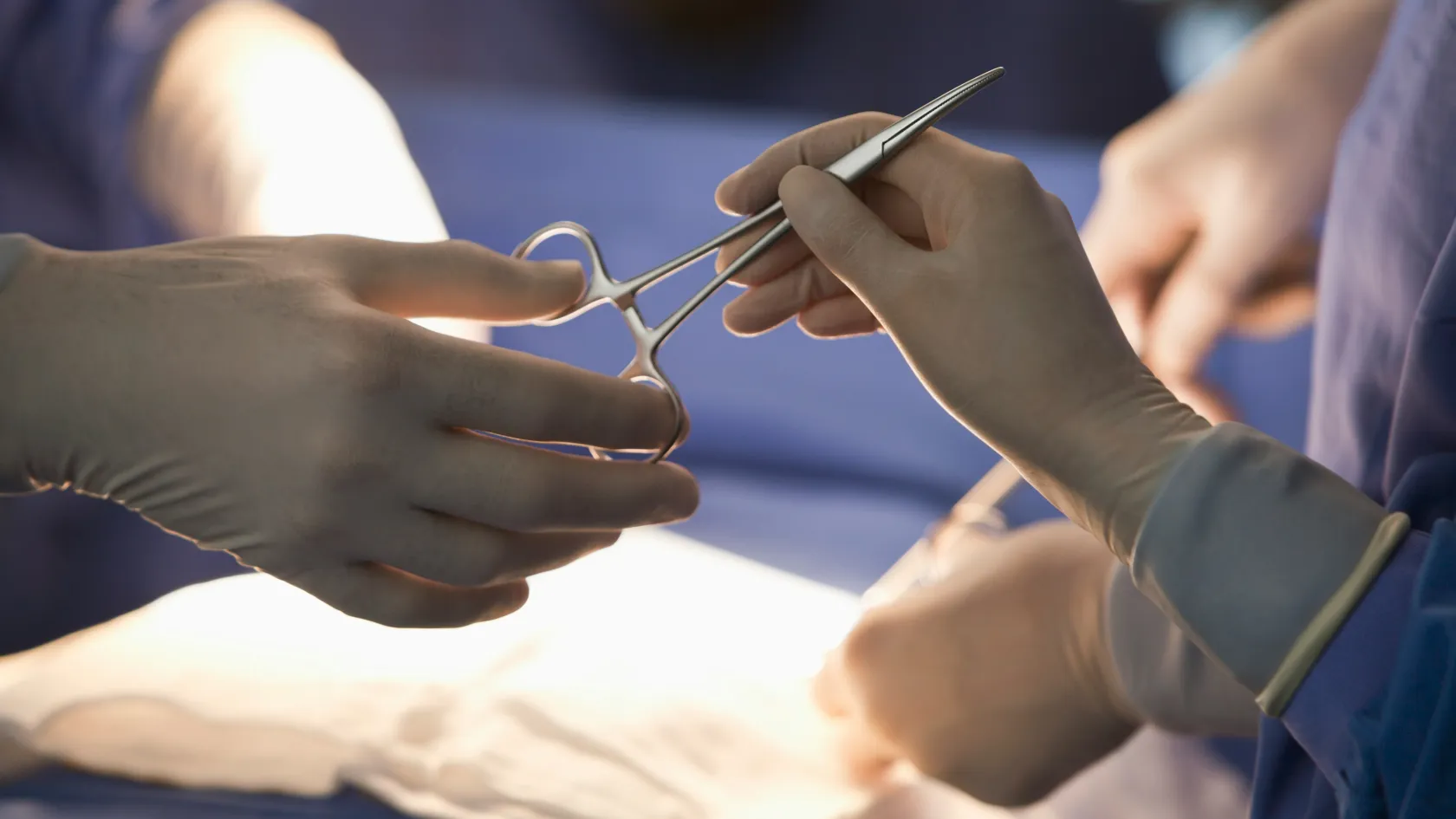
A free flap moves living tissue from one part of the body to another and reconnects its blood vessels under the microscope. In experienced centers 98 to 99 of every 100 flaps survive.
When cancer surgery, an injury or an infection leaves a wound too large or too deep to close with nearby tissue, a free flap brings skin, fat, muscle or bone from elsewhere, from the thigh, forearm, abdomen, back or lower leg. The surgeon detaches the tissue with its artery and vein, moves it to the wound and joins those vessels to vessels near the defect, each stitch finer than a hair. Free flaps rebuild the jaw and tongue after mouth cancer, the breast after mastectomy, and the leg after open fractures. The first three days decide most outcomes, so flaps are watched closely, and a flap in trouble is taken back to the operating room within hours. Send us your scans, wound photographs and medical history, and a reconstructive microsurgeon will tell you what a free flap could achieve and how long to plan.
98.8%
Free flap survival in 1,193 flaps at a cancer center
95.6%
Flap problems that appeared within the first 72 hours
A local flap stays attached to its blood supply and is swung into a nearby wound. A pedicled flap reaches a little further on a stalk of vessels. A free flap is cut loose completely, so it can travel anywhere in the body, and it lives only because the surgeon reconnects its artery and vein at the new site. That freedom lets the surgeon choose the best tissue for each wound.
What a free flap is
Every free flap is a block of tissue with a named artery and vein, called the pedicle. Matching matters. The surgeon chooses a flap whose tissue matches what is missing, thin skin for the inside of the mouth, bone for the jaw, muscle to fill a cavity, skin and fat for the breast, and whose pedicle is long and wide enough to reach healthy recipient vessels.
Under an operating microscope the artery and vein of the flap are stitched to an artery and vein near the wound, with stitches thinner than a human hair, or the veins joined with a small ring coupler. Blood flows in within minutes. The flap lives.
Microvascular free tissue transfer is a reliable reconstructive technique with low failure rates.
Review of 1,193 free flaps, Plastic and Reconstructive Surgery, 2007
Where free flaps are used
After cancer surgery of the mouth, jaw, throat and face, free flaps restore swallowing, speech and the shape of the face, with fibula or hip bone shaped into a new jaw. After mastectomy, abdominal tissue rebuilds the breast. After severe leg and arm injuries, they cover exposed bone, tendons and metal plates so fractures heal and limbs are saved. They also close chronic wounds, pressure sores and defects after infection or radiation damage.
This study confirms that free flaps are extremely reliable in achieving successful reconstruction of the head and neck, but it is essential that complications be recognized and addressed early in their course.
Review of 1,000 head and neck flaps, International Journal of Oral and Maxillofacial Surgery, 2012
Signs of trouble in the first days
A flap that turns pale, cool and loses its capillary refill points to a blocked artery. A flap that turns blue or purple, swollen and bleeds dark blood when pricked points to a blocked vein. A rapidly enlarging swelling under the skin points to bleeding. Any of these signs means the surgeon is called at once, and the patient then goes straight back to the operating room.
Common free flaps
The anterolateral thigh flap, from the outer thigh, is the workhorse for skin and fat almost anywhere. The radial forearm flap gives thin, pliable skin for the mouth and throat, and the fibula flap supplies straight, strong bone for the jaw and the long bones of the limbs, cut and shaped into segments while it is still attached to its vessels. The DIEP flap from the lower abdomen rebuilds the breast. The latissimus dorsi and gracilis muscles fill cavities and, with nerves reconnected, can even restore movement after facial paralysis.
Two teams work at once in many cases, one removing the tumor or cleaning the wound, the other raising the flap.
The recipient artery and vein are prepared under the microscope.
The flap pedicle is divided and the flap moved to the wound.
The vessels are joined, blood flow is confirmed and the flap is shaped and stitched in.
The donor site is closed directly or with a skin graft.
The anterolateral thigh flap
A Taiwanese center reported 672 anterolateral thigh flaps used for the head and neck, arms, legs and trunk between 1996 and 2000. Total failure occurred in 1.79 percent and partial failure in 2.53 percent, and every failed flap was replaced, most with a second flap. The thigh scar hides well, and the surgeon makes the flap thin or thick to match the wound.
The anterolateral thigh flap is a reliable flap that supplies a large area of skin.
Series of 672 flaps, Plastic and Reconstructive Surgery, 2002
How flaps are monitored
Nurses trained in microsurgery check the flap every hour for the first one to two days, then at longer intervals, looking at its color, warmth, swelling and the speed at which color returns after gentle pressure. A handheld Doppler probe listens to blood flow in the vessels, and some centers add implanted probes or sensors that measure oxygen in the skin.
Warm room, good hydration and pain control to keep blood vessels open.
No pressure on the flap or on its vessels, including from dressings, ties or tubes.
Blood-thinning injections to reduce the risk of clots in the legs and, in some protocols, in the flap.
When a flap needs rescue
At a New York cancer center, 6 percent of 1,193 flaps needed an emergency return to the operating room, most for a clot in the vessels, 53 percent, or bleeding, 30 percent. Venous clots were three times as common as arterial ones and easier to save, 71 against 40 percent. Flaps that were saved had been taken back four hours after the problem was noticed, against nine hours for those that failed. In the end, 98.8 percent survived.
Why do the first 72 hours matter so much?
Almost every problem shows early.
The time of presentation of flap compromise is a significant predictor of flap salvage outcome.
Review of 1,142 flaps, Plastic and Reconstructive Surgery, 2007
In a review of 1,142 free flaps in Taiwan, 9.9 percent needed re-exploration. Of those problems, 82.3 percent appeared within 24 hours and 95.6 percent within 72 hours, and 85.2 percent of flaps that ran into trouble in that window were saved. Problems present on arrival in the microsurgical intensive care unit had a lower complete salvage rate, 40.9 against 69.2 percent, and the first hours after surgery are therefore watched most closely of all.
How do free flaps rebuild the head, neck and limbs?
When a compromised flap is identified, surgical re-exploration should not be deferred.
Review of 1,000 head and neck flaps, International Journal of Oral and Maxillofacial Surgery, 2012
A German center reviewed 1,000 head and neck free flaps placed over 23 years, mostly latissimus dorsi, radial forearm, iliac crest and fibula flaps. Complications of the flap occurred in 7.6 percent, of which fewer than half meant complete loss. Clots in the veins of the neck and bleeding under the neck skin were the commonest causes, and both respond to an early return to the operating room.
Limbs after injury
For open fractures of the leg with exposed bone, the timing of the flap changes the whole course of recovery. A classic series of 532 limb reconstructions showed fewer flap failures and infections, faster bone healing and far shorter hospital stays when the flap was placed within 72 hours of injury, and modern trauma units aim to cover such wounds within the first days.
What do the results look like?
Timing changed everything in the classic trauma series.
This table scrolls sideways on a narrow screen. Swipe or drag to see every column.
Early against delayed free flaps after limb injury, 532 patients
Timing
Flap failure
Infection
Bone healing
Hospital stay
Within 72 hours
0.75 percent
1.5 percent
6.8 months
27 days
72 hours to 3 months
12 percent
17.5 percent
12.3 months
130 days
After 3 months
9.5 percent
6 percent
29 months
256 days
Careful monitoring and urgent reexploration are critical for salvage of compromised flaps.
Review of 1,193 free flaps, Plastic and Reconstructive Surgery, 2007
What is recovery like, and how long is the stay?
Recovery
The first one to three days are spent in a monitored unit with hourly flap checks. Walking begins once the surgeon allows it, within the first days for the majority. After a leg flap, the leg stays raised for a week, then is lowered for a few minutes at a time, a dangling program that lets the flap adapt to standing. The donor site heals within weeks, and the flap swells for months before thinning to its final shape. A second, smaller operation to thin or refine the flap is common months later.
1
Week one
Monitoring, then the ward. Drains removed, flap checks less frequent.
2
Weeks two to three
Stitches removed, rehabilitation, swallowing or speech therapy after head and neck surgery, dangling and gentle walking after leg flaps.
3
Months three to twelve
The flap settles.
Expect a longer stay than for most operations.
Plan two to three weeks in Istanbul for most free flaps, longer for complex head and neck or limb reconstructions. Flying waits until the flap is stable. You should walk well, and blood-thinning injections and compression stockings protect the journey.
Cost depends on the reason for reconstruction, the flap, the operating time, the intensive care and hospital days, rehabilitation and any refinement surgery. Your own figure follows the free review of your file.
The coordinator who answers your first WhatsApp message stays with you to the flight home. A visa invitation letter arrives ten days ahead, a car meets you at the airport, and a hotel close to the hospital is booked for your companion during your hospital stay and for both of you afterward. The team speaks seven languages, with interpreters for others. Halal, vegetarian and diabetic meals are prepared, a prayer room is available, and a female doctor is assigned on request when the schedule allows. Back home, send photographs of the flap and donor site at agreed intervals, and the microsurgeon advises on scar care, swelling and the timing of any refinement, while your own doctors receive a full report naming the flap, the vessels used and the medicines to continue, because a surgeon who sees you later for an unrelated problem needs to know that a vessel in your neck, groin or leg now feeds the flap and must not be tied or compressed.
Free flap surgery FAQ
How successful is free flap surgery in experienced centers?
Very. A New York series of 1,193 flaps reported 98.8 percent survival, and most experienced centers report similar figures.
What happens if the flap fails?
Most problems appear in the first 72 hours and are treated by returning to the operating room. If a flap is lost, a second flap or another method rebuilds the area.
How long does free flap surgery take?
Four to ten hours, depending on the site and whether tumor removal is part of the operation.
Will the donor site be a problem?
The donor site is chosen so that its loss causes little harm. The thigh, forearm and abdomen heal well, sometimes with a skin graft.
How long will I stay in Istanbul?
Two to three weeks for most free flaps, longer for complex cases.
Can my family come with me?
Yes. A companion stays at the hotel we book and can visit throughout the hospital stay.
References
Bui DT, Cordeiro PG, Hu QY, et al. Free flap reexploration: indications, treatment, and outcomes in 1193 free flaps. Plastic and Reconstructive Surgery. 2007;119(7):2092-2100.
Chen KT, Mardini S, Chuang DC, et al. Timing of presentation of the first signs of vascular compromise dictates the salvage outcome of free flap transfers. Plastic and Reconstructive Surgery. 2007;120(1):187-195.
Pohlenz P, Klatt J, Schön G, et al. Microvascular free flaps in head and neck surgery: complications and outcome of 1000 flaps. International Journal of Oral and Maxillofacial Surgery. 2012;41(6):739-743.
Wei FC, Jain V, Celik N, et al. Have we found an ideal soft-tissue flap? An experience with 672 anterolateral thigh flaps. Plastic and Reconstructive Surgery. 2002;109(7):2219-2226.
Godina M. Early microsurgical reconstruction of complex trauma of the extremities. Plastic and Reconstructive Surgery. 1986;78(3):285-292.
Editor's note
Written by the Biruni Hospital medical editorial team. Reviewed by Prof. Dr. Atilla ARINCI, Plastic, Reconstructive and Aesthetic Surgery.