A PET scan cannot tell a lymph node inflamed by smoking from one that holds cancer, and the difference decides whether a lung cancer is operated on. EBUS settles it with a needle through the airway wall, under sedation, with no incision. Read what the trials found, what a negative result means, and how to plan three nights or a week in Istanbul.
About This Department
Interventional Pulmonology
The lymph nodes beside the windpipe decide whether a lung cancer can be operated on. EBUS samples them through the airway wall, with no incision, in under an hour.
Reaching those nodes once meant an operation under the breastbone. In a randomized trial of 241 patients at four European centers, starting with ultrasound-guided needle sampling found more nodal spread than surgical staging alone and cut unnecessary chest operations from 18 to 7 percent. Across 1,299 patients in eleven studies, a positive result was wrong almost never. Send us your CT and PET reports and images, and a chest physician will tell you whether EBUS is the right first test and what it would settle.
93%
Sensitivity for nodal spread across 1,299 patients
14 vs 29 days
Time to a treatment decision, EBUS first against the usual route
This table scrolls sideways on a narrow screen. Swipe or drag to see every column.
Four ways of getting tissue from the lymph nodes in the center of the chest
EBUS-guided needle biopsy
EUS through the esophagus
Mediastinoscopy
CT-guided needle through the chest wall
How
A bronchoscope with an ultrasound tip, passed by the mouth into the windpipe. The needle crosses the airway wall.
An endoscope passed into the esophagus. The needle crosses the esophageal wall.
A small cut above the breastbone, and a scope passed down in front of the windpipe under general anesthetic.
A needle through the skin, guided by CT.
Reaches
Nodes beside the windpipe and at the airway branches, and masses next to the airways.
Nodes behind and below the windpipe, and the left adrenal gland.
Nodes in front of and beside the windpipe.
Peripheral masses and a few node stations.
Anesthetic
Sedation, or general anesthetic in some units.
Sedation.
General anesthetic.
Local anesthetic.
Incision
None.
None.
Yes.
None.
Same-day discharge
Yes.
Yes.
Sometimes, often one night.
Yes.
Serious complications
1.2 percent in a survey of 7,345 procedures.
Rare.
A few percent, including nerve and vessel injury.
Collapsed lung in a large minority.
Why the lymph nodes decide so much
A lung cancer is staged twice. The first stage comes from the scans, and the second from tissue, and for the nodes in the mediastinum, the space between the lungs that carries the windpipe, the heart and the large vessels, only tissue counts. A PET scan lights up nodes that contain cancer, and also nodes inflamed by infection, smoking or sarcoidosis. A CT scan measures them and misses cancer in nodes of normal size. Nobody should have a chest wall opened, or be refused an operation, on the strength of a scan alone.
That principle is the whole reason EBUS exists.
It answers three different questions, depending on who is sent for it.
Diagnosis and subtype. A mass next to an airway, or a node that has already taken cancer from a mass elsewhere in the lung, can be sampled without a needle through the chest wall. The sample serves for the diagnosis, the subtype and the molecular tests that decide drug treatment.
Nodal staging of a known cancer. Cancer confined to the lung is treated with surgery or stereotactic radiotherapy. Cancer in the mediastinal nodes is treated with chemoradiation or drugs first, and an operation that ignores the nodes is an operation the patient did not need.
Benign causes of enlarged nodes. Enlarged nodes in the chest have benign causes, above all sarcoidosis, tuberculosis and lymphoma, and EBUS reaches them for a diagnosis that once needed surgery.
What the trials show
1
ASTER, JAMA 2010, 241 patients
Patients with a lung cancer that looked operable were randomized to surgical staging by mediastinoscopy, or to endoscopic ultrasound sampling by EBUS and EUS with surgery held in reserve for a negative result. The endoscopic route found nodal spread in 46 percent of patients and the surgical route in 35, the combined strategy reached a sensitivity of 94 percent, and unnecessary chest operations fell from 18 percent to 7. Complications were the same.
2
Meta-analysis, European Journal of Cancer 2009, 1,299 patients
Across eleven studies, EBUS sampling of mediastinal nodes had a sensitivity of 93 percent and a specificity of 100 percent. In patients whose nodes were suspicious on CT or PET the sensitivity was 94 percent, and in patients scanned without any suspicious node it fell to 76 percent. Two complications occurred in 1,299 procedures.
3
Lung-BOOST, Lancet Respiratory Medicine 2015, 133 patients
Patients with suspected lung cancer were randomized to EBUS as the very first investigation after their CT scan, or to the conventional sequence of bronchoscopy, needle biopsy and surgical staging. The EBUS-first group reached a treatment decision in a median of 14 days against 29, because one procedure gave the diagnosis and the stage together.
Sarcoidosis and other benign disease
4
GRANULOMA, JAMA 2013, 304 patients
Patients with suspected sarcoidosis at fourteen centers were randomized to conventional bronchoscopy with lung biopsies or to endoscopic ultrasound sampling of the nodes. The needle route confirmed the diagnosis in 80 percent of patients against 53 percent, with one serious adverse event against two. Tuberculosis and lymphoma are diagnosed the same way, with the sample sent for culture and for flow cytometry.
What a negative result means
Trust a positive EBUS result.
A negative one is trusted only when the scans agree with it. With a sensitivity of 93 percent, about one nodal metastasis in fourteen is missed at the first sampling, and when the PET scan still shows a bright node, the guideline answer is a second test, either EUS through the esophagus in the same session or mediastinoscopy afterward. That is exactly what ASTER did, and the reason it reached 94 percent. A unit that treats a negative EBUS as the end of the road when the scan says otherwise is skipping the step that trial proved.
A staging strategy combining endosonography and surgical staging, compared with surgical staging alone, resulted in greater sensitivity for mediastinal nodal metastases and fewer unnecessary thoracotomies.
Conclusion of the ASTER trial, JAMA, 2010
This table scrolls sideways on a narrow screen. Swipe or drag to see every column.
Preparing for EBUS
When
What to do
With your first message
Send the CT and PET-CT reports with the images, any bronchoscopy or biopsy report, a full medication list with blood thinners marked, and a note of any lung disease, heart disease or previous chest surgery.
Five to seven days before
Blood thinners and antiplatelet drugs are paused or continued on a written plan agreed with the doctor who prescribed them. Aspirin alone is continued. A clotting test and a blood count are checked.
The evening before
Eat normally. Nothing to eat after midnight.
The morning of
Clear fluids until two hours before. Usual inhalers and heart tablets with a sip of water. Diabetes drugs as instructed. Bring a companion. You cannot drive or travel alone after sedation.
EBUS, EUS or both in one session
The two scopes reach different nodes. EBUS through the airway covers the stations beside the windpipe and at its branches. EUS through the esophagus covers the stations behind and below it, and the left adrenal gland, and it can be done with the same EBUS scope passed down the esophagus. In ASTER the two were combined, and for a patient whose PET shows nodes in both territories the combined session is the complete test. The reply to your file will say which is planned.
The day
Plan on half a day.
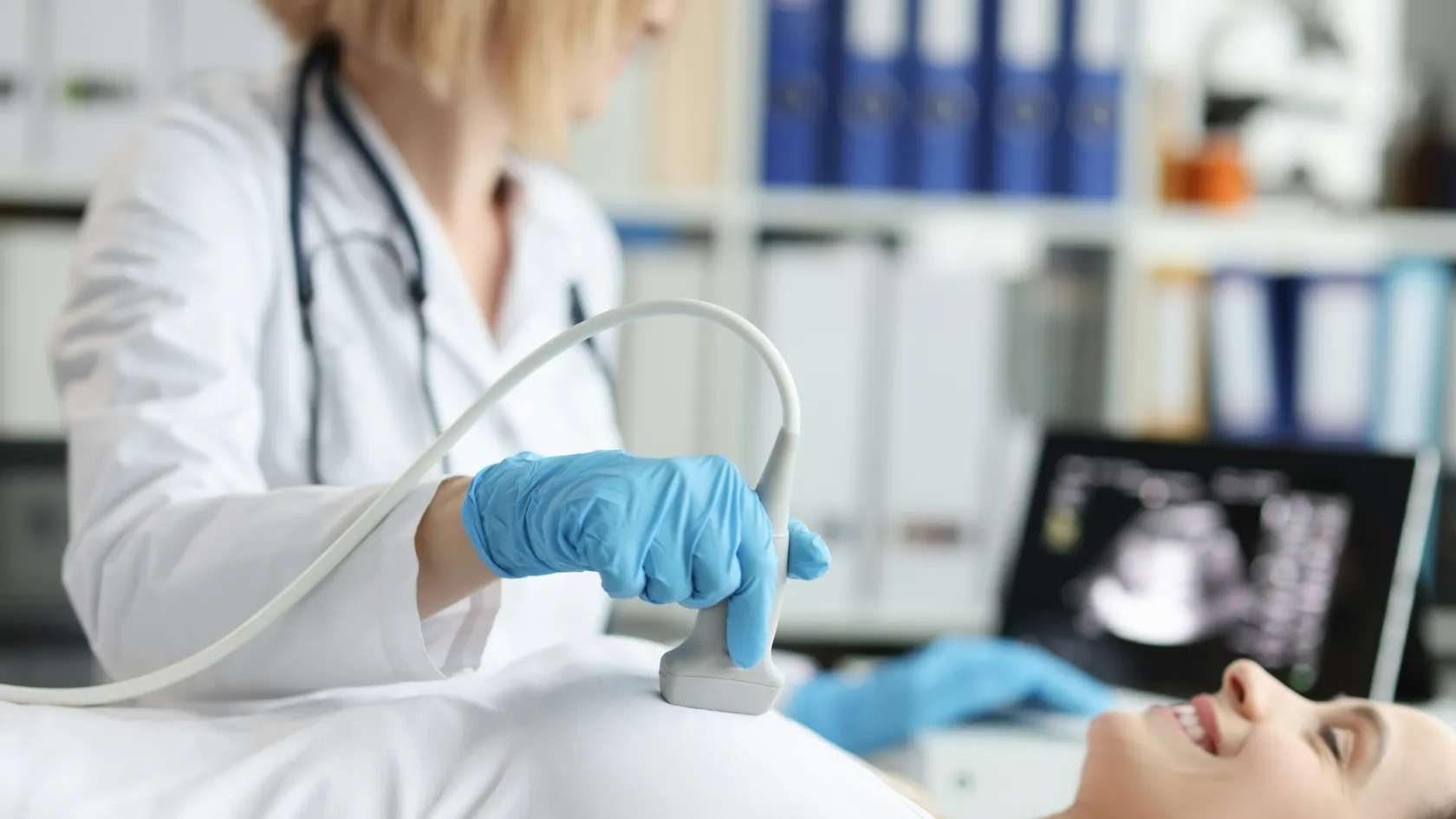
A nurse checks your blood pressure, pulse and oxygen, places a cannula, and the physician goes through the consent with your interpreter. The scope, a little thicker than a standard bronchoscope, is passed through the mouth and down the windpipe. The ultrasound tip is pressed against the airway wall at each node station, and the node appears on the screen with the vessels beside it shown in color so that the needle avoids them. The needle passes through the wall into the node and is moved back and forth a dozen times, and each node is sampled three or four times. Nodes are taken in a fixed order, the ones furthest from the tumor first, so that a needle that has been in a cancerous node never carries cells into a clean one. Twenty to forty minutes covers most examinations.
Recovery takes an hour or two. A sore throat and a little blood in the sputum for a day are expected, and nothing hot should be drunk until the throat is no longer numb.
Sedation
Two anesthetic routes exist.
EBUS is done under intravenous sedation, or under general anesthetic with a breathing tube or laryngeal mask, and the choice follows the unit's practice and your own lungs and heart. In Turkish hospitals an anesthesiologist is present for either. A rapid on-site reading by a cytopathologist, where the unit provides it, tells the physician during the procedure whether each node has yielded enough cells, which shortens the examination and reduces the number of passes.
Tell the team before sedation
Blood thinners and when you last took them. Sleep apnea. Heart disease, a pacemaker, or a recent heart attack or stroke. Severe COPD or home oxygen. Loose teeth or dental work. A cough with blood. Any allergy to anesthetic drugs. Pregnancy or any chance of it.
What are the risks?
In numbers
The Japan Society for Respiratory Endoscopy surveyed every member unit in the country and counted the complications of 7,345 EBUS procedures over eighteen months.
Ninety occurred, a rate of 1.23 percent.
Bleeding led the list, accounting for 50 of them, 0.68 percent, and was nearly always minor. Infection occurred in 14 patients, 0.19 percent, seven of them an infection of the mediastinum, which is the serious complication of the procedure and the reason a sampled cyst or a patient with fever afterward is treated promptly. Two patients had a collapsed lung. Four had a life-threatening event. One died, of a stroke, a rate of 0.01 percent. Scopes broke too, in 1.3 percent of procedures, at the unit's expense.
Those figures come from all units, skilled and new, and that is what led the authors to call for training. In the trials above, run at expert centers, serious complications were rarer still, two in 1,299 procedures in the meta-analysis, which is the difference a practiced hand makes and the reason to ask how many of these a unit does in a year.
Fever, chest pain, breathlessness or coughing up more than streaks of blood in the days after the procedure need same-day assessment. Here, that means the hospital. At home it means a local emergency department, with the procedure report in your hand, because an infection of the mediastinum is treated with intravenous antibiotics and sometimes drainage, and it does not wait.
How many days do I need in Istanbul?
Day one. Consultation with the chest physician, review of the images, a PET-CT if none has been done or the last one is more than six weeks old, blood tests, lung function tests.
Day two. The procedure in the morning, recovery, and discharge to the hotel in the afternoon.
Day three. A rest day. Flying is safe from this day on, and the physician confirms it at a short review.
Days four to seven. The cytology and tissue results arrive, followed by immune and molecular tests where a cancer is found. Staying for them allows the tumor board to meet with you in the city and a treatment plan to be made in person.
Results and the next step
Three nights covers the procedure. A week covers the answer.
The first reading, cancer or no cancer, takes two to three working days, and the subtype and molecular tests take several more. A patient who intends to be treated at home can leave on day three and receive the report by e-mail and WhatsApp, with the slides and blocks released on request. A patient who intends to be treated here should stay the week, because the result decides between surgery, stereotactic radiotherapy, chemoradiation and drug treatment, and all four are delivered on the one site, so the tumor board that reads the report can book the treatment the same afternoon.
What decides the cost, and what about the trip?
Pathology varies most. EBUS itself is priced as one procedure, and what is done with the samples afterward sets the rest. Each node station sampled adds needle passes and a specimen pot. A rapid on-site reading adds a cytopathologist's fee. Cell blocks, immune stains and the molecular panel on a cancer each add to the bill, and the panel can be the largest single line, since it decides which targeted drug or immunotherapy is used. A combined EBUS and EUS session costs more than EBUS alone. General anesthetic costs more. A PET-CT repeated for planning, and lung function tests, are separate items. Your own health moves the figure, since blood thinners, heart or lung disease and diabetes each add checks before sedation. Packages published for bronchoscopy with biopsy in Istanbul include the consultation, the procedure, sedation and transfers, with a night in hospital in some and a few hotel nights in others, and they seldom say whether the molecular panel is inside the figure.
Ask that question first.
Your own figure follows the free review of your scans.
One coordinator from the international patients team is assigned from your first message and stays with you until the report is in your hand. The team works in English, Arabic, French, Russian, Serbian, Romanian and Spanish and arranges interpreting in other languages on request, and an interpreter attends the consultation, consent and the results conversation. An invitation letter naming the hospital and the treating physician is sent about ten days before travel. The international patients office arranges the airport transfer, the hotel for you and a companion, and the rides to the hospital. EBUS needs no admission. Rooms for anyone admitted have a companion bed.
Halal, vegetarian and diabetic meals are available, a prayer room is on site, and a request for a female doctor is passed to the department and met wherever the rota allows. Once you are back home, the coordinator stays reachable on the same WhatsApp number, and the report goes to your own doctor.
EBUS-guided biopsy FAQ
Is an EBUS biopsy painful?
No. It is done under sedation or general anesthetic, and you do not feel the scope or the needle. A sore throat for a day and a little blood in the sputum are the usual after-effects.
How accurate is EBUS for lung cancer staging?
Across 1,299 patients in eleven studies, sensitivity was 93 percent and specificity 100 percent. A positive result is reliable. A negative result with a suspicious PET scan is followed by a second test, as the ASTER trial showed.
Is EBUS a replacement for mediastinoscopy?
As the first test, yes. In ASTER, starting with endoscopic sampling and reserving surgery for negative results found more nodal spread and cut unnecessary chest operations from 18 to 7 percent. Mediastinoscopy keeps its place as the second test when the scans and a negative EBUS disagree.
Can EBUS diagnose sarcoidosis?
Yes. In a randomized trial of 304 patients, needle sampling of the nodes confirmed sarcoidosis in 80 percent against 53 percent with conventional bronchoscopy and lung biopsies.
How long do I need to stay in Istanbul?
Three nights for the procedure, and a week if you want to wait for the full pathology and meet the tumor board in person. Flying is safe from the day after the procedure.
Can my family come with me?
Yes. The international patients office arranges a hotel for you and a companion, and a companion is needed on the day of the procedure because of the sedation.
References
Annema JT, van Meerbeeck JP, Rintoul RC, et al. Mediastinoscopy vs endosonography for mediastinal nodal staging of lung cancer: a randomized trial. JAMA. 2010;304(20):2245-2252.
Gu P, Zhao YZ, Jiang LY, Zhang W, Xin Y, Han BH. Endobronchial ultrasound-guided transbronchial needle aspiration for staging of lung cancer: a systematic review and meta-analysis. European Journal of Cancer. 2009;45(8):1389-1396.
Navani N, Nankivell M, Lawrence DR, et al. Lung cancer diagnosis and staging with endobronchial ultrasound-guided transbronchial needle aspiration compared with conventional approaches: an open-label, pragmatic, randomised controlled trial. Lancet Respiratory Medicine. 2015;3(4):282-289.
von Bartheld MB, Dekkers OM, Szlubowski A, et al. Endosonography vs conventional bronchoscopy for the diagnosis of sarcoidosis: the GRANULOMA randomized clinical trial. JAMA. 2013;309(23):2457-2464.
Asano F, Aoe M, Ohsaki Y, et al. Complications associated with endobronchial ultrasound-guided transbronchial needle aspiration: a nationwide survey by the Japan Society for Respiratory Endoscopy. Respiratory Research. 2013;14(1):50.
Editor's note
Written by the Biruni Hospital medical editorial team. Reviewed by Assoc. Prof. Dr. Uğur Temel, Thoracic Surgery.