
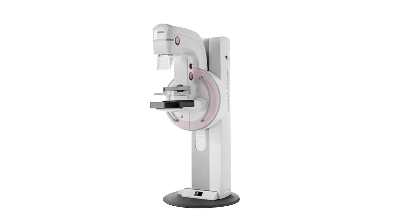
MAMMOMAT Inspiration
About This Technology
MAMMOMAT Inspiration is the digital mammography system used at Biruni Hospital for breast cancer screening and for the diagnostic imaging of breast findings. A mammogram remains the standard first examination of the breast because it shows microcalcifications and small lesions before anything can be felt by hand, and the years gained by finding a cancer early are the entire point of screening.
How the image is taken
The system records the image on a direct-conversion amorphous selenium detector, which turns X-ray energy straight into an electrical signal without an intermediate step. Fewer conversion losses mean more image from the same radiation, which is what allows fine detail such as clustered microcalcifications to stay visible at a low dose.
Compression is the part of the exam most women remember, so it deserves an honest description. The breast is compressed for a few seconds per image, because spreading the tissue is what makes small findings visible and keeps the dose down. The system's OpComp function applies compression only while the tissue yields to it and stops automatically at the optimal point. Firm pressure for a few seconds is a fair description. Pain is not the goal, and you can tell the technologist to pause at any moment.
Radiation dose, stated plainly
A mammogram uses a small amount of radiation, in the same range as a few months of natural background exposure. On this system the manufacturer's PRIME reconstruction replaces the conventional scatter grid with a software correction, which Siemens Healthineers states lowers patient dose by up to 30 percent without loss of image quality. For a test that healthy women repeat over many years of screening, dose discipline compounds, and it is a fair question to ask of any center that invites you to screen.
When this exam is used
Digital mammography on the MAMMOMAT Inspiration serves three situations at Biruni Hospital. Routine screening of women with no complaints, in most programs from around age 40 onward, on a schedule the physician sets with you. Diagnostic imaging when a lump, pain, discharge or another finding needs an answer. And guidance during breast biopsy procedures, where the image steers the needle to the exact spot that raised the question.
A mammogram does not answer everything. Dense breast tissue can hide a lesion on a standard two-dimensional image, which is why the examination is often paired with ultrasound, and why the radiologist sometimes calls a finding back for additional views rather than guessing. Biruni Hospital also operates the MAMMOMAT Revelation system for three-dimensional tomosynthesis imaging, and the radiology team decides which examination fits your case.
What the visit looks like
The examination itself takes a few minutes, most of it positioning. No preparation is needed beyond skipping deodorant and talc on the day, because their particles can mimic calcifications on the image. Results are read by a radiologist and reported to you with the images, so a physician anywhere can build on them. If you are traveling to Istanbul for a check-up or for breast diagnostics, previous mammograms are worth bringing: comparison with older images is one of the most useful tools a breast radiologist has.
Other Technologies
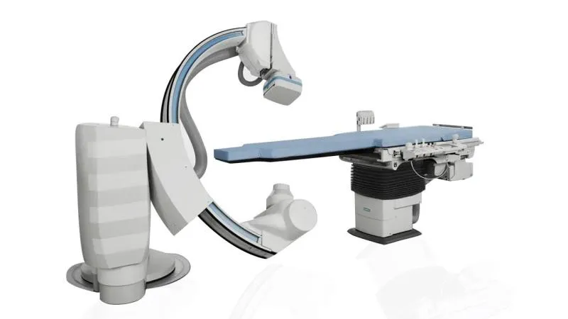
Artis zee Pure Angiography
Angiography system in the cardiac catheterization laboratory at Biruni Hospital, used to image the heart's vessels and to treat blockages in the same session where appropriate.
Learn more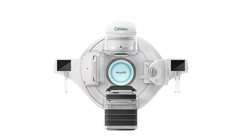
Elekta Versa HD
Linear accelerator used for external radiotherapy at Biruni Hospital, shaping high-dose radiation to the tumor while sparing the healthy tissue around it.
Learn more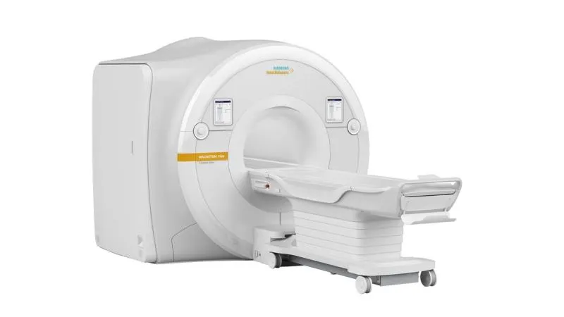
MAGNETOM Vida 3T MRI
High-field 3 Tesla MRI delivering exceptional image clarity with faster, quieter scans.
Learn more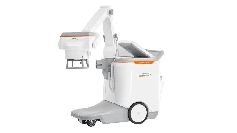
MOBILETT Elara Max
Mobile X-ray system that brings imaging to the bedside in intensive care, the operating room and the emergency department, so patients who cannot be moved are not moved.
Learn more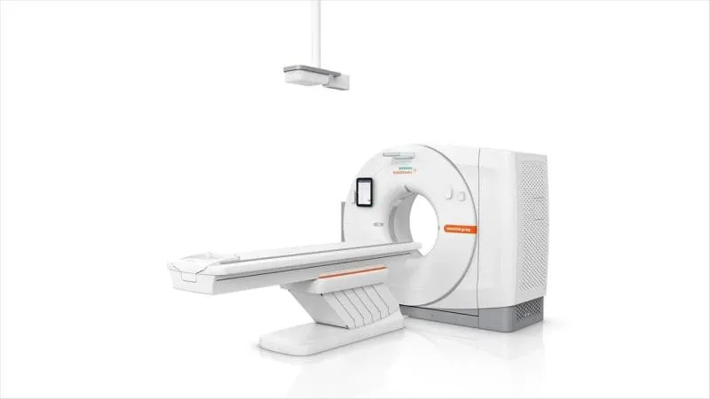
SOMATOM go.Now CT
Computed tomography scanner used at Biruni Hospital for rapid diagnostic imaging of the head, chest, abdomen and spine, with a workflow built for speed and dose control.
Learn more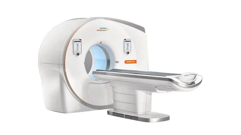
SOMATOM Force CT
Dual-source CT enabling ultra-low-dose imaging at remarkable speed and detail.
Learn more