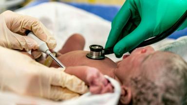
A newborn with an open back needs the closure within about 48 hours, so families who learn of spina bifida during pregnancy can plan the birth itself around the repair.
About This Department
Pediatric Neurosurgery
Closing the back early protects every nerve that still works. When the scan shows it in pregnancy, the plan starts before birth.
Among babies in the MOMS trial whose myelomeningocele was repaired after birth, 82 percent needed a shunt by the age of one, and the timing and quality of the repair shape how much a child walks, how often the shunt fails and how the bladder works for years afterward.
48 hours
Usual window for closing the back after birth
40% vs 82%
Shunt by age one, prenatal against postnatal repair, MOMS
51.3%
Walking in the community at school age after prenatal repair
Free
Review of the scans or photographs before you travel
Free consultation
The review costs nothing and carries no obligation. Send the pregnancy ultrasound or fetal MRI reports, or clear photographs of the baby's back with any MRI on disc, and a pediatric neurosurgeon will tell you which repair fits, how soon it is needed, and whether hydrocephalus needs treating too. A coordinator replies in your own language.
A scan in pregnancy or the first look at a newborn has shown an open defect over the lower back. Doctors call it spina bifida, or more precisely myelomeningocele. Spina bifida repair, also called myelomeningocele repair, closes that opening over the spinal cord, either before birth through surgery on the womb or in the first days after delivery, which protects the nerves that still work and stops infection from reaching the spinal fluid and the brain. Below you will find how the timing gets chosen, what the operation involves, how hydrocephalus is handled, what childhood with spina bifida looks like, and how families from abroad plan the trip.
What myelomeningocele means
In the first month of pregnancy, the tissue that becomes the spinal cord rolls up into a tube and closes. When the lower end of that tube fails to close, the spinal cord and its coverings stay open on the baby's back inside a thin sac, and the nerves inside are exposed to the fluid around the baby for the rest of the pregnancy. That open lesion carries the name myelomeningocele, the most severe and the most common form of open spina bifida. The level of the opening matters most. A defect high in the lower back affects more leg muscles than one near the tailbone, and almost every child has some effect on bladder and bowel control. Most babies also have a Chiari II malformation, in which the back of the brain sits low and blocks the flow of spinal fluid, which explains why hydrocephalus comes with this condition so frequently.
Folic acid taken before and in early pregnancy lowers the risk in the next pregnancy, and every family who has had one affected baby gets that advice in writing.
Chiari II malformation
The lower part of the brain sits lower than normal, in the opening at the base of the skull. It blocks the flow of spinal fluid and, in a minority of babies, affects breathing and swallowing.
Before birth or after birth
Two kinds of repair exist, and the choice depends on when the diagnosis is made.
This table scrolls sideways on a narrow screen. Swipe or drag to see every column.
Prenatal and postnatal repair compared
Question
Prenatal repair
Postnatal repair
When
Between 19 and 26 weeks of pregnancy, at a specialized fetal surgery center.
Within the first 48 to 72 hours after birth.
Shunt needed by 12 months
40 percent in the MOMS trial
82 percent in the MOMS trial
Walking at school age
Better, with more children walking without aids
Fewer children walking independently
Main cost to the family
Higher risk of preterm birth, and a thinned womb scar that affects later pregnancies.
No surgical risk to the mother.
In 2011 the New England Journal of Medicine published the Management of Myelomeningocele Study, known as MOMS, which randomly assigned women carrying a baby with myelomeningocele to surgery before 26 weeks or to standard repair after birth, and the trial stopped early because prenatal repair worked. Shunts by the age of one fell from 82 percent to 40 percent, motor function at 30 months improved, and the back of the brain sat higher on scans. The price came as earlier delivery and a thinner or separated scar on the womb at the birth. The follow-up at school age, published in Pediatrics in 2020, found 29 percent of children repaired before birth walking without braces or aids against 11 percent of those repaired after it, and shunts in 49 against 85 percent, with no clear difference in thinking and learning, so the advantage of prenatal repair lies in movement, independence and fewer operations on the shunt.
What the MOMS trial measured
The first main outcome combined fetal or infant death with the need for a shunt by 12 months. It occurred in 68 percent of babies repaired before birth and 98 percent of those repaired after it. The second main outcome, a score of mental development and motor function at 30 months, also favored prenatal repair.
Who qualifies for prenatal repair
MOMS centers took a single baby with a defect between the upper lumbar and the sacral spine, a Chiari II malformation, no other major problem, and a mother without the conditions that make uterine surgery unsafe. Ventricle size on the first scan changes the picture. A later MOMS analysis in the Journal of Neurosurgery Pediatrics found that when the ventricles already measured 15 mm or more, prenatal surgery did not reduce the need for a shunt.
Most families arrive after the window has closed.
The first 48 hours after birth
1
At delivery
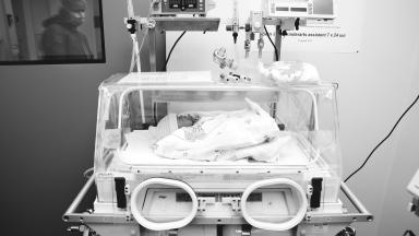
The baby goes face down in an incubator, and the sac is covered with sterile, moist, non-stick dressings. No pressure on the back, no diaper over the lesion, and gloves and equipment free of latex from the first minute.
2
The first hours
Antibiotics start to protect the exposed nerves. The neurosurgeon examines leg movement, the anus and the level of the defect, and an ultrasound or MRI looks at the brain ventricles and the kidneys.
3
The operation
Closure within 48 hours, earlier if the sac has leaked. A Colombian series of 50 babies, all closed within 48 hours and published in World Neurosurgery, reported a spinal fluid leak in 8 percent, infection in 2 percent and no deaths.
4
The neonatal unit
Feeding, wound care and daily head measurements. A bladder ultrasound and a first check of how the bladder empties come before discharge.
How the back is closed
Under the operating microscope, the neurosurgeon frees the exposed spinal cord from the surrounding skin, rolls it gently back into a tube with fine stitches so it lies smoothly inside the spinal canal, and then closes the layers over it one by one, the dura that holds the spinal fluid, the muscle and fascia beside the spine, and finally the skin. Each layer adds a seal against a fluid leak. The aim of the operation goes beyond covering the hole. Reconstructing the cord into a tube reduces the scar that later tethers it, and a watertight dura protects against meningitis. In a baby with a sharp hump in the spine, a kyphosis, the surgeon can remove part of the bent vertebrae in the same operation so the skin closes flat. Function in the legs rarely improves after the operation, because the damage happened during the pregnancy, and the operation protects what is already there.
1
The cord
Freed from the skin and rebuilt into a tube.
2
The dura
Closed watertight over the cord, with a graft if the native layer is too small.
3
Muscle and fascia
Brought together over the dura as a second seal.
4
The skin
Closed without tension, or covered with flaps when the defect is wide.
Large defects and skin cover
Wide defects need more. When the skin will not close without tension, a plastic surgeon moves flaps of skin and muscle from the back to cover it, sometimes in the same operation, and published packages from Istanbul hospitals list skin graft procedures and, where hydrocephalus is already present, a shunt in the same session as the back closure.
Hydrocephalus and the shunt question
Signs of rising pressure
A head growing faster than its curve, a tense soft spot, eyes that look downward, vomiting, poor feeding, or fluid swelling under the back wound.
Many babies need treatment for hydrocephalus in the weeks after the back closure. The closed defect no longer lets fluid escape. A ventriculoperitoneal shunt drains spinal fluid from the brain into the abdomen through a thin tube under the skin, while some babies suit an endoscopic third ventriculostomy instead, frequently combined with cauterizing the choroid plexus that makes the fluid, which spares those who respond a lifelong tube. Shunt revisions by the first birthday reached 40.2 percent after postnatal repair in the MOMS trial and 15.4 percent after prenatal repair.
Shunts can block at any age.
Risks of the operation
Spinal fluid leak from the wound. The most frequent early problem. A tight dressing and lying face down treat most, and a few need a second stitch or treatment of hydrocephalus that is pushing fluid out.
Wound breakdown and infection. More likely with wide defects closed under tension. Antibiotics and dressings treat infection, and plastic surgery closes larger gaps.
Meningitis. The reason closure happens early. Treated with intravenous antibiotics.
Breathing and swallowing problems from the Chiari II malformation. Noisy breathing, pauses in breathing or weak swallowing call for urgent assessment, and the first step usually means treating hydrocephalus.
Growing up with spina bifida
Repair begins a lifetime of care, and the coordination of that care decides how well a child does.
Bladder and bowel
Nearly every child has some effect on the nerves to the bladder. High pressure inside a bladder that does not empty can damage the kidneys silently, so a urologist follows the bladder with ultrasound and urodynamic studies from infancy, and clean intermittent catheterization, emptying the bladder with a soft tube several times a day, starts early in many children and later becomes something the child does alone. A bowel program keeps the child clean and confident at school.
Walking
Lesion level sets what the legs can do. Children with low lesions walk, many with ankle braces, and children with higher lesions use walking aids or a wheelchair for longer distances. School-age walking differed sharply in the MOMS follow-up published in JAMA Pediatrics in 2021, with 51.3 percent of children repaired before birth walking in the community against 23.1 percent after postnatal repair. Physiotherapy keeps a child moving. So does orthopedic follow-up of the hips, feet and spine.
Tethered cord
The repaired cord can stick to the scar and stretch as the child grows. New back or leg pain, worsening walking, a change in bladder control or a curve that progresses fast lead to an MRI and, when the signs fit, an untethering operation. Surgery on a scan finding alone, without symptoms, brings risk without benefit, and a seven-hospital study in Child's Nervous System found more infections and returns to theater, with no neurological gain, when the cord was untethered as a precaution before scoliosis surgery.
Latex allergy stays a lifelong precaution.
Which families travel, and when
Diagnosed during pregnancy
The family can plan the birth in Istanbul, so the closure happens within 48 hours with the neurosurgery and neonatal teams ready. Travel well before the last weeks of pregnancy, within the airline's limits for a pregnant passenger.
Baby already repaired at home
Families come for hydrocephalus that was not treated, a shunt that keeps failing, a wound that did not heal, or a second opinion on the plan.
An older child with new problems
Tethered cord symptoms, a worsening bladder, scoliosis, hip or foot deformities, or a Chiari malformation causing symptoms.
A newborn with an open back cannot safely wait days for a flight, which makes local closure the right choice for a baby already born abroad.
How long a family stays in Istanbul
A planned birth with closure of the back takes the longest. The family arrives some weeks before the due date, and after birth and the closure the baby stays in the neonatal unit until the wound has healed, feeding is established and the head size is stable, one to two weeks in most babies and longer if a shunt follows. One published Istanbul package lists four nights in a private room for the repair combined with a shunt, which suits a baby referred shortly after birth. Older children coming for untethering or shunt surgery stay around two weeks.
Flying with the baby
Flying waits for a healed back, a stable head size, steady feeding and the pediatrician's agreement. Your neurosurgeon writes the clearance date into the discharge letter, together with the shunt type and setting where one was placed.
What the discharge letter carries
The clearance date, the type and setting of any shunt, the bladder routine, latex precautions, and the signs that need a doctor the same day.
What decides the cost
Your situation sets most of the figure. A planned birth adds the delivery, the neonatal unit nights and the mother's stay, while the size and level of the defect decide whether a plastic surgeon closes the skin, and hydrocephalus adds a shunt or an endoscopic procedure, in the same operation or later. MRI of the brain and spine, urology tests and the nights in hospital all move the total.
Published packages for spina bifida surgery in Istanbul list the MRI of the brain and spine, the repair, a shunt where needed, skin graft procedures, the hospital nights with a room for a companion, airport transfers and an interpreter. Hotel or apartment nights near the hospital are billed separately, by the day.
A quote you can compare names the operation, says whether a shunt sits inside it, and counts the neonatal or ward nights. That figure comes only from a pediatric neurosurgeon looking at the scans or photographs of the back. The review costs nothing.
Arranging the trip
One coordinator follows the case from the first WhatsApp message to the flight home. The number stays open afterward. English, Arabic, French, Russian, Serbian, Romanian and Spanish are spoken by the team, with an interpreter for other languages, and a companion bed lets a parent stay in the room every night while the international patients office books the hotel for the nights before and after. The invitation letter for a medical visa, naming the hospital and the treating doctor, goes out about ten days before travel. Transfers are arranged. Halal, vegetarian and diabetic meals come from the hospital kitchen, a prayer room sits on site, and a request for a female physician goes to the department with your first message. For a planned birth, the same office books accommodation for the weeks before the due date, and the mother's pregnancy records and scans travel with her, so the obstetric and neonatal teams know the case before the first appointment.
Send the pregnancy scans or photographs of the baby's back with your first message.
Once you are back home
Spina bifida care works best through one clinic that sees the child regularly, with neurosurgery, urology, orthopedics and physiotherapy in the same place, and your local team carries that forward using the English discharge file while the coordinator passes questions and scans back to the neurosurgeon in Istanbul. Head size and development get checked frequently in the first year. Bladder tests follow the urologist's schedule.
Vomiting, drowsiness, a bulging soft spot, a headache that will not settle, fever with a red shunt track, or a sudden change in walking or bladder control. Each needs a doctor the same day. A blocked shunt or a tethered cord can be the cause.
Spina bifida repair FAQ
How soon after birth is the back closed?
Within 48 to 72 hours, earlier if the sac has leaked, to protect the nerves and prevent meningitis.
Will my child walk after the operation?
The level of the defect decides most of it. The repair protects the nerves that still work but rarely restores lost function, and children with low lesions usually walk, many with braces.
Does every child with spina bifida need a shunt?
No. In the MOMS trial, 82 percent of babies repaired after birth had a shunt by the age of one, against 40 percent after prenatal repair, and some children are treated with an endoscopic procedure instead.
Is prenatal surgery better than repair after birth?
Prenatal repair reduced shunts and improved walking in the MOMS trial, at the cost of a higher risk of preterm birth and a weaker womb scar. It suits only pregnancies that meet strict criteria before 26 weeks.
What is a tethered cord?
The repaired spinal cord sticking to its scar and stretching as the child grows. New pain, weakness or bladder changes lead to an MRI and sometimes an untethering operation.
Can both parents come?
Yes. One parent stays in the room on the companion bed, and the international patients office arranges the hotel for the other.
References
Adzick NS, Thom EA, Spong CY, et al. A randomized trial of prenatal versus postnatal repair of myelomeningocele. N Engl J Med. 2011;364(11):993-1004.
Tulipan N, Wellons JC, Thom EA, et al. Prenatal surgery for myelomeningocele and the need for cerebrospinal fluid shunt placement. J Neurosurg Pediatr. 2015;16(6):613-620.
Houtrow AJ, Thom EA, Fletcher JM, et al. Prenatal repair of myelomeningocele and school-age functional outcomes. Pediatrics. 2020;145(2):e20191544.
Houtrow AJ, MacPherson C, Jackson-Coty J, et al. Prenatal repair and physical functioning among children with myelomeningocele: a secondary analysis of a randomized clinical trial. JAMA Pediatr. 2021;175(4):e205674.
Acosta-Medina E, Zorro-Guío OF, Abdala-Vargas NJ, et al. Postnatal surgical correction of myelomeningoceles: preoperative and intraoperative risk factors associated with postoperative neurologic outcomes. World Neurosurg. 2023;170:e629-e638.
Goldstein HE, Shao B, Madsen PJ, et al. Increased complications without neurological benefit are associated with prophylactic spinal cord untethering prior to scoliosis surgery in children with myelomeningocele. Childs Nerv Syst. 2019;35(11):2187-2194.
Editor's note
Written by the Biruni Hospital medical editorial team. Reviewed by Prof. Dr. Hilmi APAK, Pediatric Hematology and Oncology.