In one recent series, 89 percent of children with a tumor in the eye socket kept the eye when surgery stopped at a biopsy, because with rhabdomyosarcoma the site decides whether an operation or radiotherapy controls the tumor.
About This Department
Pediatric Surgery
With rhabdomyosarcoma, where the tumor sits decides the operation. Sometimes the right operation is a biopsy and nothing more.
The American Cancer Society puts five-year survival in the low-risk group at about 70 percent to over 90 percent, and the way the tumor at its original site is controlled, by surgery, radiotherapy or both, helps keep a child in that range.
Over 90%
Five-year survival in the most favorable low-risk groups
94%
Five-year survival for localized tumors beside the testicle
89%
Eyes kept after biopsy-only surgery for orbital tumors
Free
Review of the scans and slides before you book
Free consultation
The review costs nothing and carries no obligation. Send the MRI and CT on disc, the biopsy report with the fusion test if it was done, and any operation note, and a pediatric surgeon and a pediatric oncologist will tell you which local treatment fits the site and whether a second operation is needed. A coordinator replies in your own language.
A biopsy has shown rhabdomyosarcoma, and the word surgery has come up alongside chemotherapy and radiotherapy. Rhabdomyosarcoma surgery covers several different operations, from a biopsy and a lymph node sample at the start to the removal of what remains of the tumor after chemotherapy, and in some places in the body no major operation at all. Where the tumor sits decides most of it. Below you will find how the team chooses between surgery and radiotherapy for each site, why the first operation matters so much, how long a family stays in Istanbul, and what follow-up looks like once you are home.
A muscle-type cancer that can start almost anywhere
Rhabdomyosarcoma grows from immature cells that would become muscle, and it accounts for half of all sarcomas in children. Those cells exist all over the body. So the tumor turns up in places that have little to do with muscle, the head and neck first, the eye socket included, then the bladder, prostate, vagina and the tissue beside the testicle, and then the arms, legs and trunk. Most children are under ten at diagnosis. Teenagers form a second, smaller peak.
Symptoms depend entirely on where the tumor grows, which explains why diagnosis sometimes takes weeks. Around the eye it shows as swelling of the lid or an eye pushed forward over a few weeks, while deeper in the head it looks like a blocked nose, an ear infection that will not settle, or bleeding from the nose or ear. A painless swelling in the scrotum, trouble passing urine, or blood in it point to the pelvis, and in small girls a grape-like mass can appear at the vaginal opening, the botryoid form, which carries a good outlook. In a limb or on the trunk the tumor presents as a firm lump that keeps growing. An MRI of the area, a CT of the chest, a PET-CT or bone scan and bone marrow samples then stage the disease, and a biopsy with the fusion test gives the type, all within the first one or two weeks. No major operation belongs in that window.
Two types matter for treatment.
On a phone, swipe the table sideways to read every column.
The two main types
Feature
Embryonal
Alveolar
Typical age
Younger children
Teenagers more than young children
Typical sites
Head and neck, bladder, prostate, vagina, beside the testicle
Arms, legs and trunk
PAX-FOXO1 gene fusion
Absent
Present in a majority
Outlook
The better of the two
Poorer, especially with the fusion
Alveolar rhabdomyosarcoma carries a gene fusion between PAX3 or PAX7 and FOXO1 in a majority of cases, and laboratories now test for that fusion directly, because the fusion predicts behavior better than the appearance under the microscope does. A 2025 surgical update in Pediatric Blood and Cancer by surgeons from the North American and European groups describes fusion status replacing histology in the newer risk systems.
The gene fusion, more than the look under the microscope, now sets the risk.
Surgery, radiotherapy or both, depending on the site
Every child with rhabdomyosarcoma receives chemotherapy. The open question for the surgeon concerns local control, meaning how the tumor at its original site gets permanently dealt with, and the answer turns on whether removing it would cost a function the child needs. A tumor in the thigh muscle can come out completely with a rim of healthy tissue and little lasting harm. Behind the eye or at the base of the skull it cannot. Radiotherapy carries the load there, and surgery stops at the biopsy.
The rule in every protocol says the same thing. Remove the tumor completely if that leaves a working child, and never take a mutilating operation when radiotherapy can do the job.
This table scrolls sideways on a narrow screen. Swipe or drag to see every column.
How local control usually works by site
Site
Main local treatment
Role of surgery
Eye socket (orbit)
Radiotherapy
Biopsy only. Removing the eye stays a rescue option for a tumor that returns.
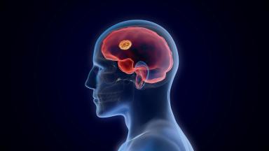
Head and neck, base of skull
Radiotherapy
Biopsy, and removal only for small tumors in accessible places.
Beside the testicle
Surgery
Removal of the testicle and cord through the groin, plus lymph node checks in older boys.
Bladder and prostate
Radiotherapy, surgery or both
Bladder-sparing removal where chemotherapy has shrunk the tumor enough.
Vagina and uterus
Chemotherapy, with local treatment for any residue
Limited surgery, often with internal radiotherapy, to keep the organs.
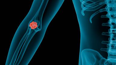
Arms, legs and trunk
Surgery, radiotherapy or both
Wide removal when it spares function, and regional lymph node sampling.
Why the first operation matters so much
Biopsy first
A lump in a child that could be a sarcoma gets a biopsy and a scan before anyone tries to take it out.
Many children with rhabdomyosarcoma arrive after a lump was removed somewhere else as if it were harmless, through a cut placed for a benign lump, with no margin of healthy tissue and no plan for what comes next. That operation leaves tumor cells behind along the cut and in the tissue it opened. A study from the German Cooperative Weichteilsarkom group, published in the Annals of Surgical Oncology in 2024, looked at 196 boys and young men with rhabdomyosarcoma beside the testicle and found protocol violations in the first operation in 42 percent of the children with localized disease. Those violations raised the rate of incomplete removal from 21 percent to 76 percent and sent most of those children back for a second operation, and after that second operation, five-year survival reached 97.6 percent when the margins came back clear and 81.8 percent when they did not. The commonest violation in that site is a cut through the scrotum, which opens a new route for spread, where the standard operation goes through the groin and ties off the cord first.
Primary re-excision
Children with an incomplete first operation do not simply move on to chemotherapy. Where the scar and the tumor bed can be removed again with a clear margin before chemotherapy starts, without harming function, the surgeon does exactly that, an operation called primary re-excision. A clean second operation can bring the child back to the risk group the original tumor deserved.
What to send first
The operation note, the pathology report and any scans from the first surgery, so the surgeon can see where the cut went and what it left.
Site by site
Each site calls for a different operation. Four situations come up again and again.
The eye socket
Orbital rhabdomyosarcoma carries one of the best outlooks of any site, and surgery stays small. A series of 27 children from the Children's Hospital of Philadelphia, published in 2026, kept the eye in 89 percent of those who had a biopsy only, against 78 percent after an attempt to debulk the tumor, and local recurrence came out lower with the smaller operation.
For a tumor in the eye socket, the biopsy is the operation.
If the tumor comes back
Removing the eye remains available as a rescue operation for a tumor that returns after radiotherapy.
The bladder and prostate
Here the aim moves from cure alone to cure with a working bladder. Chemotherapy goes first, and the team then chooses between radiotherapy, a partial removal of the bladder wall, or both, depending on how much tumor remains and where it sits relative to the bladder neck and the ureters. Removal of the whole bladder stays a last resort. Surgeons keep it for tumor that persists or returns.
Beside the testicle
Surgeons remove the tumor with the testicle and the spermatic cord through a cut in the groin. Boys aged ten or older, and any boy whose scans show enlarged nodes, also need the lymph nodes behind the abdomen checked surgically, because spread there changes the treatment.
Arms, legs and the chest wall
Limb tumors carry the alveolar type and spread to lymph nodes more frequently than tumors elsewhere, which makes node sampling part of every limb operation, and wide removal with a clear margin works well where the tumor sits in muscle the child can spare. Near a major nerve or vessel, radiotherapy takes over part of the job. For the chest wall, an international consensus from the INSTRuCT consortium, published in Pediatric Blood and Cancer, sets out when to remove ribs, when to rely on radiotherapy, and how to rebuild the wall afterward.
Lymph nodes and the sentinel node
Tumor in the lymph nodes moves a child into a higher risk group and adds radiotherapy to that node area, so the nodes get checked before treatment is planned. Enlarged nodes on a scan get a needle biopsy. For limb and trunk tumors with normal-looking nodes, a sentinel lymph node biopsy finds the first node that drains the tumor, using a tracer injected beside it, and removes that node alone through a small cut. Surgeons from both cooperative groups describe the technique spreading in the 2025 surgical update.
Where surgery sits in the treatment year
Treatment runs as a sequence, and surgery can enter it at three points.
1
Diagnosis, weeks one to two
Imaging of the tumor with MRI, a CT of the chest and a PET-CT or bone scan to look for spread, bone marrow samples, and a biopsy, often a core needle biopsy through the skin. Node sampling where needed.
2
Removal at the start
Only when the tumor can come out completely and safely without harming function, as in most tumors beside the testicle and some small limb tumors.
3
Chemotherapy, then a decision
In European protocols, ifosfamide, vincristine and actinomycin D, with doxorubicin for some children, in three-week cycles. After the first cycles a new MRI shows the response, and the team decides on surgery, radiotherapy or both for the original site.
4
Delayed removal
Surgery on what remains, timed so radiotherapy can follow without a long gap. Margins and the amount of surviving tumor guide the rest.
5
Completion and maintenance
The remaining cycles, around six months of treatment in total for standard regimens, and for high-risk tumors six further months of maintenance chemotherapy.
Why surgeons plan for fast healing
The next chemotherapy cycle waits for the wound to heal, so the operation is planned around a wound that closes quickly and a child who is back on schedule within days.
Risk group and outlook
What sets the risk group
Age, site, size, the fusion status, the completeness of the first operation, lymph node involvement and distant spread.
American Cancer Society figures put five-year survival at about 70 percent to over 90 percent for children in the low-risk group, 50 to 70 percent in the intermediate-risk group, and 20 to 30 percent in the high-risk group, which includes most children whose disease had spread at diagnosis. Those figures cover children treated years ago, and the maintenance results below came later.
Maintenance changed the picture for high-risk tumors.
In the RMS 2005 trial of the European pediatric Soft tissue sarcoma Study Group, published in the Lancet Oncology in 2019, 371 children and young people in remission after standard treatment were randomly assigned to stop or to continue with six months of vinorelbine and low-dose oral cyclophosphamide. Five-year overall survival rose from 73.7 percent to 86.5 percent with maintenance. Grade 3 or 4 neutropenia, a drop in infection-fighting white cells, affected 82 percent of the children on maintenance, and infection 31 percent, and the trial investigators judged the toxicity manageable.
Five-year survival with maintenance chemotherapy reached 86.5 percent in RMS 2005, against 73.7 percent without it.
After the operation
Hospital stay
From a night or two after a sentinel node biopsy or a small limb excision to a week or more after a bladder-sparing operation or an abdominal node dissection.
Recovery depends entirely on what was done. A urinary catheter stays in for some days after bladder surgery. One parent stays in the room on the companion bed throughout.
Wound problems. Infection or slow healing, more likely where radiotherapy came first. Dressings and antibiotics treat most, and a plastic surgeon closes the larger defects.
Swelling and stiffness after limb surgery. Physiotherapy starts in the first days and continues at home.
Bladder function after pelvic surgery. Frequency, urgency or leaking in the first months, followed with bladder scans and urodynamic tests.
Lymph fluid collections. After node surgery, managed with drainage in clinic.
When the pathology changes the plan
Clear margins, dead tumor
Radiotherapy at that site can shrink or drop out of the plan.
Tumor at the margin or still alive
Radiotherapy comes back into the plan for that site.
Pathology results on the removed tissue arrive within days. Radiotherapy and chemotherapy are both delivered at Biruni Hospital, on the same site as the surgery, so a child whose plan grows continues with the team that operated, using the same scans and the surgeon's own record of where the tumor lay and where the marker clips sit. Families who would rather continue at home leave with a written handover for their own pediatric oncologist, the slides and blocks, every scan on disc and the remaining protocol written out cycle by cycle, so the next dose lands on the right day wherever it is given.
Every change goes back to the tumor board before the next cycle.
Time in Istanbul
Families come for one of three things, and each takes a different length of time. A second opinion on the diagnosis and the plan, with a review of the slides, the scans and the fusion test, can be done within a week. Local control alone takes two to four weeks. That covers the operation and the ward days, with chemotherapy given at home before and after, and the exact length depends on the site and the size of the operation. A full course here runs for months, because standard chemotherapy lasts around six months and radiotherapy adds daily weekday sessions over several weeks, with published hospital pages in Istanbul describing four to six weeks of radiotherapy after surgery and six to eight weeks without it. A package published by one Istanbul hospital for a full treatment year lists sixteen nights in hospital across forty-eight weeks, which shows how much of the treatment runs as day care. Families who start here can split the treatment, with the planning, the operation and the radiotherapy in Istanbul and the remaining chemotherapy at home under a written protocol, and the team agrees that split before you book flights.
Flying home
Flying waits for a healed wound, blood counts that have recovered from the last cycle, and a child eating and moving normally. The surgeon writes the clearance date into the discharge letter.
What drives the cost
Two children with rhabdomyosarcoma can receive quotes that differ several times over. The treatment differs that much.
Tumor site sets the operation, from a node biopsy to pelvic surgery with reconstruction, and radiotherapy adds weeks of daily sessions and, in very young children, a short general anesthetic for each one. The number of chemotherapy cycles given in Istanbul, a central line or port, maintenance chemotherapy for high-risk tumors, repeat MRI and PET-CT scans, the fusion test, and the nights in hospital each move the total. A first operation elsewhere that needs re-excision adds one more operation to the plan.
Published packages for pediatric cancer treatment in Istanbul list the consultations, the scans and biopsies, the operation and anesthesia, the hospital stay, standard medicines, airport transfers and an interpreter. Hotel or apartment nights near the hospital are billed separately, by the day.
A figure worth comparing names the site and the operation, counts the chemotherapy cycles and the radiotherapy sessions it covers, and says whether scans, the port and the fusion test sit inside it. Only a pediatric surgeon and a pediatric oncologist reading your child's own reports can produce one. The review costs nothing.
Traveling with a sick child
A coordinator takes your case from the first message, stays with you until discharge, and keeps the same WhatsApp number after you fly home, and the team works in English, Arabic, French, Russian, Serbian, Romanian and Spanish, with interpreters arranged for other languages. One parent sleeps in the child's room on a companion bed, and the international patients office books the hotel for the nights either side of each admission. The invitation letter for a medical visa goes out about ten days before travel. Airport transfers and transport between hotel and hospital are arranged. Halal, vegetarian and diabetic meals come from the hospital kitchen, a prayer room sits on site, and a request for a female physician goes to the department with your first message.
Bring the pathology blocks or slides as well as the report.
Once you are back home
Most relapses of rhabdomyosarcoma happen in the first two to three years. The original site comes first. Follow-up therefore leans on MRI of that site and a chest CT or X-ray every few months at first, spacing out over the years, with your own pediatric oncologist arranging the scans locally and the coordinator passing the images back to the team in Istanbul for a second look.
Late effects need watching for years. Growth of the bones and soft tissue in a radiotherapy field. Hearing, the heart and the kidneys after certain drugs. Fertility after cyclophosphamide or pelvic radiotherapy. Bladder function after pelvic treatment. A survivorship clinic follows all of these into adult life.
Any new lump, pain at the original site, fever during chemotherapy, a persistent cough or blood in the urine. Each needs a doctor the same day. The local emergency department sees the child first, using the English discharge file, and the coordinator relays the details to the surgeon who operated.
Rhabdomyosarcoma surgery FAQ
Can rhabdomyosarcoma be cured with surgery alone?
No. Every child also receives chemotherapy, and many receive radiotherapy, because the tumor spreads microscopically before it shows on scans. Surgery controls the original site in the places where it can remove the tumor without costing function.
Is surgery or radiotherapy better for rhabdomyosarcoma?
Neither wins everywhere. Surgery suits tumors beside the testicle and many limb tumors, and radiotherapy suits the eye socket and the base of the skull. Bladder, prostate and some limb tumors use both.
What is the survival rate for rhabdomyosarcoma?
The American Cancer Society reports five-year survival of about 70 percent to over 90 percent in the low-risk group, 50 to 70 percent in the intermediate-risk group and 20 to 30 percent in the high-risk group.
My child's lump was removed before anyone knew it was cancer. What now?
Send the operation note, the pathology report and the scans. Many children in that position need a second operation, called primary re-excision, to remove the scar and tumor bed with a clear margin before chemotherapy.
How long is the hospital stay after rhabdomyosarcoma surgery?
From a night or two after a sentinel node biopsy or a small limb excision to a week or more after pelvic or abdominal surgery.
Can both parents come?
Yes. One parent sleeps in the room on the companion bed, and the international patients office arranges the hotel for the other.
References
Rogers T, Schmidt A, Buchanan AF, et al. Rhabdomyosarcoma surgical update. Pediatr Blood Cancer. 2025;72(Suppl 2):e31496.
Schermerhorn SMV, Routh JC, Polites SF, Dasgupta R. Management of rhabdomyosarcoma in pediatric patients. Surg Oncol Clin N Am. 2026;35(4):731-748.
Martynov I, Sparber-Sauer M, Heinz A, et al. Importance of adequate surgical local control in fusion-negative para-testicular rhabdomyosarcoma: data from the Cooperative Weichteilsarkom Studiengruppe trials (CWS-96 and CWS-2002P) and the European Soft Tissue Sarcoma Registry (SoTiSaR). Ann Surg Oncol. 2024;31(9):6209-6219.
Katowitz WR, Zhao AT. Biopsy-only versus debulking surgical approaches for pediatric orbital rhabdomyosarcoma: impact on eye preservation and oncologic outcomes. Am J Ophthalmol. 2026;286:211-217.
Schmidt A, Guillén G, Luo LY, et al. Management of pediatric and adolescent rhabdomyosarcoma of the chest wall: an expert consensus opinion from the International Soft Tissue Sarcoma Consortium (INSTRuCT). Pediatr Blood Cancer. 2026;73(1):e32117.
Bisogno G, De Salvo GL, Bergeron C, et al. Vinorelbine and continuous low-dose cyclophosphamide as maintenance chemotherapy in patients with high-risk rhabdomyosarcoma (RMS 2005): a multicentre, open-label, randomised, phase 3 trial. Lancet Oncol. 2019;20(11):1566-1575.
Editor's note
Written by the Biruni Hospital medical editorial team. Reviewed by Assistant Professor Ömer DOĞRU, Pediatric Hematology and Oncology.