A ventriculoperitoneal shunt, the VP shunt, drains excess cerebrospinal fluid from the brain's ventricles into the abdomen, where the body absorbs it. A thin catheter sits in the ventricle, a valve behind the ear controls the flow, and a tube runs under the skin of the neck and chest into the abdominal cavity. Nothing shows from outside except a small bump behind the ear.
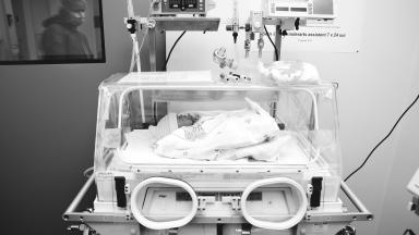
Children need one for hydrocephalus.
The causes range from bleeding in the brain of a premature baby, spina bifida and a narrowed channel between the ventricles (aqueductal stenosis) to brain tumors and meningitis. Some babies need a temporary drain or a reservoir first, while the protein and blood in the fluid clear, because a shunt placed into bloody fluid blocks early. Children with spina bifida receive their shunt in the days after the back is closed, and those with a tumor in many cases lose the need for a shunt once the tumor comes out, so the neurosurgeon times the operation to the cause. Adults with normal pressure hydrocephalus, the walking, memory and bladder problem of later life, receive the same operation with a valve set to drain more slowly. A lumbar drain trial for a few days shows first whether they will improve.
How does a VP shunt work, and who needs one?
The brain makes close to half a liter of cerebrospinal fluid every day, and in hydrocephalus that fluid cannot drain or absorb as it should, so the ventricles widen and pressure rises. In babies the head grows faster than it should and the soft spot bulges, while older children and adults get headaches that are worse in the morning, vomiting, sleepiness, blurred or double vision and, when pressure has been high for a long time, a slow slide in school work or walking that parents notice before anyone else.
Revisions belong to shunt life.
Cerebrospinal fluid shunting has a propensity for mechanical failure and patients with VP shunts should receive follow-up through the transition to adulthood.
Rochester series, 64 children followed for 20 years, Journal of Neurosurgery Pediatrics, 2013
Shunt or endoscopic third ventriculostomy
Some children, above all those with aqueductal stenosis and older than six months, can avoid a shunt altogether through an endoscopic third ventriculostomy, a small opening made inside the brain that lets fluid bypass the blockage. Babies under six months and children whose fluid is not absorbed do better with a shunt. The neurosurgeon reads the MRI and explains which route fits.
It confirms that a young age and the use of the endoscope are risk factors for first shunt failure and that valve type has no impact.
Hydrocephalus Clinical Research Network, 1,036 children, Journal of Neurosurgery Pediatrics, 2016
Choosing the valve
Valves come in two main kinds. Fixed-pressure valves open at one set pressure, and programmable valves let the surgeon change the setting with a magnet held over the skin, without surgery. The large North American study of 1,036 children found no difference in how long either kind lasted.
Programmable valves and MRI scans
An MRI magnet can reset many programmable valves. After any MRI the setting is checked with a handheld reader or an X-ray, and reset on the spot. Airport scanners, phones and headphones held away from the valve do not affect it.
Devices against over-drainage
When a tall child or an adult stands up, gravity pulls fluid down the tube faster, which can cause headaches when upright. Gravity-assisted and anti-siphon devices added to the valve slow the flow in the upright position, and the surgeon adds one for older, active patients.
Four things guide the choice.
- Age and size, since a newborn's skin is thin over the valve.
- The cause of hydrocephalus.
- Whether the child will need regular MRI scans, for example after a brain tumor.
- The surgeon's experience with the valve and the family's access to a center that can read and reset it at home.
No valve outlasts the others
Valve design made no difference. In the Hydrocephalus Clinical Research Network study the risk of an early failure came from the child, an age under six months and a heart condition, and from endoscopic placement of the catheter. Families who worry they chose the wrong valve can set that worry down.
Catheters coated with antibiotics
Many surgeons use catheters impregnated with antibiotics or silver to lower the infection risk, together with a strict theater routine. The routine, from hair clipping to double gloves and a minimum of people in the room, matters as much as the catheter.
This table scrolls sideways on a narrow screen. Swipe or drag to see every column.
Shunt routes compared
| Shunt |
Where the fluid goes |
When it is used |
| Ventriculoperitoneal (VP) |
The abdominal cavity |
First choice for children and adults |
| Ventriculoatrial (VA) |
A large vein leading to the heart |
When the abdomen cannot absorb fluid after infection or surgery |
| Ventriculopleural |
The space around the lung |
Older children and adults when the abdomen is unsuitable |
| Lumboperitoneal |
The abdomen, from the lower spine |
Some adults with normal pressure hydrocephalus or raised pressure without large ventricles |
The operation
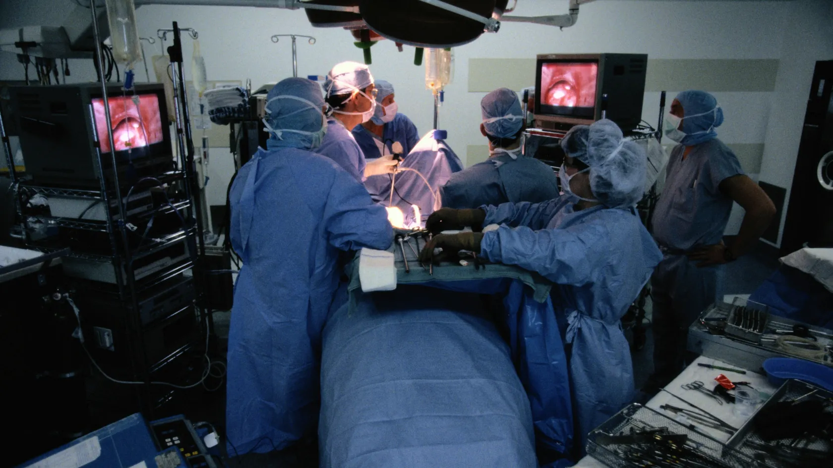
Under general anesthesia, in close to an hour, the surgeon makes a small cut behind the ear and a second on the abdomen, places the ventricular catheter through a small hole in the skull, tunnels the tube under the skin and lays the end in the abdomen. The two wounds close with dissolving stitches.
Endoscopic insertion of the initial VP shunt in children suffering from hydrocephalus did not reduce the incidence of shunt failure.
Randomized trial of 393 children, 16 centers, Journal of Neurosurgery, 2003
This table scrolls sideways on a narrow screen. Swipe or drag to see every column.
From admission to the flight home
| Stage |
Timing |
What happens |
| Admission |
The day before or the morning of surgery |
Blood tests, a fresh scan if needed, the anesthesia visit |
| Operation |
Close to one hour |
Shunt placed, child wakes in recovery |
| Ward |
Two to three nights |
Pain relief, feeding, a scan to check catheter position |
| Wound check |
Seven to ten days after surgery |
Stitches and wounds checked in clinic |
| Flight home |
After the wound check |
Air travel does not affect a working shunt |
Pain, scars and hair
Pain settles within a few days with paracetamol and ibuprofen. The surgeon shaves only a small patch, the hair grows back over the scar behind the ear, and the abdominal scar fades to a thin line.
When a shunt fails
Why shunts block
Tissue grows into the catheter holes. It comes from the brain or from the choroid plexus, the tissue that makes the fluid. The abdominal end can be blocked by tissue or pulled out as a child grows, and the tube can break or come apart at a connection.
This table scrolls sideways on a narrow screen. Swipe or drag to see every column.
Why shunts needed revision in 64 children followed for 20 years
| Cause |
Share of revisions |
What the surgeon replaces |
| Blocked ventricular catheter |
27 percent |
The catheter in the brain, and the valve if it is blocked too |
| Blocked abdominal catheter |
15 percent |
The abdominal end, through a small cut |
| Disconnection or break |
11 percent |
The broken section |
| Infection |
9 percent |
The whole shunt, after a course of antibiotics |
- A CT or rapid MRI, compared with the last scan.
- X-rays along the whole tube, to look for a break.
- A needle sample from the valve in some cases, to measure pressure and test for infection.
- Surgery to replace the failed part, the same day when the child is drowsy.
Shunt infection
In 7,071 children treated in 41 US children's hospitals, 11.7 percent had a shunt infection within two years of the first operation, most in the first months and most in babies. Fever, redness along the tube, a leaking wound or tummy pain bring a fluid test. Treatment removes the shunt, places a temporary external drain and gives antibiotics for ten to fourteen days, then a new shunt goes in.
Life with a shunt
Children with a shunt go to school, swim and play most sports.
1
First weeks
Wound care, a check at seven to ten days, and a scan in the first three months to set the baseline.
2
First year
Head measurements and development checks for babies, and a scan if symptoms appear.
3
For life
Yearly review with a neurosurgeon, an eye examination for children who cannot describe symptoms, and a planned handover to an adult neurosurgeon.
The patient's age at the time of initial shunt placement and the time interval since previous surgical revision are important predictors of repeated shunt failures in the multivariable model.
839 children, Hospital for Sick Children Toronto, Journal of Neurosurgery, 2000
Go to an emergency department the same day
Headache with vomiting, unusual sleepiness, a bulging soft spot or fast-growing head in a baby, eyes that look downward (sunset eyes), a new squint, a seizure, or fever with redness along the tube. A child with these signs and a shunt needs a scan within hours.
Travel, cost and follow-up at home
Overall unadjusted 24-month CSF shunt infection rates were 11.7% per patient and 7.2% per procedure.
41 US children's hospitals, 7,071 children, Journal of Neurosurgery Pediatrics, 2009
Infection, the complication families fear most, shows up within the first weeks after surgery in most children. The wound check therefore comes before the flight home. Babies carry the highest risk. The coordinator stays on the same WhatsApp number once you are back home, and the neurosurgeon reviews scans and photos of the wound sent from your local doctor.
Your companion, your language, your food
Each room has a companion bed, so one parent sleeps beside the child on the ward. Before you fly, the international patients office sends the letter your consulate asks for with a medical visa, ten days ahead, and it books the hotel for the nights around the admission, the pickup at the airport and the car to the hospital each day.
Messages, consultations and the consent talk run in English, Arabic, French, Russian, Serbian, Romanian or Spanish through your coordinator, with interpreters for other languages. Halal, vegetarian and diabetic food comes from the hospital kitchen, there is a prayer room, and the department arranges a female doctor on request when the rota allows.
The valve weighs most in the cost. Programmable valves, anti-siphon devices and antibiotic-coated catheters cost more than a fixed valve and a plain catheter, and a revision that replaces one section differs from a complete new shunt. Published packages for shunt surgery in Turkey include the operation, anesthesia, the valve named in the plan, two to three ward nights, the interpreter and transfers, and leave out flights, insurance and the treatment of complications.
1
Send the scans
CT or MRI and any operation notes, by WhatsApp or e-mail.
2
Neurosurgeon review
Whether a shunt, a revision or an endoscopic operation fits, and which valve.
3
Written plan and estimate
The plan, the stay and an estimate for that plan, free and in your language.