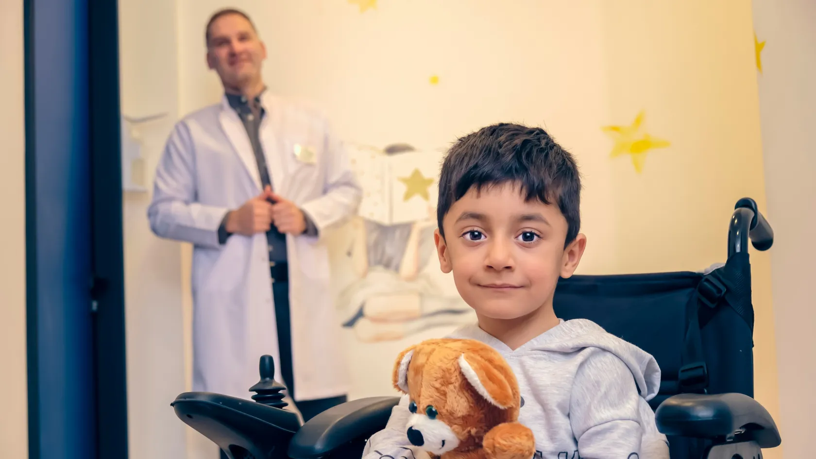
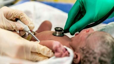
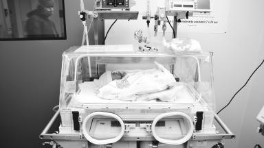
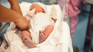
Pediatric surgical oncology is the surgery of solid tumors in children, from biopsy and port placement to removal of neuroblastoma, Wilms tumor, hepatoblastoma, sarcomas and germ cell tumors at the right moment between chemotherapy cycles. At Biruni Hospital in Istanbul, our pediatric surgeons plan every operation at the tumor board with oncologists, radiologists and pathologists, use keyhole and image-guided techniques when safe, and protect fertility before treatment.
About This Department
Pediatric Surgery
Most solid tumors in children are cured only when surgery removes them completely. Pediatric surgical oncology plans that operation with the whole cancer team.
Pediatric surgical oncology is the surgery of solid tumors in babies, children and teenagers, including neuroblastoma, Wilms tumor, hepatoblastoma, soft tissue sarcomas and germ cell tumors. The surgeon takes the biopsy that confirms the diagnosis, places the port for chemotherapy, removes the tumor at the right moment between chemotherapy cycles and protects the organs a growing child needs. Imaging decides much of this. In neuroblastoma, children whose scans showed image-defined risk factors, such as a tumor wrapped around major vessels, faced 2.45 times the risk of incomplete removal and 2.3 times the risk of surgical complications in a review of 1,132 patients (Parhar and colleagues, Pediatric Surgery International, 2020). Planning around these factors changes the timing and the approach. This page explains the tumors we treat, biopsy, neuroblastoma surgery, ports and central lines, keyhole and image-guided techniques, fertility protection, recovery, and how families from abroad arrange pediatric cancer surgery at Biruni Hospital in Istanbul.
20
Image-defined risk factors checked on scans before neuroblastoma surgery
2.3
Times higher risk of surgical complications when those risk factors are present
90
Percent five-year event-free survival for localized neuroblastoma without risk factors
Free
Review of your child's scans and pathology by our pediatric tumor board
Children's tumors differ from adult cancers. They grow fast, respond well to chemotherapy and arise in growing organs.
Who works with the surgeon
Specialist
Role
Note
Pediatric oncologist
Chemotherapy and protocol
Leads overall treatment
Pediatric radiologist
Staging scans, image-guided biopsy
Reads risk factors
Pathologist
Diagnosis, margins, molecular tests
Decides the next step
Radiation oncologist
Radiotherapy when needed
Planned before surgery
1
One tumor board
Every child is discussed at the weekly pediatric tumor board, before the biopsy and again before surgery.
International protocols
Treatment follows the international protocols of SIOP Europe and the Children's Oncology Group, the same standards used in large children's cancer centers worldwide.
Tumors we operate on
Solid tumors make up around half of childhood cancers outside the brain and blood. Each type has its own surgical rules, and the surgeon's experience with that type matters as much as general skill.
Does every child with a solid tumor need surgery?
No. Some tumors, such as certain lymphomas, are treated with chemotherapy alone after a biopsy, and some low risk neuroblastomas in infants shrink without treatment. The tumor board decides who needs an operation and when.
Common solid tumors of childhood
Tumor
Usual age
Main surgery
Neuroblastoma
Under five years
Resection after chemotherapy for most
Wilms tumor
Two to five years
Nephrectomy
Hepatoblastoma
Under three years
Liver resection or transplant
Rhabdomyosarcoma and other sarcomas
Any age
Wide excision
Germ cell tumors and teratomas
Newborns and teenagers
Excision with organ sparing
Thyroid and adrenal tumors
Teenagers
Thyroidectomy, adrenalectomy
Surgery at the right moment
Timing is part of the treatment.
A tumor removed too early may take healthy organs with it, while chemotherapy given first can shrink it away from vessels and nerves and make a safe complete operation possible, and in other tumors an early operation spares the child chemotherapy altogether, so the decision rests on the tumor type, the scan findings and the international protocol for that tumor, discussed openly with the family before the first treatment. Our surgeons attend the tumor board from the first day, so the surgical plan is part of the treatment plan from the start.
No surprises, no rushed operations.
Biopsy first
A biopsy confirms the tumor type and provides tissue for the molecular tests that decide risk and treatment. Most biopsies use a needle under ultrasound or CT guidance, through a skin opening of a few millimeters. Keyhole or open biopsy is chosen when more tissue is needed.
Biopsy methods
Method
When
Anesthesia
Image-guided needle biopsy
Most abdominal and chest tumors
General anesthesia, day case
Keyhole biopsy
Deep tumors, lymph nodes
General anesthesia, one night
Open biopsy
Large tissue needs, some sarcomas
General anesthesia
1
Same anesthetic
We combine biopsy, port placement, bone marrow tests and scans in one anesthetic whenever possible.
Some tumors should not be biopsied. A typical Wilms tumor treated by the SIOP approach, and some germ cell tumors with clear blood markers, start treatment without a biopsy.
Neuroblastoma and risk factors
Neuroblastoma arises from nerve tissue, most often in the adrenal gland, and can wrap around major blood vessels and the airway or grow into the spinal canal.
1
Image-defined risk factors
Twenty findings on MRI or CT, such as encasement of the aorta, renal vessels or superior mesenteric artery, that make surgery at diagnosis risky.
L1 and L2
In the international staging system, a localized tumor without risk factors is stage L1 and one with risk factors is L2. Among 661 European children, five-year event-free survival was 90 percent for L1 and 78 percent for L2 (Monclair and colleagues, Journal of Clinical Oncology, 2009).
How staging guides surgery
Stage
First step
Surgery
L1, no risk factors
Surgery alone for most
Resection at diagnosis
L2, risk factors present
Chemotherapy or observation
Resection when risk factors resolve
M, metastatic
Intensive chemotherapy
Resection of the primary after induction
MS, infants
Observation for most
Rarely needed
The neuroblastoma operation
1
The aim
Remove as much tumor as possible while keeping the kidneys, vessels and nerves intact. A small piece left on a vessel is safer than losing an organ.
After chemotherapy
Scans after chemotherapy show whether risk factors have resolved. The surgeon peels the tumor away from the vessels it surrounds, working along each artery, a technique that takes hours and rewards experience.
When the tumor cannot be removed completely
In intermediate and high risk neuroblastoma, removing most of the tumor while protecting organs gives results close to complete removal. Radiotherapy, chemotherapy and immunotherapy treat what remains. We explain the expected extent of surgery before the operation.
2
Risks we watch for
Bleeding, kidney injury from vessel damage, diarrhea after nerve injury around the mesenteric artery, and chyle leak. Each has a plan, and most settle within weeks.
Kidney and liver tumors
Kidney tumors
Tumor
Treatment
Our page
Wilms tumor
Chemotherapy, then nephrectomy
Nephrectomy in children
Tumors in both kidneys
Partial nephrectomy on both sides
Same page
Other kidney tumors
Surgery by type
Tumor board
Wilms tumor and hepatoblastoma each have their own detailed page on this site, covering staging, operations and follow-up.
Liver tumors
Tumor
Treatment
Our page
Hepatoblastoma
Cisplatin, then resection
Hepatoblastoma surgery
Unresectable liver tumors
Living donor transplant
Pediatric liver transplant
Benign liver tumors
Observation or resection
Tumor board
Why links matter
A child whose liver tumor may need a transplant is referred to the transplant team at diagnosis, and a child with tumors in both kidneys is planned for kidney sparing surgery from the first scan.
Sarcomas and germ cell tumors
Rhabdomyosarcoma, the most common soft tissue sarcoma of childhood, arises in the head and neck, bladder and prostate, limbs or genital area. Surgery removes the tumor with a margin of normal tissue when this is possible without disabling loss of function, and radiotherapy controls what surgery cannot.
Sarcoma and germ cell tumor surgery
Tumor
Surgery
Note
Rhabdomyosarcoma
Wide excision, sentinel node biopsy for limbs
Organ sparing first
Non-rhabdomyosarcoma soft tissue sarcoma
Wide excision
Surgery is the main treatment
Ovarian germ cell tumor
Removal of the ovary, keeping the other
Fertility sparing
Testicular tumor
Inguinal orchiectomy
Through the groin
Sacrococcygeal teratoma
Removal with the coccyx
In the newborn period
1
Margins
The pathologist checks every edge of the specimen under the microscope, and a clear margin decides whether more surgery or radiotherapy follows.
Blood markers
Germ cell tumors release AFP and beta-hCG into the blood. Falling markers after surgery confirm removal, and a rise points to return.
Bone tumors
2
Shared care
Osteosarcoma and Ewing sarcoma of bone are operated on by orthopedic oncology surgeons, with the same tumor board and chemotherapy team.
Ports and central lines
Central venous devices in 40 studies of children with cancer
Finding
Rate
Note
Any complication
31.4 percent of devices
Infection, blockage, clot, dislodgement
Device failure before treatment ended
14.8 percent
Needed replacement
Fewest complications
Totally implanted ports
Against tunneled lines and PICC lines
A port sits under the skin of the chest and connects to a large vein. Nurses reach it with a fine needle for chemotherapy, blood tests and transfusions, and between treatments nothing shows outside, so children bathe and swim. A review of 40 studies found fewer complications with ports than with tunneled lines or PICC lines (Nunn and colleagues, Supportive Care in Cancer, 2024).
Which device for which child
When is a tunneled line better than a port?
Intensive treatments that need several infusions at once, stem cell collection and transplant use a tunneled line with two or three channels. For most solid tumor protocols, a single port is enough, and we decide with the oncologist before placement.
Caring for the port at home
Item
What to do
When to call
Skin over the port
Keep clean, no special care
Redness or swelling
Fever
Never ignore during chemotherapy
At once, any temperature over 38 degrees
Arm or neck swelling
Possible clot
The same day
Keyhole and image-guided surgery
1
Keyhole surgery for selected tumors
Small, well defined tumors without risk factors, such as some adrenal neuroblastomas, ovarian tumors and lung nodules, can be removed by laparoscopy or thoracoscopy, with smaller scars and a faster return to chemotherapy.
Indocyanine green, a fluorescent dye given before surgery, lights up liver tumor deposits and lymph nodes under a special camera.
Radioguided surgery with MIBG helps find neuroblastoma deposits.
Three-dimensional models built from MRI or CT plan complex resections.
Evidence
A 2025 review by international pediatric surgical oncologists described fluorescence, radioguided techniques and three-dimensional planning as promising aids for finding small deposits and defining margins (Fusco and colleagues, Pediatric Blood and Cancer, 2025).
Our rule
The approach never changes the cancer operation. Keyhole surgery is used only when the same complete, intact removal and lymph node sampling are possible.
Protecting fertility
Before treatment starts
Fertility protection options
Patient
Option
Timing
Girls before puberty
Ovarian tissue freezing
Before chemotherapy, by keyhole surgery
Girls after puberty
Egg freezing or ovarian tissue freezing
Before treatment, when time allows
Boys after puberty
Sperm banking
Before chemotherapy
Pelvic radiotherapy
Moving an ovary out of the field
During surgery
Some chemotherapy and pelvic radiotherapy can damage fertility. The discussion belongs before the first treatment.
1
Combined with other procedures
Ovarian tissue can be removed by keyhole surgery under the same anesthetic as port placement or biopsy.
Our reproductive medicine team stores tissue and cells for the future, and the family receives written information on each option.
After the operation
A typical recovery
Stage
Timing
Note
Intensive care
One to two days after major surgery
Pediatric intensive care unit
Ward
Five to ten days
Parent in the room
Return to chemotherapy
Two to three weeks after surgery
Once the wound has healed
Pain relief starts in the operating room with epidural or regional blocks, and play specialists and physiotherapists help children move and eat early. Pathology results arrive within one to two weeks and go back to the tumor board for the next step.
1
Nutrition
Dietitians support children who lose weight during treatment, with high calorie feeds and, when needed, a feeding tube.
Growing up after cancer
Survivors deserve long-term care.
Children treated for solid tumors return to school and sport, and most grow into healthy adults, but chemotherapy, radiotherapy and surgery leave marks that can appear years later, in hearing, kidney function, the heart, growth, puberty and fertility, so our long-term follow-up clinic checks each of these on a schedule set by the treatment the child received and passes a written summary to the family doctor at the transition to adult care. Psychologists support children and their brothers and sisters throughout.
Scars fade, follow-up stays.
For families from abroad
1
Length of stay
A biopsy and port placement take one to two weeks, and a major tumor operation two to three weeks before fitness to fly. Full treatment in Istanbul lasts months.
Families can choose full treatment in Istanbul, or chemotherapy at home with surgery at Biruni Hospital following the same protocol.
2
What to send
MRI, CT or PET images with reports, pathology reports and slides, blood markers and a summary of chemotherapy given so far.
One coordinator from the international patients office handles arrangements from the first message through discharge and answers the same WhatsApp number after you return. The office works in English, Arabic, French, Russian, Serbian, Romanian and Spanish and books interpreters for other languages, and it arranges the visa invitation letter, airport transfers, daily transport and accommodation near the hospital. Halal, vegetarian and diabetic meals, a prayer room on site, and a female physician on request wherever the rota allows.
Our pathologists can review slides and tissue blocks before travel. Sometimes this changes the diagnosis or the plan.
After you return home, scans and blood tests continue with your local oncologist, and our tumor board reviews results by video and message.
Cost
The quote follows the plan the tumor board proposes, and lists surgery, chemotherapy, radiotherapy and hospital stay separately.
We send a written quote after reviewing scans and pathology, before you travel.
What a quote for pediatric cancer surgery covers
Item
Included
Priced separately
Surgery
Surgeon, anesthesia, operating room, pathology
Second operations
Hospital
Intensive care and ward stay as planned
Extra days
Aftercare
Medicines at discharge, video follow-up
Chemotherapy, radiotherapy
Factors that change the quote
Factor
Effect
Example
Tumor type and stage
Extent of surgery
Neuroblastoma with risk factors
Approach
Keyhole or open
Small adrenal tumor
Extra procedures
Port, fertility protection
Same anesthetic when possible
Compare quotes for the same plan, and check that tumor board review, pathology and follow-up are part of it.
References
Monclair T, Brodeur GM, Ambros PF, et al. The International Neuroblastoma Risk Group (INRG) staging system, an INRG Task Force report. J Clin Oncol. 2009;27(2):298-303.
Parhar D, Joharifard S, Lo AC, Schlosser MP, Daodu OO. How well do image-defined risk factors (IDRFs) predict surgical outcomes and survival in patients with neuroblastoma? A systematic review and meta-analysis. Pediatr Surg Int. 2020;36(8):897-907.
Nunn JL, Takashima MD, Wray-Jones EM, et al. Central venous access device adverse events in pediatric patients with cancer, a systematic review and meta-analysis. Support Care Cancer. 2024;32(10):662.
Fusco JC, Abdelhafeez AH, Krauel L, et al. Imaging adjuvants in pediatric surgical oncology. Pediatr Blood Cancer. 2025;72(Suppl 2):e31241.
Editor's note
Written by the Biruni Hospital medical editorial team. Reviewed by Assist. Prof. Abdullah SERT, Pediatrics.