
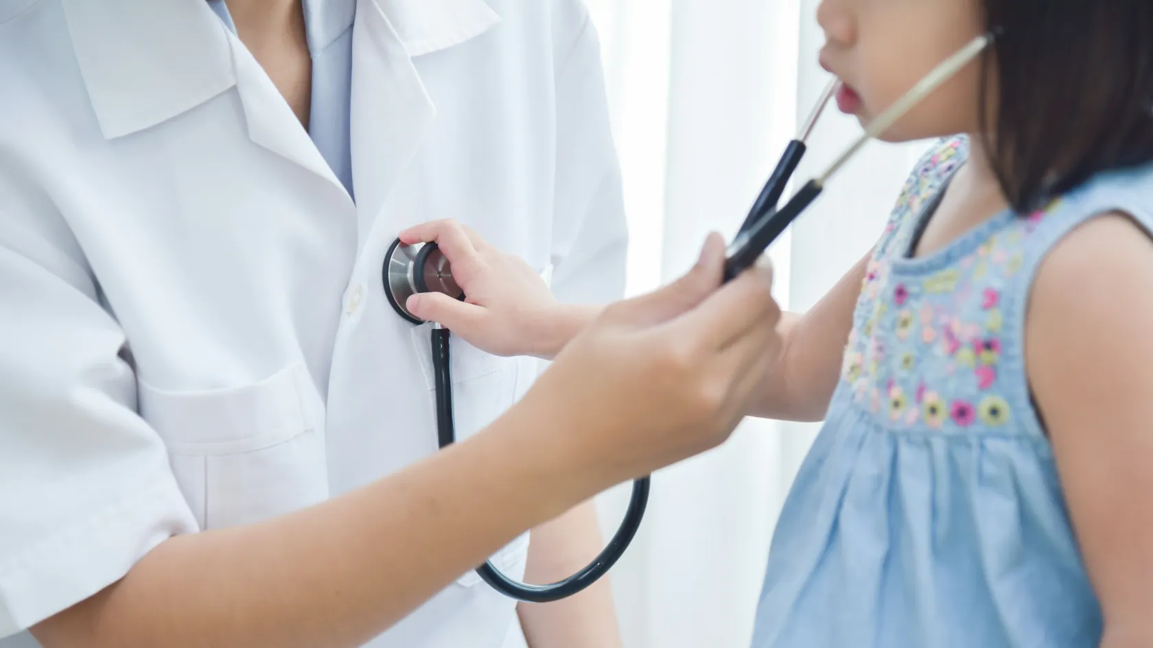
Pediatric Glioma Surgery
Where a glioma sits in a child's brain decides what surgery can do. A cerebellar pilocytic astrocytoma removed completely often needs no further treatment, while a tumor in the optic pathway or the pons calls for a biopsy, drugs or radiotherapy. This page sets out each situation, the operation, the molecular tests that now guide treatment, and how long a family stays in Istanbul.
About This Department
Most childhood gliomas are low grade. Removing all of it is often the cure.
A scan has shown a glioma in your child's brain or spinal cord, and the word that keeps coming up is surgery. Low-grade gliomas make up more than 40 percent of all childhood brain and spinal cord tumors, and in a Children's Oncology Group study of 518 children treated with surgery and then watched, 97 percent were alive five years later. This page explains what pediatric glioma surgery can and cannot achieve depending on where the tumor sits, what the operation and the first weeks involve, why the molecular result now matters as much as the scan, how long a family stays in Istanbul, and what to send us for a free review by a pediatric neurosurgeon.
Location decides the goal
Children with the same diagnosis on the pathology report can need entirely different operations, because a glioma in the cerebellum and a glioma wrapped around the optic nerves are different surgical problems.
Pediatric glioma surgery means removing as much of a glial tumor from the brain or spinal cord as can be taken without causing a new permanent deficit. Neurosurgeons call this maximal safe resection. Where the tumor sits next to tissue a child can do without, such as much of the cerebellum or the front of a cerebral hemisphere, the aim is to take all of it, because complete removal of a low-grade glioma is frequently the only treatment the child ever needs, whereas where it sits inside the brainstem, the thalamus or the hypothalamus, the same aim would cost the child movement, sight, hormones or life, and the operation changes into one that takes a sample, relieves pressure or removes only the part that can safely come out.
This table scrolls sideways on a narrow screen. Swipe or drag to see every column.
| Location | Usual tumor | Surgical goal |
|---|---|---|
| Cerebellum | Pilocytic astrocytoma, often a cyst with a nodule in its wall | Complete removal. Most children need nothing further. |
| Cerebral hemisphere | Low-grade glioma, ganglioglioma, DNET, or a high-grade glioma | Complete removal where speech and movement areas allow, which also controls seizures in many children. |
| Thalamus, midbrain | Low-grade or high-grade glioma | Selected low-grade tumors can be removed by an experienced team. Others are biopsied for diagnosis. |
| Optic pathway, hypothalamus | Low-grade glioma, often with neurofibromatosis type 1 | Rarely complete removal. Surgery drains cysts, relieves pressure or takes tissue, and drug treatment carries the rest. |
| Pons, diffuse | Diffuse midline glioma, H3 K27 altered | No removal. Radiotherapy leads. A biopsy is offered only where it opens a clinical trial or a targeted drug for the child. |
| Spinal cord | Low-grade astrocytoma or ganglioglioma | The largest safe removal under continuous nerve monitoring. |
What the largest study found
Location also predicts how well the child will do.
In the Children's Oncology Group natural history study, published in Neurosurgery in 2011, 518 children with low-grade gliomas were treated with surgery alone and then observed. Five-year overall survival was 97 percent and five-year progression-free survival was 80 percent. In the multivariate analysis complete removal with no visible residual tumor was the strongest single predictor of staying free of progression. Children left with a small residual still did reasonably well, with 56 percent free of progression at five years. Leaving tumor behind meant more regrowth. Nearly every series since has found the same.
Low grade and high grade
Low-grade gliomas, WHO grade 1 and 2, grow slowly, rarely spread and allow a long life even when some tumor stays. Pilocytic astrocytoma leads the group. Surgery cures these tumors more often than any other treatment.
High-grade gliomas, grade 3 and 4, form a smaller and much harder group, and the pediatric versions differ from the adult glioblastoma a family may read about. Surgery still matters for them. A 2022 meta-analysis in JAMA Network Open pooled 1,387 children with high-grade gliomas from 37 studies and found that, compared with complete removal, a partial removal nearly doubled the risk of death, with a hazard ratio of 1.91, and a biopsy alone gave a hazard ratio of 2.10. The benefit of complete removal was largest for tumors in the cerebral hemispheres, where the hazard ratio fell to 0.29, and for tumors below the tentorium, and it was not significant for midline tumors, where complete removal is rarely safe in any case. Where a high-grade tumor can be reached, the operation comes first and matters most, followed by radiotherapy and drug treatment.
Scans do not always show the grade.
On MRI a pilocytic astrocytoma can look very characteristic, and a diffuse pontine glioma has a near-diagnostic appearance, but between those two ends the pathologist and the molecular laboratory make the diagnosis, and the treatment plan after surgery depends on what they find.
Is surgery the first step?
For most children referred to a neurosurgeon with a glioma, yes, and for some the right first step is a biopsy, a drain for raised pressure, or no operation at all.
Optic and hypothalamic gliomas show where surgery's limits lie. In a 2024 two-center German series of 63 children with chiasmatic hypothalamic gliomas followed for nine years on average, 43 had debulking operations and 20 had biopsies, most had two operations over the years, and 69.8 percent also received drug treatment or radiotherapy. Three quarters had stable disease at the end of follow-up, and 30.2 percent needed hormone replacement. Surgery formed one part of a long plan in those children, with drugs and repeated scans doing most of the work.
The day of the operation
Removing a glioma from the brain means a craniotomy, in which a window of skull bone is lifted, the tumor is removed under the operating microscope and the bone is fixed back in place. It takes three to eight hours.
Planning scans the day before
Anesthesia and positioning
Opening and finding the tumor
Removal under monitoring
Closing and waking
Within 48 hours of the operation, an MRI measures what was removed. That early scan, read against the planning scan, decides whether the resection is called gross total, near total or subtotal, and it becomes the baseline every later scan is compared with.
Monitoring that protects function
Continuous nerve monitoring during the operation is standard for any glioma near the movement pathways, the brainstem or the spinal cord. Motor evoked potentials test the pathway from the brain to the muscles by sending a small current and recording the muscle response, sensory evoked potentials do the same in the other direction, and for a brainstem tumor the cranial nerves controlling swallowing, the face and eye movement are mapped as well. A spinal cord series from London reported that with this monitoring 26 of 30 children walked independently at last follow-up, and all 30 were alive. Awake surgery, in which the patient is woken during the operation to speak while the surgeon maps language areas, works well in adults and in some cooperative teenagers. Younger children rarely tolerate it. Fluorescent dyes can help the surgeon see tumor edges. Sodium fluorescein, given into a vein, stained 84 percent of pediatric low-grade gliomas in a 2022 literature review, though the evidence is small case series and the dye is an aid to judgment. An MRI scanner inside the operating room, available in some centers, lets the surgeon check for residual tumor before closing and go back in if something is left. Without one, the early postoperative MRI does the same job a day later, and a second operation follows only if a removable residual shows.
What can go wrong, and how often
America's pediatric National Surgical Quality Improvement Program holds the largest dataset. Between 2012 and 2021 it recorded 9,845 children who had a craniotomy for a tumor. Within 30 days, 9.8 percent were readmitted unplanned, 10.8 percent had a second operation and 0.8 percent died, and the deaths clustered in emergency operations and in children who were already on a ventilator or needed nutritional support before surgery. A single-center series from Alder Hey Children's Hospital in Liverpool graded every complication in 477 operations over ten years. The 30-day mortality was 1.26 percent and none of those deaths was a direct result of a surgical complication. A new neurological deficit was still present at 30 days in 8.39 percent of cases, and 78 percent of returns to the operating room were for the fluid drainage problems described below.
This table scrolls sideways on a narrow screen. Swipe or drag to see every column.
| Risk | What it looks like | How it is handled |
|---|---|---|
| New weakness or speech change | Weakness on one side, unsteady walking, slurred speech, double vision | Swelling around the cavity causes most of it, and steroids and time resolve much of it. Physiotherapy starts on the ward. |
| Fluid buildup | Headache, vomiting, sleepiness, a soft swelling under the wound | A temporary drain, an endoscopic third ventriculostomy or a permanent shunt, depending on the cause. |
| Seizures | A new seizure in the first month, in 7.8 percent of children without previous seizures in a Dutch series of 306 | Sodium levels are watched closely, since a low sodium raises the risk, and seizure medication is started if one occurs. |
| Cerebellar mutism | Loss of speech and irritability a day or two after a cerebellar operation near the midline | Speech therapy and time. It is less common with cerebellar gliomas than with midline tumors such as medulloblastoma. |
| Hormone or vision loss | After surgery near the hypothalamus or optic nerves | Endocrine tests before and after, and hormone replacement where needed. This risk is why these tumors are rarely removed completely. |
| Infection or wound leak | Fever, redness, clear fluid from the wound | Antibiotics, and repair of the wound if fluid is leaking. |
Where the child is treated changes these numbers. An American national readmission study of 4,003 pediatric tumor craniotomies found that children operated on in designated children's hospitals were a third less likely to be readmitted within 30 days, with an odds ratio of 0.68. Experience with small children, from the anesthetic to the intensive care night, is a large part of what a family is choosing.
Why the molecular result matters
Tissue removed at surgery now does a second job. Beyond the microscope diagnosis, it is tested for the genetic changes that drive the tumor, and those changes increasingly decide the next treatment. A change in the BRAF gene drives most pediatric low-grade gliomas. The commonest, a fusion called KIAA1549-BRAF, belongs to cerebellar pilocytic astrocytomas and carries a good outlook, while the other, a point mutation called BRAF V600E, behaves very differently.
BRAF V600E predicts a poor response to the older treatments and a good response to the newer ones. In a Toronto study published in the Journal of Clinical Oncology in 2017, the mutation was found in 17 percent of 405 pediatric low-grade gliomas, and after chemotherapy or radiotherapy the ten-year progression-free survival was 27 percent with the mutation against 60.2 percent without it. Incomplete removal and a second change called CDKN2A deletion made the outlook worse again. For those children the 2023 trial of dabrafenib plus trametinib, two oral drugs that block the BRAF pathway, changed practice. Among 110 children with BRAF V600 low-grade glioma needing first treatment, the tumor shrank in 47 percent of those given the two drugs against 11 percent given standard chemotherapy, median progression-free survival was 20.1 months against 7.4, and serious side effects were half as common. For children with other BRAF changes whose tumor has come back, tovorafenib, a once-weekly oral drug, shrank tumors in 51 to 67 percent of 77 children in the FIREFLY-1 trial depending on the response criteria used.
In high-grade gliomas the molecular result separates very different tumors. A histone H3 K27 alteration marks a diffuse midline glioma, a tumor with a poor outlook and no role for extended surgery. An H3 G34 change, an IDH mutation or a fusion involving NTRK or ALK each points to a different group, and some of those have targeted drugs of their own.
Two to three weeks pass before the molecular report comes back from most laboratories. By then the family is home, and the result follows with a written plan.
If tumor remains, or the grade is high
A low-grade glioma that has been removed completely needs scans and nothing else.
Small residuals get the same watchful approach, because many stay still for years, and treatment starts only if it grows or causes symptoms. Drug treatment comes first for growth in a place where a second operation is unsafe, either chemotherapy with carboplatin and vincristine or, for a BRAF V600E tumor, dabrafenib and trametinib. Radiotherapy waits in young children with low-grade tumors, because of its long-term effects on learning and growth. A high-grade glioma moves on to radiotherapy three to five weeks after surgery, given in daily sessions over six weeks, with chemotherapy alongside in most protocols and with targeted drugs where the molecular result allows. Radiotherapy and chemotherapy are both delivered at Biruni Hospital, so a child whose pathology calls for further treatment continues with the same team, on the same site, without a transfer of records. Families who prefer to have it at home leave with a written handover to their own oncologist containing the operation note, the pathology, the molecular report and both MRIs.
A second operation has two triggers. The early MRI shows a removable residual that the surgeon judged unsafe to chase in the first operation but can reach by a different route, or a low-grade tumor grows back later in a place where it can again come out, which in a slow tumor can be many years after the first operation. Neither means the first operation failed.
The first six weeks
Day one passes in intensive care.
Nurses check the pupils, movement and speech every hour. Steroids keep the swelling down. Most children move to the ward the next day, eat on the second or third day and walk with help by the third or fourth, and the steroid dose comes down over a week or two, during which a child's appetite and mood swing in ways that settle once the course ends. Published series and the pediatric neurosurgery units treating international patients in Turkey report four to seven nights in hospital after an uncomplicated craniotomy, longer after a brainstem or spinal cord operation or if a drain or shunt is needed. Ten days after surgery the wound gets checked. Swelling of the face around the eye on the side of the operation is common in the first days and fades.
Weakness or unsteadiness present after surgery improves fastest in the first six weeks and keeps improving for months, so physiotherapy, and speech or occupational therapy where needed, begins on the ward and continues at home. School resumes four to six weeks after a simple resection.
Contact sports and swimming wait until the neurosurgeon clears them, at around three months.
Weeks in Istanbul and the flight home
Two to three weeks covers most families whose child has a low-grade glioma removed, made up of two or three days for the outpatient assessment and the planning MRI, four to seven nights in hospital, and one more week near the hospital for the wound check and a final review before flying.
Flying after a craniotomy is cleared ten to fourteen days after surgery, once any air left inside the skull has been absorbed and the wound is healed. Airlines differ in their rules, so the discharge letter states the operation, the date and the clearance to fly in writing. A child with a shunt placed during the admission flies on the same timeline.
High-grade glioma changes the timeline. A family who chooses to have radiotherapy here stays ten to twelve weeks in total, because the six weeks of daily sessions start three to five weeks after surgery, and many families go home in that gap between the operation and the first session, with the coordinator planning both trips together so the planning scan, the mask fitting and the first session fall in the same week of the return trip.
How a quote is built
No single figure fits pediatric glioma surgery, because the location of the tumor changes the operation more than any other factor. A cerebellar cyst with a small nodule is a three-hour operation and a short stay. A thalamic tumor approached between the two hemispheres with full nerve monitoring, a planning tractography study and two nights in intensive care is a different admission altogether. A spinal cord tumor adds spinal monitoring and a longer rehabilitation. A drain or a shunt, a second operation for residual tumor, extra intensive care nights and the molecular panel each add to the total, and molecular testing is quoted separately by many hospitals because the range of tests varies by tumor. Radiotherapy and drug treatment, where needed, are separate courses with their own planning.
The child matters too, since infants under a year need more anesthetic and intensive care support, hydrocephalus at presentation sometimes has to be treated before the tumor operation, and neurofibromatosis type 1 adds eye and skin assessments.
Packages published by Turkish private hospitals and medical travel agencies for brain tumor surgery include the neurosurgeon and anesthesia fees, the operating room, intraoperative monitoring, the planning and postoperative MRIs, a set number of intensive care and ward nights, the pathology report, an interpreter, airport transfers and hotel nights for the family. They leave out flights, travel insurance, extra hospital nights, a second operation, shunt hardware unless listed, molecular testing unless listed and any later radiotherapy or chemotherapy.
Four questions separate a real quote from a headline. Which approach and which monitoring does the quote assume, and are the MRI within 48 hours and the frozen section inside it. Is the molecular panel inside it, and which genes does it cover. What happens financially if a shunt is needed.
Real figures come only after a pediatric neurosurgeon has read your child's own MRI, and that review is free.
The family during the admission
One parent stays. Every patient room has a companion bed, and that parent sleeps beside the child through the whole admission. Siblings and the other parent stay in accommodation the international patients office arranges for the nights either side, and the same office handles the airport transfer and the daily transport between hotel and hospital. From the first message a coordinator stays with your family through discharge. The team works in English, Arabic, French, Russian, Serbian, Romanian and Spanish, and interpreting in other languages is arranged on request, so the consent conversation before surgery, the morning ward rounds and the pathology result are all explained in your language. A request for a female physician goes to the department and is met wherever the rota allows, so say so in the first message. Halal, vegetarian and diabetic meals come from the hospital kitchen, and a prayer room is on site. Ten days before travel, an appointment confirmation and an invitation letter naming the hospital and the treating neurosurgeon go out. Most consulates ask for it.
Bring the MRI discs from diagnosis and any later scans, the radiology reports, any previous pathology or biopsy slides, the list of current medications including seizure medication and the dose of any steroid, and the vaccination record.
Scans and school after you go home
You leave with the operation note, the pathology report, the early postoperative MRI on disc with its report, a medication plan including the steroid taper, and a letter to your child's doctor at home. The coordinator sends the molecular report and the plan that goes with it when it arrives.
Once you are back home, follow-up means MRI. For a low-grade glioma the schedule in most protocols is a scan at three months, then every three to six months for the first few years, then yearly, with the interval set by the extent of removal and the tumor type. Recurrence of a low-grade glioma can come late.
Five or ten years on is not unusual, so the scans continue into the teenage years. High-grade gliomas need a scan every two to three months during and after treatment. Each new scan can be sent by WhatsApp, and the neurosurgeon who operated compares it with the baseline at no charge.
School and development deserve their own attention.
Children who had surgery for a cerebellar tumor, a hemispheric tumor near language areas or a tumor near the hypothalamus benefit from a neuropsychology assessment at around a year, and children with tumors near the pituitary need endocrine checks of growth and puberty as they grow.
Call the coordinator the same day, and go to your local emergency department, for a severe or worsening headache, repeated vomiting, unusual sleepiness, a new seizure, new weakness, a fever above 38 degrees, or clear fluid or swelling at the wound. The neurosurgeon here speaks to the doctor treating your child with the operation note and the scans in front of them.
Pediatric glioma surgery FAQ
What is the survival rate for a child with a glioma?
Can a low-grade glioma be cured with surgery alone?
How long will we stay in Turkey for the operation?
Will my child need chemotherapy or radiotherapy after surgery?
Can one parent stay with the child in hospital?
What should we send for a free review?
References
- Wisoff JH, Sanford RA, Heier LA, et al. Primary neurosurgery for pediatric low-grade gliomas: a prospective multi-institutional study from the Children's Oncology Group. Neurosurgery. 2011;68(6):1548-1554.
- Hatoum R, Chen JS, Lavergne P, et al. Extent of tumor resection and survival in pediatric patients with high-grade gliomas: a systematic review and meta-analysis. JAMA Netw Open. 2022;5(8):e2226551.
- Lassaletta A, Zapotocky M, Mistry M, et al. Therapeutic and prognostic implications of BRAF V600E in pediatric low-grade gliomas. J Clin Oncol. 2017;35(25):2934-2941.
- Bouffet E, Hansford JR, Garre ML, et al. Dabrafenib plus trametinib in pediatric glioma with BRAF V600 mutations. N Engl J Med. 2023;389(12):1108-1120.
- Kilburn LB, Khuong-Quang DA, Hansford JR, et al. The type II RAF inhibitor tovorafenib in relapsed/refractory pediatric low-grade glioma. The phase 2 FIREFLY-1 trial. Nat Med. 2024;30(1):207-217.
- Stanton AN, DeYoung C, Still MEH, et al. Unplanned readmission after cranial tumor surgery in the pediatric National Surgical Quality Improvement Program. J Neurosurg Pediatr. 2025;36(4):439-443.
- Foster MT, Hennigan D, Grayston R, et al. Reporting morbidity associated with pediatric brain tumor surgery. Are the available scoring systems sufficient? J Neurosurg Pediatr. 2021;27(5):556-565.
Editor's note
Written by the Biruni Hospital medical editorial team. Reviewed by Assistant Professor Kamil Değer DEVECİOĞLU, Pediatric Surgery.
Medically reviewed by
Assistant Professor Kamil Değer DEVECİOĞLU
Pediatric Surgery
Related Treatments
View All-
Appendectomy For Children - Pediatric Appendix Surgery
Appendicitis is the commonest reason for emergency abdominal surgery in children, and a child with the warning signs needs an emergency department the same day. This guide covers the signs to watch for, diagnosis with ultrasound, keyhole surgery against antibiotics alone, perforated appendicitis, recovery and what families in Istanbul need to know.
-
Biliary Atresia Surgery
Biliary atresia blocks the bile ducts of a newborn, and the Kasai operation restores bile flow in about half of babies, best when done in the first weeks of life. This guide covers the warning signs of pale stools and lasting jaundice, diagnosis, the Kasai, results in expert centers, liver transplant, stool color screening and the stay.
-
Circumcision Surgery
Circumcision removes the foreskin for religious, cultural or medical reasons, in babies, boys and adults. With a trained surgeon and proper pain relief, complications are rare and lowest in the first year of life. This guide covers the reasons, the best age, pain relief, techniques, health effects from the main studies, healing and the short stay in Istanbul.
-
Ewing Sarcoma Surgery
Unlike most bone cancers, Ewing sarcoma responds to radiotherapy, yet removing the tumor still cuts the chance of it returning at the same site by more than half. This guide explains how the team chooses between surgery and radiotherapy, how the limb or pelvis is rebuilt, what the pathology decides and how follow-up continues at home.
-
Hepatoblastoma Surgery
A child can lose three quarters of the liver to remove a hepatoblastoma, and the remainder regrows within weeks. This page walks through the operation, when a transplant replaces it, how many weeks a family spends in Istanbul and what the follow-up at home involves.